
 Pentoxifylline mitigates CCl4-induced liver damage in rats: integrated biochemical, hematological, histopathological, and correlation heatmap analysis
Abstract
Pentoxifylline (PTX), a methylxanthine-derived phosphodiesterase inhibitor, has demonstrated anti-inflammatory and hepatoprotective potential. This study evaluated the effects of PTX on carbon tetrachloride (CCl4)-induced liver injury in rats, with clear differentiation between treatment conditions. Sixty Wistar rats were allocated into four groups: control, CCl4-only, PTX-only, and CCl4 followed by PTX (post-treatment model). CCl4 (1 mL/kg, i.p.) induced hepatic injury, while PTX was administered orally (50 mg/kg/day) for seven consecutive days either alone or after CCl4 exposure. CCl4 administration resulted in significant hepatotoxicity, evidenced by elevated serum ALT, AST, and LDH levels (p < 0.001), along with marked reductions in hepatic antioxidant markers (GSH, GPX, and CAT; p < 0.001), and alterations in organ weights and hematological parameters. PTX administered after CCl4 significantly ameliorated liver injury, as demonstrated by reductions in ALT (66.3 U/L, p < 0.001) and AST (184.7 U/L, p < 0.001), and restoration of antioxidant defenses, including GSH (6.70 µmol/g, p < 0.001). In contrast, PTX administered alone did not enhance antioxidant status and was associated with reductions in GSH, GPX, and CAT compared to control (p < 0.05), indicating that its protective effects are context-dependent. Correlation heatmap analysis revealed strong associations between liver enzymes and oxidative stress markers, supporting the mechanistic link between hepatocellular damage and redox imbalance. Hematological alterations induced by CCl4, including monocytosis and neutropenia, were partially normalized following PTX post-treatment. Histopathological findings corroborated the biochemical results, showing improved hepatocyte architecture and reduced inflammatory infiltration in PTX-treated rats. These findings suggest that PTX exerts significant hepatoprotective effects when administered after toxic insult, likely through modulation of oxidative stress and inflammation. Further studies are warranted to optimize dosing strategies and clarify its therapeutic window.
Keywords:
pentoxifylline; carbon tetrachloride; oxidative stress; hepatoprotection; rats
Resumo
A pentoxifilina (PTX), um inibidor não seletivo da fosfodiesterase derivado da metilxantina, apresenta propriedades anti-inflamatórias e hepatoprotetoras. O presente estudo avaliou os efeitos da PTX na lesão hepática induzida por tetracloreto de carbono (CCl4) em ratos, com diferenciação clara entre as condições de tratamento. Sessenta ratos Winstar foram alocados em quatro grupos: controle, CCl4, PTX isolado e CCl4 seguido de PTX (modelo de pós-tratamento). A hepatotoxicidade foi induzida por administração intraperitoneal de CCl4 (1 mL/kg), enquanto a PTX foi administrada por via oral na dose de 50 mg/kg/dia durante sete dias, isoladamente ou após a exposição ao CCl4. A exposição ao CCl4 promoveu alterações hepáticas significativas, caracterizadas pelo aumento das atividades séricas de ALT, AST e LDH (p < 0,001), redução dos marcadores antioxidantes hepáticos (GSH, GPX e catalase; p < 0,001), além de alterações no peso dos órgãos e no perfil hematológico. A administração de PTX após o CCl4 atenuou significativamente a lesão hepática, evidenciada pela redução de ALT (66,3 U/L; p < 0,001) e AST (184,7 U/L; p < 0,001), e pela restauração das defesas antioxidantes, incluindo GSH (6,70 µmol/g; p < 0,001). Em contraste, a PTX isolada não melhorou o status antioxidante, estando associada à redução de GSH, GPX e catalase em comparação ao controle (p < 0,05), indicando que seus efeitos são dependentes do contexto experimental. A análise da matriz de correlação (heatmap) revelou fortes associações entre enzimas hepáticas e marcadores de estresse oxidativo, corroborando a relação entre dano hepatocelular e desequilíbrio redox. Alterações hematológicas induzidas pelo CCl4, incluindo monocitose e neutropenia, foram parcialmente normalizadas após o tratamento com PTX. A análise histopatológica confirmou a melhora da arquitetura hepática e a redução da inflamação nos grupos tratados com PTX. Em conjunto, os resultados indicam que a PTX exerce efeitos hepatoprotetores significativos quando administrada após a agressão tóxica, provavelmente por meio da modulação do estresse oxidativo e da inflamação. Estudos adicionais são necessários para otimizar estratégias de dose e esclarecer sua janela terapêutica.
Palavras-chave:
pentoxifilina; tetracloreto de carbono; estresse oxidativo; hepatoproteção; ratos
1. Introduction
Degenerative diseases have had a marked increase in incidence during the last decade. They occur due to aging-related molecular and cellular alterations, such as mitochondrial dysfunction, oxidative stress, chronic low-grade inflammation, and a senescence-associated secretory phenotype that disrupts tissue homeostasis and drives fibrotic remodeling across multiple organs (Miwa et al., 2022). These mechanisms support the growing frequency of nonalcoholic fatty liver disease (NAFLD), which rose from 25.3% in 1990-2006 to 38.2% in 2016-2019 in the liver (Manikat et al., 2023). Also, numerous causes of liver damage finally compromise the critical metabolic and detoxifying activities of the liver, including viral infections, alcohol addiction, non-alcoholic fatty liver disease, chemical exposure, and autoimmune diseases. Nowadays, liver injury caused by drugs is a major concern that has become the focus of researchers, medical professionals, and pharmaceutical companies (Weber and Gerbes, 2022). Similarly, chronic kidney disease (CKD) incidence climbed from 192.45 to 233.65 cases per 100,000 population between 1990 and 2019 (average annual percent change 1.82%) (Qin et al., 2024). Heart failure with preserved ejection fraction (HFpEF) experienced a >50% greater incidence in 2000-2009 than in 1990-1999 in the cardiovascular system, and HFpEF hospitalizations more than doubled from 189,260 in 2008 to 495,095 in 2018 (Borlaug et al., 2023).
Although less often discussed, the spleen exhibits notable age-associated alterations, including expansion of the white pulp, stromal fibrosis, and an endothelial-to-immune-like cell Transition that results in immunosenescence and compromised phagocytic function (Huang et al., 2024). Reactive oxygen species (ROS) play a significant role in the progression of degenerative diseases, including various liver disorders (Ding and Zhuo, 2013). The excessive generation of reactive oxygen species (ROS), leading to oxidative stress, can be triggered by multiple factors, such as exposure to xenobiotics like acetaminophen and carbon tetrachloride (CCl4) (Guo et al., 2013).
In the area of experimental models for liver injury, the CCl4-induced model stands out as the most thoroughly examined in terms of the changes it brings about at the histological, biochemical, cellular, and molecular levels as fibrosis advances. Frequent exposure to CCl4 can result in liver fibrosis and potentially cirrhosis, influenced by the amount and length of exposure (Marques et al., 2012). Recent findings suggest that hepatic fibrosis, and in some cases cirrhosis, may be reversible (Li et al., 2011).
Antioxidants are significant in protecting the liver from damage. A variety of natural and synthetic agents have shown protective benefits against liver toxicity (Ghate et al., 2019; Lyons and Brennan, 2017). Pentoxifylline (PTX), a non-selective phosphodiesterase inhibitor, acts as a vasodilator, improving peripheral and hepatic blood circulation (Bektas et al., 2016). In addition to its anti-inflammatory and immune-modulatory properties, PTX enhances liver perfusion, inhibits fibroblast proliferation, promotes interstitial collagenase activity, and reduces the accumulation of fibrillar collagens (types I and III), proteoglycans, and fibronectin (Li et al., 2011). By controlling transforming growth factor-beta and tissue inhibitor of metalloproteinase-1, PTX has been shown to guard against bile duct ligation-induced hepatic fibrosis (Babaei and Bayat, 2015). Beyond its effects on blood cell rheology, PTX also exerts anti-inflammatory actions by inhibiting cytokine production. It inhibits the production of tumor necrosis factor-alpha (TNF-α) in monocytes and T-cells triggered by lipopolysaccharide and stops leukocyte adhesion induced by interleukin-2 (IL-2) (Palafox Mariscal et al., 2023). Moreover, evidence indicates PTX’s capacity to rebalance antioxidants and reduce inflammation (Zaitone et al., 2011; Speer et al., 2017; Mostafa-Hedeab et al., 2022).
Self-nano-emulsifying drug delivery systems are well-documented for enhancing drug bioavailability by forming nano-sized globules upon contact with biological fluids following oral administration (Shailendrakumar et al., 2020).
Incorporating polyethylene glycol onto the surface of lipid nanoparticles increases their hydrophilicity while reducing recognition by the mononuclear phagocyte system, also known as the reticuloendothelial system. This modification decreases drug clearance from circulation, ultimately prolonging its half-life (Santos, 2015). Lipid nanoparticles, typically ranging from 100 to 200 nm in size, facilitate the crossing of barriers owing to the enhanced permeation and retention effect. These nanoparticles may target areas of inflammation and decrease macrophage activity in phagocytosis (Wang et al., 2014). Furthermore, lipid nanoparticles show a gradual dissociation rate, which enhances drug retention, increases accumulation at the target site, and facilitates extended drug action (Dhiman et al., 2016).
The study aims to investigate the hepatoprotective effects of pentoxifylline (PTX) against carbon tetrachloride (CCl4)-induced liver injury in rats, focusing on biochemical markers, antioxidant status, hematological parameters, and histopathological changes. Additionally, correlation analyses are integrated to explore relationships between liver enzymes, oxidative stress markers, and hematological alterations.
2. Materials and Methods
2.1. Animal model and experimental design
The present study included 60 adult Wistar albino male rats, each weighing 300 ± 60 g (range: 200-400 g). They were maintained in standard laboratory settings, featuring a 12-hour light-dark cycle, a regulated temperature of 22 ± 2 °C, and unrestricted access to food and water. The animals were randomly assigned to control and treatment groups to guarantee unbiased distribution. Animals were anesthetized using ketamine (80 mg/kg, intraperitoneally) combined with xylazine (10 mg/kg, intraperitoneally) to achieve deep anesthesia. Euthanasia was performed by exsanguination via cardiac puncture while under deep anesthesia, followed immediately by cervical dislocation to ensure death, in accordance with the AVMA Guidelines for the Euthanasia of Animals (AVMA, 2020).
2.2. Hepatic injury induction and treatment
An intraperitoneal injection of CCl4 was used to induce hepatic injury, simulating oxidative stress and liver fibrosis. The treatment groups received PTX (50 mg/kg/day) orally, either in its conventional or nanoparticle formulation, in conjunction with CCl4 for a chosen duration.
2.3. Sample collection and tissue preparation
After the experimental phase, the animals were euthanized under anesthesia, and liver tissue, together with blood samples, were collected for histological and biochemical analyses. Blood samples were taken using a heart puncture, then spun at 3000 rpm for ten minutes at 4 °C to let serum separate for biochemical testing. To assess histological changes, liver tissues were fixed in 10% formalin, paraffin-embedded, cut for hematoxylin and eosin (H&E), and Masson's trichrome staining.
2.4. Statistical analysis
The data were collected, checked, revised, and organized in tables and figures using Microsoft Excel 2016. The data were subjected to outlier detection and normality statistical tests to determine whether the data are parametric or non-parametric. Data were analyzed for descriptive statistics, both graphical and numerical descriptions. Inferential statistics for evaluating and comparing treatments were performed using one-way analysis of variance (ANOVA) or corresponding nonparametric tests at the 0.05 significance level. Tukey's HSD was used to follow up on ANOVA comparisons between treatment groups. Data analyses were carried out using the computer software Statistical Package for Social Science SPSS (IBM-SPSS ver. 26.0 for Mac OS).
2.5. Ethical approval
All experimental procedures adhered to institutional and national guidelines for the ethical treatment of animals and received approval from the relevant Animal Ethics Committee. The study was approved by the Imam Abdulrahman Bin Faisal University IRB after it satisfied the standards set by the Institutional Animal Care and Use Committee (IACUC) and the Institutional Ethics Committee (IEC), bearing No. IRB-A-2025-10-0202).
3. Results
The experimental results demonstrate significant alterations in hepatic function markers, antioxidant status, organ weights, and hematological parameters across the four treatment groups (p < 0.001 for most parameters). Carbon tetrachloride (CCl4) administration induced characteristic hepatotoxic effects, as evidenced by elevated liver enzymes ALT (123.7±1.89 U/L) and AST (261.3±2.53 U/L) compared to control values (82.5±1.71 and 233.4±3.03 U/L, respectively), confirming successful induction of liver injury. Interestingly, CCl4 treatment resulted in decreased alkaline phosphatase (ALP) levels (323.7±5.56 U/L) compared to controls (413.8±6.62 U/L), while lactate dehydrogenase (LDH) showed marked elevation (5,114.8±72.39 U/L versus 4,320.5±71.33 U/L), indicating cellular damage and membrane integrity compromise (Figure 1).

Bar chart presenting (a) Alt, (b) AST, (c) ALP, (d) LDH, and (e) serum albumin of experimental rats treated with CCl4, PTX, and both CCL4 and PTX.
The pentoxifylline (PTX) treatment group exhibited distinct biochemical profiles, with substantially reduced ALT (52.0±1.03 U/L) and ALP (151.9±2.62 c) levels compared to controls, suggesting potential hepatoprotective or metabolic modulatory effects. However, PTX administration alone resulted in compromised antioxidant status, as demonstrated by reduced glutathione (GSH) levels (5.0±0.16 µmol/g tissue) compared to controls (8.5±0.40 µmol/g tissue), and decreased activities of antioxidant enzymes glutathione peroxidase (GPX) and catalase. The combination therapy (CCl4+PTX) demonstrated partial amelioration of CCl4-induced hepatotoxicity, with intermediate ALT (66.3±1.15 U/L) and significantly reduced AST (184.7±4.43 U/L) levels compared to CCl4 alone, suggesting protective effects of PTX against CCl4-induced liver damage (Figure 2).

Bar chart presenting (a) GSH, (b) GPX, and (c) Catalase of experimental rats treated with CCl4, PTX and both CCL4 and PTX.
Organ weight analysis revealed significant treatment-related changes, with CCl4 administration causing liver weight reduction (3.4±0.19 g versus 4.3±0.12 g in controls), while the combination treatment resulted in increased organ weights across liver (5.0±0.36 g), heart (0.8±0.06 g), spleen (1.2±0.08 g), and kidney (1.5±0.10 g), possibly indicating compensatory responses or treatment-related hypertrophy (Figure 3).

Bar chart presenting organs weight of (a) liver, (b) heart, (c) Spleen, and (d) Kidney, of experimental rats treated with CCl4, PTX and both CCL4 and PTX.
Hematological parameters showed significant alterations in leukocyte differential counts, with PTX treatment causing monocyte elevation (4.0±0.22%) and neutrophil increase (31.9±0.41%), while the combination treatment resulted in the highest lymphocyte percentage (72.5±0.35%). These findings suggest complex interactions between CCl4-induced toxicity and PTX-mediated protective mechanisms, with the combination therapy demonstrating both beneficial hepatoprotective effects and some concerning systemic alterations that warrant further investigation into optimal dosing and treatment duration strategies (Figure 4).

Bar chart presenting different blood parameters (a) monocytes, (b) Neutrophils, (c) Lymphocytes, (d) hemoglobin, and (e) packed cell volume of experimental rats treated with CCl4, PTX, and both CCL4 and PTX.
3.1. Histopathological evaluation
3.1.1. CCl4-treated group
Liver sections from the CCl4-treated group showed profound histopathological changes, underscoring extensive hepatic damage. The central vein (CV) was significantly enlarged and encircled by degenerated hepatocytes (H) exhibiting pyknotic nuclei. Furthermore, the blood sinusoids (S) exhibited significant dilation, indicating congestion and potential vascular injury (Figure 5).

A photomicrograph of the liver of the CCl4-treated group showing an enlarged central vein surrounded by degenerated hepatocytes (H) and dilated blood sinusoids (S). H&E stain X400.
3.1.2. PTX-treated group
Conversely, liver sections from the PTX-treated group exhibited a maintained hepatic architecture. CV was seen to be healthy, and the hepatocytes (H) next to it had intact nuclei (arrows) and well-defined cytoplasm. The observation of normal blood sinusoids (S) indicates a potential protective role of PTX in mitigating hepatic damage, providing hope for potential therapeutic applications (Figure 6).

Photomicrograph of the liver of the PTX-treated group showing the central vein surrounded by normal hepatocytes (H) with normal nuclei (arrows) and between them blood sinusoid (S). H&E X 400.
3.1.3. CCl4 then PTX-treated group
Liver slices from the CCl4 then PTX-treated group revealed signs of partial hepatic recovery. Although CV was substantially engorged with blood (B), hepatocytes showed almost normal cytoplasm and nuclei; some showed visible nucleoli. The general tissue architecture seemed better, but blood sinusoids (S) stayed swollen (Figure 7a). Furthermore, a portal vessel (P) encircled by inflammatory cells (I), with almost normal hepatocytes and blood sinusoids (S), suggested ongoing tissue healing (Figure 7b).
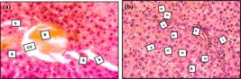
(a) Photomicrograph of liver of group treated with CCL4 then PTX showing highly dilated central vein, which is highly engorged with blood (B) surrounded by hepatocytes (H) with nearly normal cytoplasm and nuclei, some of them with clear nucleoli. Note the enlarged blood sinusoids (S); (b) Photomicrograph of liver of group CCl4 then PTX showing the portal vessel (P) surrounded by inflammatory cells (I). Nearly normal hepatocytes and blood sinusoids (S). H&E X 400.
3.1.4. PTX then CCl4-treated group
Nuclear anomalies in liver sections taken from the PTX and then CCl4-treated group suggested cellular stress. The hepatocytes had black nuclei (arrows) devoid of distinct nucleoli, and several of their nuclei seemed pyknotic, indicative of apoptotic changes (Figure 8a). Significant liver damage was shown by the group treated with PTX and CCl4. The major vein showed marked dilation with notable lymphocytic infiltration and blood engorgement. Further proof of hepatic damage came from hepatocytes showing dark nuclei (arrows) marked by noticeable nuclear abnormalities; blood sinusoids (S) were also greatly dilated (Figure 8b).

(a) Photomicrograph of the liver in treated group PTX then CCl4, showing dark nuclei (arrows) with no clear nucleoli and some nuclei are pyknotic (arrowhead); (b) Photomicrograph of liver of group treated with PTX + CCL4 showing enlarged central vein engorged with blood and surrounded by apparent lymphocytic infiltration (L). Note the dark nuclei of hepatocytes (arrows) and the highly dilated blood sinusoids (S). H&E X 400.
The correlation heatmap reveals intricate interdependencies among the measured parameters, providing insights into the systemic physiological responses to the experimental treatments. The analysis demonstrates several significant positive correlations (indicated by blue coloration with black borders), most notably among liver function enzymes ALT, AST, and LDH, suggesting coordinated responses during hepatocellular injury. Strong positive correlations are also evident between organ weights, particularly among heart, spleen, and kidney weights, indicating potential systemic growth or compensatory responses across multiple organ systems during treatment interventions (Figure 9).

Correlation heatmap matrix displaying Pearson correlation coefficients between all measured biochemical, physiological, and hematological parameters across treatment groups. Blue circles indicate positive correlations, red circles indicate negative correlations, and white areas represent no significant correlation. Black-bordered circles denote statistically significant correlations (p<0.05). Correlation strength is represented by color intensity, with darker shades indicating stronger correlations (correlation coefficients ranging from -1 to +1 as shown in the scale bar). Parameters include liver function markers (ALT, AST, ALP, LDH), serum albumin, antioxidant enzymes (GSH, GPX, catalase), organ weights (liver, heart, spleen, kidney), and complete blood count differentials (monocyte, neutrophil, lymphocyte percentages, hemoglobin, PCV).
Significant negative correlations (red coloration with black borders) are prominently observed between antioxidant enzymes (GSH, GPX, and catalase) and liver damage markers, confirming the inverse relationship between antioxidant capacity and hepatocellular injury. Additionally, strong negative correlations exist between ALP levels and several other parameters, reflecting its distinct regulatory pattern compared to other liver enzymes. The hematological parameters display complex correlation patterns, with lymphocyte percentages showing significant negative correlations with neutrophil counts, consistent with expected leukocyte differential relationships. Notably, the correlation matrix reveals that organ weights demonstrate significant positive intercorrelations, suggesting coordinated systemic responses rather than isolated organ-specific effects. The absence of correlation (white areas) between certain parameters, such as PCV with most biochemical markers, indicates independent regulatory mechanisms for these physiological processes, highlighting the multifactorial nature of the experimental responses observed across the treatment groups (Figure 9).
4. Discussion
This study examined the biochemical and histopathological effects of PTX and its nanoparticle formulations on the liver, kidney, and spleen in rat models with CCl4-induced degenerative disease. The hepatotoxic effects of CCl4 arise from the generation of free radicals through its metabolism by cytochrome P450, specifically through the production of trichloromethyl radicals, leading to membrane disruption and lipid peroxidation (Unsal et al., 2020).
Among PTX's other preventive actions is inhibiting the generation of pro-inflammatory cytokines, especially TNF-α, which is essential for liver damage and fibrosis. Its preventive actions include its capacity to lower NF-kappa B activation and block TNF-alpha expression. Additionally, as an antioxidant, PTX reduces malondialdehyde (MDA) levels and increases superoxide dismutase and glutathione (GSH) levels. It also inhibits phosphodiesterase, thereby raising intracellular cyclic monophosphate (cAMP) levels and modulating the inflammatory response. PTX helps fight fibrosis by reducing the expression of procollagen I and profibrogenic cytokines, which is aided by the methyl xanthine derivative. PTX also enhances hepatic microcirculation, supporting recovery (Aboelez et al., 2024; Das and Vasudevan, 2007).
The isolated ALT elevation in the PTX-alone group suggests that, in the absence of concurrent liver injury, prolonged PTX exposure may exert mild hepatocyte stress in certain species, potentially through mechanisms unrelated to its anti-fibrotic or anti-inflammatory pathways (van Wagner et al., 2006; Du et al., 2014). However, the partial restoration of ALP in the CCl4-PTX group suggests that PTX’s efficacy is attenuated after established injury, possibly due to irreversible fibrotic changes or persistent oxidative stress overwhelming its antifibrotic pathways (Vial et al., 2006; Mohamed et al., 2014).
There were no significant differences in serum albumin levels across the experimental groups. Numerous studies stated similar findings (El-Boshy, 2015; Hassanein et al., 2014).
According to biochemical parameters, our study findings revealed a significant increase in ALT and AST levels following CCl4 exposure, suggesting substantial liver cell damage. This increase is attributed to CCl4-induced damage to liver cell membranes, specifically through the disruption of phospholipids and proteins, leading to the leakage of enzymes into the bloodstream.
ALT and AST levels significantly decreased with PTX. Also, ALP activity was significantly elevated in the CCl4 group compared to the control group. However, PTX administration alone significantly decreased ALP activity, and the CCl4 then PTX treatment showed partial restoration. These results align with a study by Hendawy (2017), which also observed similar outcomes in their Experimental Autoimmune Hepatitis group; serum ALT and AST levels increased significantly compared to controls. However, compared to the other group, PTX treatment at 100 mg/kg/d and 200 mg/kg/d markedly reduced these enzyme levels (Hendawy, 2017). Hassanein et al. (2014) and Hamid (unpublished data) found a significant reduction in ALT and AST in the PTX+CCL4-treated group. Hamid reported that the combined treatment group showed a significant decrease in ALP levels.
El-Boshy (2015) found elevated ALT and AST serum levels, with a decrease in ALP levels at the 8th week post-treatment with PTX in guinea pig models. Also, a previous study suggested that PTX alone might cause a slight elevation in liver enzymes; these differences may arise from various factors in the experimental model, such as the treatment duration, animal strain, method of PTX administration, subject age, health status, diet, or environmental conditions during the study (Jiménez-Luévano et al., 2024).
Regarding antioxidant biomarkers, PTX significantly reduced GSH, GPX, and CAT. At the same time, co-administered PTX-CCL4 revealed a partial restoration of GSH levels, decreased GPX, and a modest increase in CAT levels. Luo et al. (2015) investigated PTX’s effects on thioacetamide-induced acute liver injury; PTX significantly increased GSH and superoxide dismutase levels while reducing MDA. Similarly, Vircheva et al. (2010) examined carrageenan-induced inflammation and found that PTX protected against GSH depletion but had no significant effect on antioxidant enzymes, such as GPX and SOD. Surprisingly, small elevations in CAT levels seen in the co-administered group could be a response to residual oxidative stress, which is adaptive. This is in line with the results of studies on non-alcoholic fatty liver disease. PTX highlighted its selective control of oxidative paths by lowering oxidative stress indicators but not uniformly increasing all antioxidant enzymes (Massart et al., 2012; Acedo et al., 2015).
Not only does the liver degenerate in certain conditions, but many organs, such as the heart, kidney, and spleen, also have degenerative disorders. While studying organ weight, we found that CCl4 exposure caused a significant increase in spleen, kidneys, and heart weights, while there was a slight but non-significant reduction in liver weight. Rats given both CCl4 and PTX showed remarkably higher liver, heart, and kidney weight; PTX only lowered heart weight with little or no effect on other organs. Previous investigations have shown contradicting findings about changes in liver weight after CCl4 exposure. For instance, a study on acute CCl4 poisoning showed temporary increases in liver weight resulting from fatty degeneration and necrosis during the early phases of exposure, followed by decreases as fibrosis advances and liver function declines (Uemitsu and Nakayoshi, 1984). Furthermore, observed by Fortea et al. (2018) were portal hypertension, ascites, and a notable rise in the spleen-to-body weight ratio in rats at increasing dosages of CCl4. However, they found liver atrophy in CCl4 models. Moreover, CCl4‑induced cirrhosis is known to provoke cardiac hypertrophy and heavier heart weights, reflecting both direct oxidative injury and compensatory remodeling in response to systemic inflammation and altered hemodynamics (Chang et al., 2014). Raetsch et al. (2002) maintained that by downregulating profibrogenic cytokines and blocking stellate cell activation, PTX has been proven to prevent fibrosis in CCl4‑injured rats, conserving hepatic architecture and mass. Lin et al. (2002) found that PTX attenuates the progression of chronic renal disease, reducing interstitial inflammation, fibrosis, and functional decline in subtotal nephrectomy models, suggesting a capacity to stabilize the renal parenchymal mass under injurious conditions. Furthermore, Sridharan et al. (2013) found that PTX significantly reduced heart weight in our rats, indicating an anti-hypertrophic effect.
Finally, regarding Hematological and Immune Cell Analysis, we observed a significant increase in monocyte percentage in the PTX-treated and CCl4-treated rats compared to the control. Also, we found that neutrophil counts significantly decreased in CCl4-treated rats compared to the control. Moreover, lymphocyte counts increased dramatically in the CCL4 than in the PTX group compared to the control group. Moreover, we noted no significant changes in hemoglobin concentration or packed cell volume were observed between the experimental groups. This aligns with previous studies, indicating that CCl4-induced hepatotoxicity involves the activation of Kupffer cells and the production of pro-inflammatory mediators. However, no significant change in neutrophils was noted previously, which warrants further investigation (Luckey and Petersen, 2001; Sato et al., 2014; Sipes et al., 1991). Although the evidence mainly emphasizes PTX's anti-inflammatory properties, the rise in monocytes in the PTX-treated group alone is intriguing and could indicate a baseline immunomodulatory effect of PTX. Consistent with its prevention of T-cell hyperactivation and cytokine production (e.g., IL-6, TNF-α), Chae et al. (2012) showed that PTX alone lowered lymphocyte numbers relative to CCl4 (Luckey and Petersen, 2001). Histologically speaking, the CCl4-Treated group showed notable alterations suggestive of serious liver damage. These results fit earlier studies. CCl4 produces free radicals that cause lipid peroxidation, hepatocellular necrosis, and activation of Kupffer cells, which release pro-inflammatory cytokines, including TNF-α and IL-6, hence generating fibrosis of the tissues. Dong et al. (2016) and Luo et al. (2015) observed.
In the PTX-Treated Group, we found a preserved hepatic architecture with a normal central vein, hepatocytes exhibiting intact nuclei and well-defined cytoplasm, and normal blood sinusoids. This suggests a protective effect of PTX against hepatic damage when administered alone. Previous studies have supported these findings, stating that PTX attenuates hepatic steatosis, inflammatory cell infiltration, and apoptosis in models of induced liver injury (Luo et al., 2015; Chae et al., 2012). Notably, while PTX alone preserved hepatic structure in our study, its efficacy in fibrosis prevention remains context-dependent. For instance, as mentioned in Vial et al. (2006) PTX failed to inhibit early fibrogenic events in non-alcoholic steatohepatitis models.
This study has some limitations. The relatively short experimental duration may not fully reflect the long-term effects of pentoxifylline (PTX) on chronic liver injury and fibrosis progression. In addition, molecular and gene expression analyses (e.g., pro-inflammatory cytokines and signaling pathways) were not assessed, limiting the mechanistic interpretation of the observed effects. Only a single PTX dose and treatment regimen were evaluated, precluding conclusions on optimal dosing strategies and therapeutic windows. Furthermore, the use of a single hepatotoxicity model may limit the generalizability of the findings to other liver injury models and clinical settings. Finally, the inclusion of only male rats does not account for potential sex-related differences. Future studies incorporating longer follow-up periods, molecular investigations, and diverse experimental designs are warranted to validate and expand upon these findings.
5. Conclusions
Our findings indicate that CCl4 induces systemic inflammation and hematological abnormalities, including monocytosis and neutropenia, without significantly affecting total liver mass. Whether given alone or in response to CCl4 exposure, pentoxifylline either increases monocyte activation or regulates these immunological and blood-related alterations in a context-dependent manner. It softened unless directly measured; only when provided following toxin exposure does it significantly restore lymphocyte balance and help hepatic recovery. Histological and biochemical findings further support PTX's ability to reduce oxidative damage and encourage tissue regeneration. These findings highlight PTX's dual anti-inflammatory and cytoprotective properties, suggesting its potential as an adjuvant therapy for acute and chronic liver damage. To effectively utilize PTX, future research should investigate optimal dosages, long-term effects, and the underlying molecular mechanisms.
Acknowledgements
This research received no external funding.
Data Availability Statement
The data that support the findings of this study are available from the corresponding author upon reasonable request.
References
-
ABOELEZ, M.O., EZELARAB, H.A.A., ALOTAIBI, G. and ABOUZED, D.E.E., 2024. Inflammatory setting, therapeutic strategies targeting some pro-inflammatory cytokines and pathways in mitigating ischemia/reperfusion-induced hepatic injury: a comprehensive review. Naunyn-Schmiedeberg’s Archives of Pharmacology, vol. 397, no. 9, pp. 6299-6315. https://doi.org/10.1007/s00210-024-03074-y PMid:38643452.
» https://doi.org/10.1007/s00210-024-03074-y -
ACEDO, S.C., CARIA, C.R.E.P., GOTARDO, E.M.F., PEREIRA, J.A., PEDRAZZOLI, J., RIBEIRO, M.L. and GAMBERO, A., 2015. Role of pentoxifylline in non-alcoholic fatty liver disease in high-fat diet-induced obesity in mice. World Journal of Hepatology, vol. 7, no. 24, pp. 2551-2558. https://doi.org/10.4254/wjh.v7.i24.2551 PMid:26523207.
» https://doi.org/10.4254/wjh.v7.i24.2551 - AMERICAN VETERINARY MEDICAL ASSOCIATION – AVMA, 2020. AVMA guidelines for the euthanasia of animals Schaumburg, IL.
-
BABAEI, S. and BAYAT, M., 2015. Pentoxifylline accelerates wound healing process by modulating gene expression of MMP-1, MMP-3, and TIMP-1 in normoglycemic rats. Journal of Investigative Surgery, vol. 28, no. 4, pp. 196-201. https://doi.org/10.3109/08941939.2014.1002642 PMid:26087281.
» https://doi.org/10.3109/08941939.2014.1002642 -
BEKTAS, S., KARAKAYA, K., CAN, M., BAHADIR, B., GUVEN, B., ERDOGAN, N. and OZDAMAR, S.O., 2016. The effects of tadalafil and pentoxifylline on apoptosis and nitric oxide synthase in liver ischemia/reperfusion injury. The Kaohsiung Journal of Medical Sciences, vol. 32, no. 7, pp. 339-347. https://doi.org/10.1016/j.kjms.2016.05.005 PMid:27450022.
» https://doi.org/10.1016/j.kjms.2016.05.005 -
BORLAUG, B.A., SHARMA, K., SHAH, S.J. and HO, J.E., 2023. Heart failure with preserved ejection fraction. Journal of the American College of Cardiology, vol. 81, no. 18, pp. 1810-1834. https://doi.org/10.1016/j.jacc.2023.01.049 PMid:37137592.
» https://doi.org/10.1016/j.jacc.2023.01.049 -
CHAE, M.K., PARK, S.G., SONG, S.-O., KANG, E.S., CHA, B.S., LEE, H.C. and LEE, B.-W., 2012. Pentoxifylline attenuates methionine- and choline-deficient-diet-induced steatohepatitis by suppressing TNF-alpha expression and endoplasmic reticulum stress. Experimental Diabetes Research, vol. 2012, pp. 762565. https://doi.org/10.1155/2012/762565 PMid:22319522.
» https://doi.org/10.1155/2012/762565 -
CHANG, H.C., CHIU, Y.W., LIN, Y.M., CHEN, R.J., LIN, J.A., TSAI, F.J., TSAI, C.H., KUO, Y.C., LIU, J.Y. and HUANG, C.Y., 2014. Herbal supplement attenuation of cardiac fibrosis in rats with CCl4-induced liver cirrhosis. The Chinese Journal of Physiology, vol. 57, no. 1, pp. 41-47. https://doi.org/10.4077/CJP.2014.BAB147 PMid:24621337.
» https://doi.org/10.4077/CJP.2014.BAB147 -
DAS, S.K. and VASUDEVAN, D.M., 2007. Alcohol-induced oxidative stress. Life Sciences, vol. 81, no. 3, pp. 177-187. https://doi.org/10.1016/j.lfs.2007.05.005 PMid:17570440.
» https://doi.org/10.1016/j.lfs.2007.05.005 -
DHIMAN, S., MISHRA, N. and SHARMA, S., 2016. Development of PEGylated solid lipid nanoparticles of pentoxifylline for their beneficial pharmacological potential in pathological cardiac hypertrophy. Artificial Cells, Nanomedicine, and Biotechnology, vol. 44, no. 8, pp. 1901-1908. https://doi.org/10.3109/21691401.2015.1111234 PMid:26631531.
» https://doi.org/10.3109/21691401.2015.1111234 -
DING, Z. and ZHUO, L., 2013. Attenuation of hepatic fibrosis by an imidazolium salt in thioacetamide-induced mouse model. Journal of Gastroenterology and Hepatology, vol. 28, no. 1, pp. 188-201. https://doi.org/10.1111/j.1440-1746.2012.07265.x PMid:22989190.
» https://doi.org/10.1111/j.1440-1746.2012.07265.x -
DONG, S., CHEN, Q.-L., SONG, Y.-N., SUN, Y., WEI, B., LI, X.-Y., HU, Y.Y., LIU, P. and SU, S.B., 2016. Mechanisms of CCl4-induced liver fibrosis with combined transcriptomic and proteomic analysis. The Journal of Toxicological Sciences, vol. 41, no. 4, pp. 561-572. https://doi.org/10.2131/jts.41.561 PMid:27452039.
» https://doi.org/10.2131/jts.41.561 -
DU, J., MA, Y.Y., YU, C.H. and LI, Y.M., 2014. Effects of pentoxifylline on nonalcoholic fatty liver disease: a meta-analysis. World Journal of Gastroenterology, vol. 20, no. 2, pp. 569-577. https://doi.org/10.3748/wjg.v20.i2.569 PMid:24574727.
» https://doi.org/10.3748/wjg.v20.i2.569 -
EL-BOSHY, M.E., 2015. Studies on hepatoprotective effect of pentoxifylline on CCl4 induced liver fibrosis in guinea pigs. International Clinical Pathology Journal, vol. 1, no. 5. https://doi.org/10.15406/icpjl.2015.01.00021
» https://doi.org/10.15406/icpjl.2015.01.00021 -
FORTEA, J.I., FERNANDEZ-MENA, C., PUERTO, M., RIPOLL, C., ALMAGRO, J., BANARES, J., BELLÓN, J.M., BAÑARES, R. and VAQUERO, J., 2018. Comparison of two protocols of carbon tetrachloride-induced cirrhosis in rats - improving yield and reproducibility. Scientific Reports, vol. 8, no. 1, pp. 9163. https://doi.org/10.1038/s41598-018-27427-9 PMid:29907790.
» https://doi.org/10.1038/s41598-018-27427-9 -
GHATE, V., KODOTH, A., SHAH, A., VISHALAKSHI, B. and LEWIS, S., 2019. Colloidal nanostructured lipid carriers of pentoxifylline produced by microwave irradiation ameliorates imiquimod-induced psoriasis in mice. Colloids and Surfaces. B, Biointerfaces, vol. 181, pp. 389-399. https://doi.org/10.1016/j.colsurfb.2019.05.074 PMid:31170645.
» https://doi.org/10.1016/j.colsurfb.2019.05.074 -
GUO, X.-L., LIANG, B., WANG, X.-W., FAN, F.-G., JIN, J., LAN, R., YANG, J.H., WANG, X.C., JIN, L. and CAO, Q., 2013. Glycyrrhizic acid attenuates CCl4-induced hepatocyte apoptosis in rats via a p53-mediated pathway. World Journal of Gastroenterology, vol. 19, no. 24, pp. 3781-3791. https://doi.org/10.3748/wjg.v19.i24.3781 PMid:23840116.
» https://doi.org/10.3748/wjg.v19.i24.3781 - HASSANEIN, N.M.A., SAYD ELAHL, H.M., HEGAB, A.M. and HASSAN, E.S.G., 2014. Possible hepatoprotective effects of pentoxifylline-glycyrrhizin combined treatment against CCl4-induced liver injury in rats. Current Science International, vol. 3, pp. 454-467.
-
HENDAWY, N., 2017. Pentoxifylline attenuates cytokine stress and Fas system in syngeneic liver proteins induced experimental autoimmune hepatitis. Biomedicine & Pharmacotherapy = Biomédecine & Pharmacothérapie, vol. 92, pp. 316-323. https://doi.org/10.1016/j.biopha.2017.05.084 PMid:28551553.
» https://doi.org/10.1016/j.biopha.2017.05.084 -
HUANG, Y., LIU, Z., LI, M., WANG, D., YE, J., HU, Q., ZHANG, Q., LIN, Y., CHEN, R., LIANG, X., LI, X. and LIN, X., 2024. Deciphering the impact of aging on splenic endothelial cell heterogeneity and immunosenescence through single-cell RNA sequencing analysis. Immunity & Ageing, vol. 21, no. 1, pp. 48. https://doi.org/10.1186/s12979-024-00452-1 PMid:39026350.
» https://doi.org/10.1186/s12979-024-00452-1 -
JIMÉNEZ-LUÉVANO, M.A., JIMENEZ-PARTIDA, A.E., SIERRA-DIAZ, E., OROZCO-ALONSO, E., VILLASENOR-GARCIA, M., BRAVO-HERNANDEZ, A., GUTIÉRREZ-ORTÍZ, J.A., BRAVO-CUELLAR, A. and HERNÁNDEZ-FLORES, G., 2024. Prolonged use of pentoxifylline increases the life expectancy of patients with compensated cirrhosis: a 20-year retrospective study. Biomedical Reports, vol. 21, no. 6, pp. 173. https://doi.org/10.3892/br.2024.1861 PMid:39355527.
» https://doi.org/10.3892/br.2024.1861 -
LI, W., ZHENG, L., SHENG, C., CHENG, X., QING, L. and QU, S., 2011. Systematic review on pentoxifylline treatment in patients with non-alcoholic fatty liver disease. Lipids in Health and Disease, vol. 10, no. 1, pp. 49. https://doi.org/10.1186/1476-511X-10-49 PMid:21477300.
» https://doi.org/10.1186/1476-511X-10-49 -
LIN, S.-L., CHEN, Y.-M., CHIEN, C.-T., CHIANG, W.-C., TSAI, C.-C. and TSAI, T.-J., 2002. Pentoxifylline attenuated the renal disease progression in rats with remnant kidney. Journal of the American Society of Nephrology, vol. 13, no. 12, pp. 2916. https://doi.org/10.1097/01.ASN.0000034909.10994.8A PMid:12444210.
» https://doi.org/10.1097/01.ASN.0000034909.10994.8A -
LUCKEY, S.W. and PETERSEN, D.R., 2001. Activation of Kupffer cells during carbon tetrachloride-induced liver injury and fibrosis in rats. Experimental and Molecular Pathology, vol. 71, no. 3, pp. 226-240. https://doi.org/10.1006/exmp.2001.2399 PMid:11733948.
» https://doi.org/10.1006/exmp.2001.2399 - LUO, M., DONG, L., LI, J., WANG, Y. and SHANG, B., 2015. Protective effects of pentoxifylline on acute liver injury induced by thioacetamide in rats. International Journal of Clinical and Experimental Pathology, vol. 8, no. 8, pp. 8990-8996. PMid:26464641.
-
LYONS, A.J. and BRENNAN, P.A., 2017. Pentoxifylline: a review of its use in osteoradionecrosis. The British Journal of Oral & Maxillofacial Surgery, vol. 55, no. 3, pp. 230-234. https://doi.org/10.1016/j.bjoms.2016.12.006 PMid:28034471.
» https://doi.org/10.1016/j.bjoms.2016.12.006 -
MANIKAT, R., AHMED, A. and KIM, D., 2023. Up-to-date global epidemiology of nonalcoholic fatty liver disease. Hepatobiliary Surgery and Nutrition, vol. 12, no. 6, pp. 956-959. https://doi.org/10.21037/hbsn-23-548 PMid:38115930.
» https://doi.org/10.21037/hbsn-23-548 -
MARQUES, T., CHAIB, E., FONSECA, J., LOURENCO, A., SILVA, F., RIBEIRO JUNIOR, M., GALVÃO, F.H.F. and D’ALBUQUERQUE, L.A.C., 2012. Review of experimental models for inducing hepatic cirrhosis by bile duct ligation and carbon tetrachloride injection. Acta Cirurgica Brasileira, vol. 27, no. 8, pp. 589-594. https://doi.org/10.1590/S0102-86502012000800013 PMid:22850713.
» https://doi.org/10.1590/S0102-86502012000800013 -
MASSART, J., ROBIN, M.A., NOURY, F., FAUTREL, A., LETTERON, P., BADO, A., ELIAT, P.A. and FROMENTY, B., 2012. Pentoxifylline aggravates fatty liver in obese and diabetic ob/ob mice by increasing intestinal glucose absorption and activating hepatic lipogenesis. British Journal of Pharmacology, vol. 165, no. 5, pp. 1361-1374. https://doi.org/10.1111/j.1476-5381.2011.01580.x PMid:21740407.
» https://doi.org/10.1111/j.1476-5381.2011.01580.x -
MIWA, S., KASHYAP, S., CHINI, E. and VON ZGLINICKI, T., 2022. Mitochondrial dysfunction in cell senescence and aging. The Journal of Clinical Investigation, vol. 132, no. 13, pp. e158447. https://doi.org/10.1172/JCI158447 PMid:35775483.
» https://doi.org/10.1172/JCI158447 -
MOHAMED, D.I., ELMELEGY, A.A.S.M., EL-AZIZ, A., ABDEL KAWY, H.S., ABDEL-SAMAD, A.A. and EL-KHARASHI, O.A., 2014. Hepatoprotective effects of early pentoxifylline administration on hepatic injury induced by concanavalin A in rat. Canadian Journal of Physiology and Pharmacology, vol. 92, no. 6, pp. 490-497. https://doi.org/10.1139/cjpp-2014-0085 PMid:24886185.
» https://doi.org/10.1139/cjpp-2014-0085 -
MOSTAFA-HEDEAB, G., AL-KURAISHY, H., AL-GAREEB, A., JEANDET, P., SAAD, H. and BATIHA, G., 2022. A raising dawn of pentoxifylline in management of inflammatory disorders in Covid-19. Inflammopharmacology, vol. 30, no. 3, pp. 799. https://doi.org/10.1007/s10787-022-00993-1 PMid:35486310.
» https://doi.org/10.1007/s10787-022-00993-1 -
PALAFOX MARISCAL, L., ORTIZ-LAZARENO, P., JAVE SUAREZ, L., AGUILAR-LEMARROY, A., VILLASENOR GARCIA, M., CRUZ-LOZANO, J., GONZÁLEZ-MARTÍNEZ, K.L., MÉNDEZ-CLEMENTE, A.S., BRAVO-CUELLAR, A. and HERNÁNDEZ-FLORES, G., 2023. Pentoxifylline inhibits TNF-alpha/TGF-beta1-induced epithelial-mesenchymal transition via suppressing the NF-kappaB pathway and SERPINE1 expression in CaSki cells. International Journal of Molecular Sciences, vol. 24, no. 13, pp. 10592. https://doi.org/10.3390/ijms241310592 PMid:37445768.
» https://doi.org/10.3390/ijms241310592 -
QIN, K., QING, J., WANG, Q. and LI, Y., 2024. Epidemiological shifts in chronic kidney disease: a 30-year global and regional assessment. BMC Public Health, vol. 24, no. 1, pp. 3519. https://doi.org/10.1186/s12889-024-21065-9 PMid:39695543.
» https://doi.org/10.1186/s12889-024-21065-9 -
RAETSCH, C., JIA, J.D., BOIGK, G., BAUER, M., HAHN, E.G., RIECKEN, E.-O. and SCHUPPAN, D., 2002. Pentoxifylline downregulates profibrogenic cytokines and procollagen I expression in rat secondary biliary fibrosis. Gut, vol. 50, no. 2, pp. 241-247. https://doi.org/10.1136/gut.50.2.241 PMid:11788567.
» https://doi.org/10.1136/gut.50.2.241 -
SANTOS, S., 2015. The use of coating agents to enhance liposomes blood circulation time. Journal of Molecular Pharmaceutics & Organic Process Research, vol. 3, no. 1. https://doi.org/10.4172/2329-9053.1000e120
» https://doi.org/10.4172/2329-9053.1000e120 -
SATO, A., NAKASHIMA, H., NAKASHIMA, M., IKARASHI, M., NISHIYAMA, K., KINOSHITA, M. and SEKI, S., 2014. Involvement of the TNF and FasL produced by CD11b Kupffer cells/macrophages in CCl4-induced acute hepatic injury. PLoS One, vol. 9, no. 3, pp. e92515. https://doi.org/10.1371/journal.pone.0092515 PMid:24667392.
» https://doi.org/10.1371/journal.pone.0092515 -
SHAILENDRAKUMAR, A., GHATE, V., KINRA, M. and LEWIS, S., 2020. Improved oral pharmacokinetics of pentoxifylline with palm oil and Capmul MCM containing self-nano-emulsifying drug delivery system. AAPS PharmSciTech, vol. 21, no. 4, pp. 118. https://doi.org/10.1208/s12249-020-01644-w PMid:32318890.
» https://doi.org/10.1208/s12249-020-01644-w -
SIPES, I.G., EL SISI, A.E., SIM, W.W., MOBLEY, S.A. and EARNEST, D.L., 1991. Reactive oxygen species in the progression of CCl4-induced liver injury. In: C.M. WITMER, R.R. SNYDER, D.J. JOLLOW, G.F. KALF, J.J. KOCSIS and I.G. SIPES, eds. Biological reactive intermediates IV: molecular and cellular effects and their impact on human health Boston: Springer, pp. 489-497. https://doi.org/10.1007/978-1-4684-5877-0_65
» https://doi.org/10.1007/978-1-4684-5877-0_65 -
SPEER, E.M., LIN, X., MURTHY, A., HOU, W., ISLAM, S. and HANNA, N., 2017. Pentoxifylline inhibits lipopolysaccharide-induced inflammatory mediators in human second-trimester placenta explants. Placenta, vol. 58, pp. 60-66. https://doi.org/10.1016/j.placenta.2017.08.006 PMid:28962697.
» https://doi.org/10.1016/j.placenta.2017.08.006 -
SRIDHARAN, V., TRIPATHI, P., SHARMA, S., CORRY, P.M., MOROS, E.G., SINGH, A., COMPADRE, C.M., HAUER-JENSEN, M. and BOERMA, M., 2013. Effects of late administration of pentoxifylline and tocotrienols in an image-guided rat model of localized heart irradiation. PLoS One, vol. 8, no. 7, e68762. https://doi.org/10.1371/journal.pone.0068762 PMid:23894340.
» https://doi.org/10.1371/journal.pone.0068762 -
UEMITSU, N. and NAKAYOSHI, H., 1984. Evaluation of liver weight changes following a single oral administration of carbon tetrachloride in rats. Toxicology and Applied Pharmacology, vol. 75, no. 1, pp. 1-7. https://doi.org/10.1016/0041-008X(84)90069-3 PMid:6464015.
» https://doi.org/10.1016/0041-008X(84)90069-3 -
UNSAL, V., CICEK, M. and SABANCILAR, I., 2020. Toxicity of carbon tetrachloride, free radicals and role of antioxidants. Reviews on Environmental Health, vol. 36, no. 2, pp. 279-295. https://doi.org/10.1515/reveh-2020-0048 PMid:32970608.
» https://doi.org/10.1515/reveh-2020-0048 -
VAN WAGNER, L.B., KOPPE, S.W.P., BRUNT, E.M., GOTTSTEIN, J., GARDIKIOTES, K., GREEN, R.M. and RINELLA, M.E., 2006. Pentoxifylline for the treatment of non-alcoholic steatohepatitis: a randomized controlled trial. Annals of Hepatology, vol. 5, no. 3, pp. 277-286. https://doi.org/10.1016/S1665-2681(19)31539-X PMid:21677329.
» https://doi.org/10.1016/S1665-2681(19)31539-X -
VIAL, P., RIQUELME, A., PIZARRO, M., SOLIS, N., ANDRES MADARIAGA, J., AGUAYO, G., DUARTE, I., SOZA, A., ACCATINO, L. and ARRESE, M., 2006. Pentoxifylline does not prevent either liver damage or early profibrogenic events in a rat model of non-alcoholic steatohepatitis. Annals of Hepatology, vol. 5, no. 1, pp. 25-29. https://doi.org/10.1016/S1665-2681(19)32035-6 PMid:16531961.
» https://doi.org/10.1016/S1665-2681(19)32035-6 -
VIRCHEVA, S., ALEXANDROVA, A., GEORGIEVA, A., MATEEVA, P., ZAMFIROVA, R., KUBERA, M. and KIRKOVA, M., 2010. In vivo effects of pentoxifylline on enzyme and non-enzyme antioxidant levels in rat liver after carrageenan-induced paw inflammation. Cell Biochemistry and Function, vol. 28, no. 8, pp. 668-672. https://doi.org/10.1002/cbf.1705 PMid:21104934.
» https://doi.org/10.1002/cbf.1705 -
WANG, Z., LI, J., CHO, J. and MALIK, A.B., 2014. Prevention of vascular inflammation by nanoparticle targeting of adherent neutrophils. Nature Nanotechnology, vol. 9, no. 3, pp. 204-210. https://doi.org/10.1038/nnano.2014.17 PMid:24561355.
» https://doi.org/10.1038/nnano.2014.17 -
WEBER, S. and GERBES, A.L., 2022. Challenges and future of drug-induced liver injury research - laboratory tests. International Journal of Molecular Sciences, vol. 23, no. 11, pp. 6049. https://doi.org/10.3390/ijms23116049 PMid:35682731.
» https://doi.org/10.3390/ijms23116049 -
ZAITONE, S., HASSAN, N., EL-ORABI, N. and EL-AWADY, E.-S., 2011. Pentoxifylline and melatonin, in combination with pioglitazone, ameliorate experimental non-alcoholic fatty liver disease. European Journal of Pharmacology, vol. 662, no. 1-3, pp. 70-77. https://doi.org/10.1016/j.ejphar.2011.04.049 PMid:21549113.
» https://doi.org/10.1016/j.ejphar.2011.04.049
Edited by
-
Editor:
Takako Matsumura Tundisi


