
 Production and partial characterization of a new fibrinolytic protease from salmon oyster mushroom from Amazonia
Abstract
Edible mushrooms are excellent sources of enzymes, especially fibrinolytic proteases, which work to dissolve blood clots and can be obtained through different fermentative processes. This research evaluated the production of fibrinolytic protease from a specie of edible mushroom in different formulations of liquid cultures. Pleurotus ostreatoroseus was provided by the DPUA culture collection at the Federal University of Amazonas and cultivated on PDA agar supplemented with 0.5% (w/v) yeast extract. Liquid fermentation was carried out in an Erlenmeyer flask in 50 mL of GYP, MGYP, or Malt medium under stirring at 150 rpm at 30 °C, eight days. The extracts were recovered and dialyzed, and the liquid culture medium was selected based on the activity of fibrinolytic enzymes. In the recovered extracts, qualitative activity (fibrin plate) and quantitative activity of fibrinolytic proteases were determined, and the effect of pH, temperature, stability, ions, and inhibitors on enzymatic activity was evaluated. Pleurotus ostreatoroseus excreted proteases in all culture media tested. However, the translucent halo (12.59 ± 0.7 mm) and significant activity of fibrinolytic enzymes (449.32 ± 0.01 U/mL) was determined in GYP. In the dialyzed extract, P. ostreatoroseus had an increase in the excretion of fibrinolytic proteases (1,361.73 ± 0.09). Results indicated that in GYP extracts, proteases showed optimal activity at pH 8.0 and at 30 °C of the serine and metallo protease types. Thus, these biocatalysts have strong potential for use in the pharmaceutical, detergent sectors and food industries.
Keywords:
proteases; submerged fermentation; fibrin; fibrinolytic; Pleurotus
Resumo
Os cogumelos comestíveis são excelentes fontes de enzimas, especialmente proteases fibrinolíticas, que atuam na dissolução de coágulos sanguíneos e podem ser obtidas por meio de diferentes processos fermentativos. Esta pesquisa avaliou a produção e a caracterização da protease fibrinolítica de uma espécie de cogumelo comestível em diferentes formulações de cultivo líquido. Pleurotus ostreatoroseus foi fornecido pela Coleção de Culturas DPUA da Universidade Federal do Amazonas e cultivado em ágar PDA suplementado com 0,5% (p/v) de extrato de levedura. A fermentação líquida foi realizada em frascos Erlenmeyer em 50 mL de meio GYP, MGYP ou Malte sob agitação a 150 rpm a 30 °C, 8 dias. Os extratos foram recuperados e dialisados, e o meio de cultura líquido foi selecionado com base na atividade das enzimas fibrinolíticas. Nos extratos recuperados, foram determinadas a atividade qualitativa (placa de fibrina) e a atividade quantitativa das proteases fibrinolíticas, foi avaliado o efeito do pH, da temperatura, da estabilidade, dos íons e dos inibidores na atividade enzimática. Pleurotus ostreatoroseus excretou proteases em todos os meios de cultura testados. No entanto, o halo translúcido (12,59 ± 0,7 mm) e a atividade significativa de enzimas fibrinolíticas (449,32 ± 0,01 U/mL) foram determinados em GYP. No extrato dialisado, P. ostreatoroseus teve um aumento na excreção de proteases fibrinolíticas (1361,73 ± 0,09). Os resultados indicaram que, nos extratos de GYP, as proteases apresentaram atividade ótima em pH 8,0 e a 30 °C dos tipos serina e metalo proteases. Assim, esses biocatalisadores têm grande potencial para utilização na indústria farmacêutica, setores de detergentes e indústria alimentícia.
Palavras-chave:
proteases; fermentação submersa; fibrina; fibrinolítico; Pleurotus
1. Introduction
Proteases are biocatalysts found in plants, animals, and microorganisms that have applications in various industrial, chemical, and biochemical sectors, with several advantages due to their high specificity and biodegradability (Naveed et al., 2021; Troncoso et al., 2022). Fibrinolytic proteases are specific biocatalysts that act in the biological system, mainly in blood coagulation, in the process of dissolving blood clots. This mechanism, known as fibrinolysis, triggers the activation of the fibrinolytic system, which is regulated by proteolytic enzymes with fibrinolytic action, such as serine proteases, metalloproteases, and protease inhibitors (Lin et al., 2020).
When the hemostatic system does not function as expected, hemostasis does not occur, and blood clots, or “thrombi” are formed by the accumulation of a serine protease, plasmin, in the blood. This enzyme blocks veins or arteries, limiting blood flow, causing swelling and pain in the region, and causing various vascular diseases (Hu et al., 2019; Roth et al., 2020; Sondag et al., 2023).
During the period of COVID-19 caused by the “Severe Acute Respiratory Syndrome Coronavirus-2” (SARS-CoV-2), several patients were affected by different pathophysiologies, particularly thrombosis. Resulting from this pathology, hypercoagulation and obstruction of blood vessels, inducing an imminent prothrombotic state, contribute significantly to the mortality and morbidity rate (Hanff et al., 2020; Mandel et al., 2022; Kim et al., 2022).
For the treatment of these pathophysiologies, fibrinolytic enzymes are required due to their ability to lyse clots through the fibrinolysis of thrombi and are, therefore, fundamental compounds in the treatment of thrombosis, a disease responsible for 29% of total patient mortality (Silva et al., 2016; Kumar and Sabu, 2019; Ji et al., 2020). Globally, the commercialization of fibrinolytic proteases indicates that these enzymes are valued at US$5.5 billion and, by 2023, US$7.0 billion (Tarafdar et al., 2021).
Among the sources of fibrinolytic enzymes, fungi stand out due to their ability to synthesize extracellular enzymes. Due to this property, several species are being cultivated on different substrates to obtain environmentally friendly enzymes, many of which may have antithrombotic action in cardiovascular diseases (Salim et al., 2021; Barzee et al., 2021).
In studies with edible mushroom, the fibrinolytic activity of the complex of significant proteolytic enzymes was reported by Flammulina velutipes, which gave rise to research with other edible mushroom species. Thus, several studies have discovered fibrinolytic proteases in the fruiting bodies of mushrooms such as Pleurotus djamor, Auricularia polytricha, Pleurotus eryngii, Lentinula edodes (Ali et al., 2018; Petraglia et al., 2022; Santana et al., 2024). In this context, studies with Pleurotus species revealed promising results regarding proteolytic activity; however, there is still a need for studies that verify the fibrinolytic enzymatic capacity of these organisms (Santana et al., 2024; Supit et al., 2024).
Among the mushroom species, Pleurotus ostreatoroseus Singer. stands out in the synthesis of proteases in various cultivation media and substrates (Machado et al., 2017; Barbosa et al., 2020, 2023). However, there are no current studies revealing the fibrinolytic potential of this mushroom. In this way, this research contains new discoveries regarding the enzymatic synthesis capacity of P. ostreatoroseus. Therefore, this study evaluated the production and characterization of fibrinolytic protease from a species of edible mushroom P. ostreatoroseus in cultures of different formulations for industrial purposes.
2. Materials and Methods
2.1. Mushroom cultivation and maintenance
In this research, Pleurotus ostreatoroseus DPUA 1720 was used, provided by the DPUA Culture Collection from the Federal University of Amazonas-UFAM, Manaus, Amazonas, Brasil. To obtain a pure and viable culture, the mushroom was cultivated on Potato Dextrose agar (PDA) with 0.5% yeast extract (w/v). The cultures were maintained at 28 °C for eight days in the absence of light and 60% humidity. Species registered in the National Genetic Heritage and Associated Traditional Knowledge Management System (SisGen) under registration number AC60E82.
2.2. Preparation of the matrix culture
The matrix culture was prepared on GYP agar (glucose, yeast extract, and peptone), supplemented with 0.5% (w/v) yeast extract, in 90 mm × 12 mm Petri dishes. The culture media were sterilized at 121 °C for 15 minutes. The cultures were maintained at 25 °C, in the absence of light, for eight days, prepared in triplicate (Coelho et al., 2021).
2.3. Submerged fermentation
2.3.1. Inoculum preparation in liquid medium
For the selection of the liquid culture medium, the inoculum culture was prepared in the following media: GYP (glucose, yeast extract, and peptone), MGYP (glucose, yeast extract, and peptone), and Malt, supplemented with 0.5% (w/v) yeast extract. The culture media were sterilized at 121 °C for 15 minutes, in triplicate. The liquid media were maintained at 150 rpm for 8 days.
2.4. Recovery, filtration and partial purification of crude extract
The separation of the mycelial mass was carried out under vacuum on sterilized Whatman No. 1 filter paper. The recovered extracts were again vacuum filtered through a 0.45 µm Millipore filter. Subsequently, to eliminate interferents, the recovered extracts were subjected to dialysis. Every 10 mL of crude extract was transferred to a 33 × 21 mm dialysis bag with 25 Å porosity, INLAB®. Dialysis was carried out in 500 mL Tris-HCL-NaCl buffer, kept at 4 °C under agitation on an orbital shaker, with three changes of the buffer. After the last replacement, the enzyme extracts remained under the same conditions for four hours (Nascimento et al., 2016). After dialysis, the filtrates were placed in test tubes with screw caps and stored at 4 °C until enzymatic analysis.
2.5. Determination of quantitative activity of proteases
The quantitative activity of proteases was determined according to a quote from Machado et al. (2017). A volume of 150 µL of crude extract was added to 250 µL of 1% (w/v) azocasein solution in 0.1 M Tris-HCl buffer, pH 7.2. The reaction tubes were kept for one hour at 25 °C in a dark chamber at room temperature. The reaction was stopped by adding 1.2 mL of 10% (w/v) trichloroacetic acid.The sample was then subjected to centrifugation for 10 minutes at 10000 rpm at 4 °C. Subsequently, 0.8 mL was removed from each supernatant and transferred to test tubes containing 1.4 mL of 1 M sodium hydroxide. A 1% (w/v) azocasein solution in Tris-HCl buffer 0 was used as a blank. 1 M, pH 7.2. The reading was performed at 440 nm. One unit of proteolytic activity was defined as the amount of enzyme capable of producing an increase in absorbance of 0.1 in one hour.
2.6. Determination of total proteins
The total protein content obtained in the enzyme extracts was estimated according to the method described by Bradford (1976), using Coomassie brilliant blue G-250 and bovine serum albumin (BSA) as standard solutions. In the tests, 100 µL of the enzyme extract was used, added to 2.5 mL of Bradford reagent, and kept in a dark chamber for 10 minutes. Protein concentration was determined based on the BSA standard curve (Sigma-Aldrich®). The tests were carried out in triplicate. Samples were read at 595 nm (Santana et al., 2024).
2.7. Determination of qualitative fibrinolytic activity in fibrin plate
Fibrinolytic activity was determined according to the method cited by Santos et al. (2016), with modifications. Modifications consist of using solutions in different concentrations. In each 60 mm × 15 mm Fibrin Petri dish was added: (a) 0.025 mg of 0.01% (w/v) bovine plasma fibrinogen solution (Sigma-Aldrich®) in 0.1 Phosphate Buffer M, pH 7.4; (b) 0.015 mM NaCl solution; (c) 0.074 mg of 0.01% (w/v) agarose solution in 7.4 mL of 0.1 M Phosphate Buffer, pH 7.4; These solutions were homogenized, and then (d) 100 µL of thrombin 101 NIH aqueous solution (100 U/mL) Sigma-Aldrich, St. Louis, MO, USA® was added. The plates were left to rest for 60 minutes for fibrin polymerization, and 100 µL of the dialyzed enzymatic extract was added to each eight-millimeter-diameter pit on the surface of the Fibrin Plate. The plates were kept at 37 °C for 18 hours. Activity was assessed by halo diameter in millimeters (Petraglia et al., 2022).
2.8. Determination of quantitative fibrinolytic activity
Quantitative fibrinolytic activity was evaluated according to the method of Wang et al. (2011), the formation of an artificial blood clot in an Eppendorf-type tube, and fibrin degradation in the presence of fibrinolytic protease. To prepare the artificial clot, 500µL of fibrinogen solution (w/v) was added to 100µL of thrombin, 20 U/mL. After clot formation, 100 µL of dialyzed enzyme extract was added. The reaction was maintained at 37 °C in a water bath for 60 minutes. The reaction was stopped by adding 700 µL of Tricloacetic Acid (0.2 M at 10%). The samples were centrifuged at 15,000 xg for 10 minutes at 4 °C. All samples were prepared in triplicate. The supernatant from each assay was read on an ultraviolet-visible (UV-Vis) spectrophotometer at 275 nm. One unit of fibrinolytic activity (FU) was defined as the amount of enzyme responsible for a 0.01 increase in absorbance per minute.
2.9. Biochemical characterization of fibrinolytic proteases
2.9.1. Effect of pH and fibrinolytic protease temperature
To determine the enzyme's optimal pH, different buffers were used in a pH range between 4.0 and 10.0, namely: sodium acetate (pH 4.0-5.0), sodium phosphate (pH 6.0-7.0), Tris-HCl (pH 8.0-9.0), and Carbonate-Bicarbonate (pH 10.0). The effect of temperature was determined in the range of 30 °C to 80 °C for 60 minutes. At the end of the incubation time, fibrinolytic enzyme activity was determined as described in the previously mentioned items (Cardoso et al., 2022).
2.9.2. Effect of pH and temperature on the stability of fibrinolytic protease
To check pH stability, the enzyme was incubated in the buffers mentioned in the previous items for 120 minutes and 24 hours, and its residual enzymatic activity was analyzed. The effect of temperature on enzyme stability was evaluated by incubating the enzyme extract at temperatures ranging from 10 °C to 70 °C. For this purpose, aliquots were removed every 30 minutes over a period of 180 minutes and subjected to determination of fibrinolytic activity (Cardoso et al., 2022).
2.9.3. Effect of ions and inhibitors on fibrinolytic enzyme activity
The effect of different metal ions on enzymatic activity was evaluated at ion concentrations equivalent to 10.0 mM. The dialyzed enzyme extract was incubated at 37 °C for 60 minutes with the following salts: ZnSO4, MgSO4, CuSO4, FeSO4, CaCl2, NaCl and KCl. To determine the effect of inhibitors on protease activity, ethylenediaminetetraacetic acid (EDTA 10.0 mM), phenylmethylsulfonyl fluride (PMSF 10.0 mM), 10.0 mM iodine acetic acid, 10 mM pepstatin A, and 10.0 mM iodoacetic acid were used. The reaction was maintained for 60 minutes at 37 °C in the presence of inhibitors. The reaction mixture, including the control, was incubated for 30 minutes at optimal temperature and pH conditions. Fibrinolytic activity was measured as mentioned in the previous items. All samples were prepared in triplicate. The residual enzymatic activity was compared with the control incubated without inhibitors or metal ions, which corresponds to 100% activity (Martim et al., 2017).
2.10. Statistical analysis
The data were subjected to descriptive mean statistical analysis, standard deviation, graphs, and enzymatic activity calculations (R2 ≥ 95%) by analysis of variance (Anova) and Tukey test (p > 0.05) to compare means using the Minitab® software version 19.0.
3. Results
3.1. Qualitative fibrinolytic activity in fibrin plate
In this research, it was observed that P. ostreatoroseus synthesized fibrinolytic enzymes. Table 1 demonstrates the activity of fibrinolytic proteases synthesized by P. ostreatoroseus. In the fibrin plate, the translucent halo of significant value was determined in GYP (12.59 ± 0.70 mm). In the other fermentation media, Malt and MGYP, halo values varied from 8.98 ± 0.51 mm to 10.00 ± 0.30 mm, respectively (Table 1).
Qualitative and quantitative activity of fibrinolytic proteases from P. ostreatoroseus produced by submerged fermentation in different culture media.
Malt extract was added into the different fermentation media formulations to evaluate the enzyme excretion, as malt provides the carbon, protein and nitrogen necessary for the growth of organisms. In addition, recent research with fungi has obtained promising results when malt was added to liquid culture media, incresing the excretion of fibrinolytic enzymes (Sharma et al., 2021; Acosta et al., 2022).
However, in this study, the presence of malt did not have influence in the increase in fibrinolytic synthesis of P. ostreatoroseus.
3.2. Determination of quantitative fibrinolytic activity
The specific proteolytic and fibrinolytic activity of P. ostreatoroseus proteases determined in different culture media is represented in Table 1. Under the conditions tested, P. ostreatoroseus excreted fibrinolytic proteases in all culture media. The significant value of proteolytic activity (1,361.73 ± 0.09 U/mL) and specific fibrinolytic activity (449.32 ± 0.01 U/mL) of Pleurotus ostreatoroseus was observed in GYP medium, a value 80.75% higher than those obtained in MGYP extract (Table 1). The activity of fibrinolytic enzymes in Malt (348.52 ± 0.05 U/mL) was 30% lower than that determined in GYP.
Therefore, of the culture media evaluated in this research, GYP was promising for the synthesis of fibrinolytic enzymes. In purified GYP extracts, total protein activity was 0.04 ± 0.05 U/mL, and specific fibrinolytic and proteolytic activity were equivalent to 611.12 ± 0.04 U/mL and 2,302.41 ± 0.06 U/mL, respectively. Data reveals a significant increase of 41% in the activity of proteases, followed by the production of fibrinolytic enzymes by 22.39%.
3.3. Biochemical characterization of fibrinolytic proteases
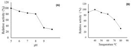
Figure 1 shows the effect of pH and temperature on the activity of P. ostreatoroseus proteases determined in the dialyzed GYP extract. The results showed the activity of proteases at all pH and temperatures tested. However, optimal activity was observed at pH 8.0 (158.61 ± 0.13 U/mL), followed by a decrease in activity at higher pH values, with a maximum reduction (53.72%) at alkaline pH.

Effect of pH (A) and temperature (B) on the fibrinolytic activity of P. ostreatoroseus in GYP medium.
The effect of temperature on the activity of P. ostreatoroseus fibrinolytic proteases was observed at all temperatures tested (Figure 1), with maximum activity at 30 °C and a reduction at the other temperatures evaluated (40 °C to 80 °C).
Figure 2 shows the effect of pH and temperature stability on the activity of P. ostreatoroseus fibrinolytic proteases determined in GYP extracts. P. ostreatoroseus proteases showed high stability, retaining significant activity from pH 5.0 to pH 8.0. With a decrease in activity of 66.08% and 71.22% at pH 9.0 and pH 10.0, respectively. Regarding temperature, fibrinolytic proteases exhibited greater stability at temperatures between 30 °C and 60 °C. At temperatures of 70 °C and 80 °C, with a marked decrease in catalytic action, the enzyme retained only 32.68% of its activity.
Effect of pH (A) and temperature (B) stability on the fibrinolytic activity of P. ostreatoroseus in GYP medium.
The results regarding the effect of metal ions on the activity of fibrinolytic proteases are shown in Table 2. Mn+2 ions completely inhibited enzymatic activity. Zn+2 and Cu+2 reduced the catalytic action by 90.38% and 97.38%, respectively.
Effect of metal ions and inhibitor substances on the activity of fibrinolytic proteases of P. ostreatoroseus.
The effect of inhibitory substances and metal ions on the activity of fibrinolytic proteases in P. ostreatoroseus is shown in Table 2. In the stability test against inhibitors, it was found that the serine protease inhibitor (PMSF) and metalloprotease (EDTA) caused a reduction in activity of 80.15% and 92.45%, respectively. Proteases were not affected by Pepstatin and iodoacetic acid; in this condition, activity retention was greater than 70%.
4. Discussion
In the scientific literature, little research has been done on the synthesis of fibrinolytic enzymes by P. ostreatoroseus. Although mushrooms are considered the most valuable resources to produce fibrinolytic enzymes, studies on the fibrinolytic potential of edible mushrooms are still scarce (Santana et al., 2024).
In studies with Pleurotus species carried out by Petraglia et al. (2022), similar results were obtained with basiodiome extracts of P. eryngii and P. ostreatus, which showed degradation halos of 4.91 cm and 6.15 cm, respectively. Liu et al. (2014) and Choi et al. (2017) verified enzymatic action in fibrin plates of P. ostreatus and P. ferulae when cultivated in PDA medium added with glucose. In crude extracts of Lentinus edodes, Ali et al. (2018) obtained lithic degradation halo of 19 mm, data that corroborates the current research.
In studies by Ali et al. (2018), evaluating the extract extracted from 10 basidiomas of mushroom species, it was observed that fibrinolytic proteases were active in all of them, highlighting the highest fibrinolytic activity (48.06 U/mL) in extracts from the basidiomas of L. edodes; however, in the other species of mushrooms evaluated, this activity varied from 9.0 to 17.0 U/mL.
Research focusing on Pleurotus species developed by Petraglia et al. (2022) verified the fibrinolytic activity of extracts from the basidiomes of P. ostreatus and P. eryngii, with a significant value of 39.14 ± 1.01 U/mL and 35.09 ± 0.40 U/mL, respectively. In other studies, with P. eryngii and P. ostreatus, the significant activity of fibrinolytic enzymes in submerged fermentation media [Malt (226.47 ± 7.26 U/mL) and Sabouraud (100.14 ± 0.28 U/mL)] showed promise regarding the fibrinolytic activity of the mushrooms evaluated by Santana et al. (2024), respectively.
Fibrinolytic enzymes act in the degradation of fibrin; however, the synthesis of these biocatalysts by microorganisms depends on several factors, such as the quality of nutrients in the fermentation medium, sources of carbon and nitrogen, the time and age of the inoculum, the genetic characteristics of the species, agitation, pH, and temperature. In this way, each organism can achieve maximum enzyme production under specific conditions, according to the physiological needs of each microorganism (Gimenes et al., 2021; Cardoso et al., 2022; Pessoa et al., 2023).
Understanding the biochemical characteristics of proteases is an important factor in verifying the viability of commercial applications of these biocatalysts (Barzkar et al., 2022). Fungi can produce acidic, neutral, and alkaline proteases. Proteases that exhibit optimal catalytic activity at pH 5 to 8 have applications in various industrial segments, such as the food, textile, pharmaceutical, and detergent industries (Barzee et al., 2021; Petraglia et al., 2022).
Similar data about effect of pH were obtained by Braga et al. (2020), Petraglia et al. (2022), and Santana et al. (2024), whose maximum protease activity was observed at pH 7.0 to 8.0 when analyzing species of P. eryngii, L. villosus, and P. ostreatus, respectively. The study of the effect of pH is one of the important parameters in processes determining the optimal conditions for enzyme synthesis, as it directly influences the activity of biocatalysts essential for the transport of nutrients as well as the growth and maintenance of the morphological structure of filamentous fungi (Brito et al., 2019; Pimenta et al., 2021).
The data obtained on the optimum temperature are in accordance with those reported by Machado et al. (2017), Barbosa et al. (2020) and Petraglia et al. (2022) in studies with P. ostreatoroseus and P. ostreatus, respectively. Most fibrinolytic enzymes have an optimum temperature between 35 °C and 50 °C (Cardoso et al., 2022). In addition, the data on pH stability were similar with the studies by Pimenta et al. (2021) and Santana et al. (2022) found that P. albidus and P. eryngii had reduced protease activity at pH 8.0 to 10.0. Pimenta et al. (2021) described a similar stability regarding temperature to P. albidus proteases.
In other words, extremely high temperatures can cause the enzyme to denature and stop working. Each enzyme has an optimum pH range, changing the pH to a value outside this range will cause enzyme activity to decrease. Consequently, extreme pH values can cause enzymes to denature. Conventional and statistical optimization of nutritional components and physicochemical parameters, such as pH and temperature, are fundamental approaches for the significant fermentative production of fibrinolytic enzymes, as these parameters enable their efficient use in catalytic action (Sharma et al., 2021).
In the presence of other ions, peptidase retained a catalytic action greater than 40%. Barbosa et al. (2020) reported that Zn+2 and Cu+2 reduced enzymatic action by 59.32% and 52.0%, respectively. Machado et al. (2017) reported that Cu+2 and Zn+2 caused a 95% reduction in the proteolytic activity of P. ostreatoroseus. Pimenta et al. (2021) found that Zn+2 reduced the activity of P. albidus proteases by 87.37%, data that corroborates those obtained in this research.
Ions and inhibitors can affect protease activity mainly by binding to the enzyme's active site, thus preventing binding to the substrate, or by binding to another site on the enzyme so that catalysis of the enzyme's reaction is blocked, thus inhibiting the process of substrate binding to the active site (Nelson et al., 2022).
Abdel-Rahman (2018) stated that protease inhibitors are used to identify the group in the active site of the enzyme. The results suggest that in the fibrinolytic proteases of the extracts obtained from P. ostreatoroseus, there is a predominance of metalloproteases and serine proteases (Table 2). Barbosa et al. (2020), Pimenta et al. (2021) and Santana et al. (2022) verified the significant production of serine and metalloproteases by P. ostreatoroseus, P. albidus, and P. eryngii, respectively. Meshram et al. (2016) studies with endophytic fungus Xylaria curta was reported that a metalloprotease (with 8.0 pH optima) produced appreciable fibrinolytic properties and thus has the potential of being used for therapeutic purposes.
Alkaline serine and metalloproteases are economical sources for hydrolyzing different types of proteins in various industrial applications. The detergent sector consumes the most alkaline proteases, which are generally serine proteases with an alkaline pH range. These properties of alkaline proteases make them suitable for use in the detergent industry (Razzaq et al., 2019; Gurumallesh et al., 2019; Miranda et al., 2022).
In the food industry, commercial alkaline serine proteases are mainly involved in the hydrolysis of proteins to prepare hydrolysates with high nutritional value. The protein hydrolysates produced are widely used in the fortification of baby food formulations, dietary products, and processed foods. In the pharmaceutical industry, they are used to treat thrombosis (urokinase, fibrinolytic), hemophilia (factor VIIa), and other pathologies (Matkawala et al., 2021; Solanki et al., 2021).
5. Conclusion
Pleurotus ostreatoroseus, an edible mushroom, synthesizes fibrinolytic enzymes under submerged cultivation conditions, showing significant fibrinolytic protease activity in fibrin plate and in the crude extract from fermentation in GYP (liquid), demonstrating optimal activity at alkaline pH and at 30 °C, of the serine type and metalloproteases. These biocatalysts have strong potential for use in the pharmaceutical, detergent sectors, and food industries. Therefore, this research has produced promising and innovative results about the enzymatic synthesis capacity of fibrinolytic enzymes from P. ostreatoroseus.
Acknowledgements
To Federal University of Amazonas (UFAM) for technical and scientific support. To the Culture Collection DPUA/UFAM. To the Postgraduate Program in Biotechnology – PPGBIOTEC/UFAM. To fellow Masters and Doctors collaborators. To the Coordination for the Improvement of Higher Education Personnel – CAPES for the financial support.
References
- ABDEL-RAHMAN, T.M., 2018. Biological and chemical sciences purification and characterization of milk -clotting enzyme from the edible mushroom Pleurotus albidus. Research Journal of Pharmaceutical, vol. 9, no. 49, pp. 49-63.
-
ACOSTA, G.A., FONSECA, M.I., FARIÑA, J.I. and ZAPATA, P.D., 2022. Exploring Agaricomycetes from the Paranaense rainforest (Misiones, Argentina) as an unconventional source of fibrinolytic enzymes. Mycologia, vol. 114, no. 2, pp. 242-253. http://doi.org/10.1080/00275514.2022.2035148 PMid:35394849.
» http://doi.org/10.1080/00275514.2022.2035148 -
ALI, S.M., RAMAN, J., LAKSHMANAN, H., LING, T.C., PHAN, C.-W., TAN, Y.S. and SABARATNAM, V., 2018. Surface decoration of selenium nanoparticles by proteins from the culinary-medicinal shiitake mushroom, Lentinus edodes (Agaricomycetes), for enhanced fibrinolytic activity. International Journal of Medicinal Mushrooms, vol. 20, no. 11, pp. 1021-1030. http://doi.org/10.1615/IntJMedMushrooms.2018028307
» http://doi.org/10.1615/IntJMedMushrooms.2018028307 -
BARBOSA, E., PIMENTA, L., BRITO, A.K., BATISTA, S., MARTIM, S. and TEIXEIRA, M., 2023. Salmon oyster mushroom: growth characteristics and protease production on a laboratory scale. Concilium, vol. 23, no. 2, pp. 367-379. http://doi.org/10.53660/CLM-801-23A54
» http://doi.org/10.53660/CLM-801-23A54 -
BARBOSA, E.E.P., PIMENTA, L., BRITO, A.K.P., MARTIM, S.R. and TEIXEIRA, M.F.S., 2020. Cultivo de cogumelo comestível em resíduos lignocelulósicos de floresta tropical para produção de proteases. Brazilian Journal of Development, vol. 6, no. 11, pp. 92475-92485. http://doi.org/10.34117/bjdv6n11-598
» http://doi.org/10.34117/bjdv6n11-598 -
BARZEE, T.J., CAO, L., PAN, Z. and ZHANG, R., 2021. Fungi for future foods. Journal of Future Foods, vol. 1, no. 1, pp. 25-37. http://doi.org/10.1016/j.jfutfo.2021.09.002
» http://doi.org/10.1016/j.jfutfo.2021.09.002 -
BARZKAR, N., JAHROMI, S.T. and VIANELLO, F., 2022. Marine microbial fibrinolytic enzymes: an overview of source, production, biochemical properties and thrombolytic activity. Marine Drugs, vol. 20, no. 1, pp. 56. http://doi.org/10.3390/md20010046 PMid:35049901.
» http://doi.org/10.3390/md20010046 -
BRADFORD, M.M., 1976. A rapid and sensitive method for the quantitation of microgram quantities of protein utilizing the principle of protein-dye binding. Analytical Biochemistry, vol. 72, no. 1-2, pp. 248-254. http://doi.org/10.1016/0003-2697(76)90527-3 PMid:942051.
» http://doi.org/10.1016/0003-2697(76)90527-3 -
BRAGA, R.S., BRITO, É.C.M., SOUZA, R.Á.T., TEIXEIRA, M.F.S. and MARTIM, S.R., 2020. Lentinus Villosus Klotzsch (1833) Am 169: a natural and renewable source of alkaline protease. Brazilian Journal of Development, vol. 6, no. 11, pp. 85867-85883. http://doi.org/10.34117/bjdv6n11-127
» http://doi.org/10.34117/bjdv6n11-127 -
BRITO, E.C.D.M., BRAGA, R.D.S., TEIXEIRA, M.F.S. and MARTIM, S.R., 2019. Produção e caracterização parcial de proteases aspárticas sintetizadas por Lentinus crinitus (L.) Fr. 1825 DPUA 1693 (Polyporaceae). Boletim do Museu Paraense Emílio Goeldi. Ciências Naturais, vol. 14, no. 3, pp. 463-472. http://doi.org/10.46357/bcnaturais.v14i3.232
» http://doi.org/10.46357/bcnaturais.v14i3.232 -
CARDOSO, K.B.B., NASCIMENTO, M.C., BATISTA, A.C., OLIVEIRA, V.M., NASCIMENTO, T.P., BATISTA, J.M.S., COSTA, R.M.P.B., PASTRANA, L. and PORTO, A.L.F., 2022. View of systematic analysis on the obtaining of fibrinolytic fungi enzymes. Research, Society and Development, vol. 11, no. 2, e13611225449. http://doi.org/10.33448/rsd-v11i2.25449
» http://doi.org/10.33448/rsd-v11i2.25449 -
CHOI, J.H., KIM, D.W., KIM, S. and KIM, S.J., 2017. Purification and partial characterization of a fibrinolytic enzyme from the fruiting body of the medicinal and edible mushroom Pleurotus ferulae. Preparative Biochemistry & Biotechnology, vol. 47, no. 6, pp. 539-546. http://doi.org/10.1080/10826068.2016.1181083 PMid:27136080.
» http://doi.org/10.1080/10826068.2016.1181083 -
COELHO, M.P.S.L.V., BARBOSA, E.E.P., PIMENTA, L., BATISTA, S.C.P., PRADO, F.B., MARTIM, S.R., CRUZ FILHO, R.F. and TEIXEIRA, M.F.S., 2021. Alternativa fontes nutricionais para desenvolvimento da fase micelial e produção de hidrolases por cogumelo comestível de floresta tropical. Brazilian Journal of Development, vol. 7, no. 3, pp. 22890-22907. http://doi.org/10.34117/bjdv7n3-145
» http://doi.org/10.34117/bjdv7n3-145 -
GIMENES, N.C., SILVEIRA, E. and TAMBOURGI, E.B., 2021. An overview of proteases: production, downstream processes and industrial applications. Separation and Purification Reviews, vol. 50, no. 3, pp. 223-243. http://doi.org/10.1080/15422119.2019.1677249
» http://doi.org/10.1080/15422119.2019.1677249 -
GURUMALLESH, P., ALAGU, K., RAMAKRISHNAN, B. and MUTHUSAMY, S., 2019. A systematic reconsideration on proteases. International Journal of Biological Macromolecules, vol. 128, pp. 254-267. http://doi.org/10.1016/j.ijbiomac.2019.01.081 PMid:30664968.
» http://doi.org/10.1016/j.ijbiomac.2019.01.081 -
HANFF, T.C., MOHAREB, A.M., GIRI, J., COHEN, J.B. and CHIRINOS, J.A., 2020. Thrombosis in COVID-19. American Journal of Hematology, vol. 95, no. 12, pp. 1578-1589. http://doi.org/10.1002/ajh.25982 PMid:32857878.
» http://doi.org/10.1002/ajh.25982 -
HU, Y., YU, D., WANG, Z., HOU, J., TYAGI, R., LIANG, Y. and HU, Y., 2019. Purification and characterization of a novel, highly potent fibrinolytic enzyme from Bacillus subtilis DC27 screened from Douchi, a traditional Chinese fermented soybean food. Scientific Reports, vol. 9, no. 1, pp. 9235. http://doi.org/10.1038/s41598-019-45686-y PMid:31239529.
» http://doi.org/10.1038/s41598-019-45686-y -
JI, H.L., ZHAO, R., MATALON, S. and MATTHAY, M.A., 2020. Elevated plasmin (Ogen) as a common risk factor for COVID-19 susceptibility. Physiological Reviews, vol. 100, no. 3, pp. 1065-1075. http://doi.org/10.1152/physrev.00013.2020 PMid:32216698.
» http://doi.org/10.1152/physrev.00013.2020 -
KIM, A.Y., WOO, W., YON, D.K., LEE, S.W., YANG, J.W., KIM, J.H., PARK, S., KOYANAGI, A., KIM, M.S., LEE, S., SHIN, J.I. and SMITH, L., 2022. Thrombosis patterns and clinical outcome of COVID-19 vaccine-induced immune thrombotic thrombocytopenia: a systematic review and meta-analysis. International Journal of Infectious Diseases, vol. 119, pp. 130-139. http://doi.org/10.1016/j.ijid.2022.03.034 PMid:35339716.
» http://doi.org/10.1016/j.ijid.2022.03.034 -
KUMAR, S.S. and SABU, A., 2019. Fibrinolytic enzymes for thrombolytic theraphy. therapeutic enzymes: function and clinical implications. Advances in Experimental Medicine and Biology, vol. 1148, pp. 345-381. http://doi.org/10.1007/978-981-13-7709-9_15
» http://doi.org/10.1007/978-981-13-7709-9_15 -
LIN, H., XU, L., YU, S., HONG, W., HUANG, M. and XU, P., 2020. Therapeutics targeting the fibrinolytic system. Experimental & Molecular Medicine, vol. 52, no. 3, pp. 367-379. http://doi.org/10.1038/s12276-020-0397-x PMid:32152451.
» http://doi.org/10.1038/s12276-020-0397-x -
LIU, X.L., ZHENG, X.Q., QIAN, P.Z., KOPPARAPU, N.K., DENG, Y.P., NONAKA, M. and HARADA, N., 2014. Purification and characterization of a novel fibrinolytic enzyme from culture supernatant of Pleurotus ostreatus Journal of Microbiology and Biotechnology, vol. 24, no. 2, pp. 245-253. http://doi.org/10.4014/jmb.1307.07063 PMid:24248089.
» http://doi.org/10.4014/jmb.1307.07063 -
MACHADO, A.G.M., MARTIM, S.R., ALECRIM, M.M. and TEIXEIRA, M.F.S., et al, 2017. Production and characterization of proteases from edible mushrooms cultivated on amazonic tubers. African Journal of Biotechnology, vol. 16, no. 46, pp. 2160-2166. http://doi.org/10.5897/AJB2017.16154
» http://doi.org/10.5897/AJB2017.16154 -
MANDEL, J., CASARI, M., STEPANYAN, M., MARTYANOV, A. and DEPPERMANN, C., 2022. Beyond hemostasis: platelet innate immune interactions and thromboinflammation. International Journal of Molecular Sciences, vol. 23, no. 7, pp. 3868. http://doi.org/10.3390/ijms23073868 PMid:35409226.
» http://doi.org/10.3390/ijms23073868 -
MARTIM, S.R., SILVA, L.S.C., ALECRIM, M.M., SOUZA, B.C., OLIVEIRA, I.M.D.A. and TEIXEIRA, M.F.S., 2017. Proteases ácidas de cogumelo comestível da Amazônia para aplicabilidade industrial. Boletim do Museu Paraense Emílio Goeldi. Ciências Naturais, vol. 12, no. 3, pp. 353-362. http://doi.org/10.46357/bcnaturais.v12i3.86
» http://doi.org/10.46357/bcnaturais.v12i3.86 -
MATKAWALA, F., NIGHOJKAR, S., KUMAR, A. and NIGHOJKAR, A., 2021. Microbial alkaline serine proteases: production, properties and applications. World Journal of Microbiology & Biotechnology, vol. 37, no. 4, pp. 63. http://doi.org/10.1007/s11274-021-03036-z PMid:33730214.
» http://doi.org/10.1007/s11274-021-03036-z -
MESHRAM, V., SAXENA, S. and PAUL, K., 2016. Xylarinase: a novel clot busting enzyme from an endophytic fungus Xylaria curta Journal of Enzyme Inhibition and Medicinal Chemistry, vol. 31, no. 6, pp. 1502-1511. http://doi.org/10.3109/14756366.2016.1151013 PMid:27033431.
» http://doi.org/10.3109/14756366.2016.1151013 -
MIRANDA, V.M.A.M., BARBOSA FILHO, J.P.M., COSTA, R.M.P.B., LEITE, A.C.L., OLIVEIRA, V.M., BATISTA, J.M.S., PASTRANA, L., NASCIMENTO, T.P. and PORTO, A.L.F., 2022. Evaluation of partial thromboplastin time, thrombin time and prothrombin time over treated plasma using a fibrinolytic protease. Research, Society and Development, vol. 11, no. 2, e15311225439. http://doi.org/10.33448/rsd-v11i2.25439
» http://doi.org/10.33448/rsd-v11i2.25439 - NASCIMENTO, T.P., SALES, A.E., PORTO, C.S., BRANDÃO, R.M., CAMPOS-TAKAKI, G.M., TEIXEIRA, J.A., PORTO, T.S., PORTO, A.L. and CONVERTI, A., 2016. Purification of a fibrinolytic protease from Mucor subtilissimus UCP1262 by aqueous two-phase systems (PEG/sulfate). Journal of Chromatography. A, vol. 1025, pp. 16-24. PMid:27183214.
-
NAVEED, M., NADEEM, F., MEHMOOD, T., BILAL, M., ANWAR, Z. and AMJAD, F., 2021. Protease: a versatile and ecofriendly biocatalyst with multi-industrial applications: an updated review. Catalysis Letters, vol. 151, no. 2, pp. 307-323. http://doi.org/10.1007/s10562-020-03316-7
» http://doi.org/10.1007/s10562-020-03316-7 - NELSON, D.L., COX, M.M. and HOSKINS, A.A., 2022. Princípios de bioquímica de Lehninger. 8ª ed. Porto Alegre: ArtMed, pp. 183-187.
-
PESSOA, V.A., SOARES, L.B.N., SILVA, G.L., VASCONCELOS, A.S., SILVA, J.F., FARIÑA, J.I., OLIVEIRA-JUNIOR, S.D., SALES-CAMPOS, C. and CHEVREUIL, L.R., 2023. Production of mycelial biomass, proteases and protease inhibitors by Ganoderma lucidum under different submerged fermentation conditions. Brazilian Journal of Biology = Revista Brasileira de Biologia, vol. 83, e270316. http://doi.org/10.1590/1519-6984.270316 PMid:37162094.
» http://doi.org/10.1590/1519-6984.270316 -
PETRAGLIA, T., LATRONICO, T., LIUZZI, G.M., FANIGLIULO, A., CRESCENZI, A. and ROSSANO, R., 2022. Edible mushrooms as source of fibrin(ogen)olytic enzymes: comparison between four cultivated species. Molecules, vol. 27, no. 23, pp. 8145. http://doi.org/10.3390/molecules27238145 PMid:36500238.
» http://doi.org/10.3390/molecules27238145 -
PIMENTA, L., BARBOSA, E.E.P., BRITO, A.K.P., MARTIM, S.R. and TEIXEIRA, M.F.S., 2021. Processo Eco-amigável para selecionar substrato lignocelulósico para produção de peptidases ácidas. Brazilian Journal of Development, vol. 7, no. 1, pp. 3469-3479. http://doi.org/10.34117/bjdv7n1-234
» http://doi.org/10.34117/bjdv7n1-234 - RAZZAQ, A., SHAMSI, S., ALI, A., ALI, Q., SAJJAD, M., MALIK, A. and ASHRAF, M., 2019. Microbial proteases applications. Frontiers in Bioengineering and Biotechnology, vol. 7, pp. 110. PMid:31263696.
-
ROTH, G.A., MENSAH, G.A., JOHNSON, C.O., ADDOLORATO, G., AMMIRATI, E., BADDOUR, L.M., BARENGO, N.C., BEATON, A.Z., BENJAMIN, E.J., BENZIGER, C.P., BONNY, A., BRAUER, M., BRODMANN, M., CAHILL, T.J., CARAPETIS, J., CATAPANO, A.L., CHUGH, S.S., COOPER, L.T., CORESH, J., CRIQUI, M., DECLEENE, N., EAGLE, K.A., EMMONS-BELL, S., FEIGIN, V.L., FERNÁNDEZ-SOLÀ, J., FOWKES, G., GAKIDOU, E., GRUNDY, S.M., HE, F.J., HOWARD, G., HU, F., INKER, L., KARTHIKEYAN, G., KASSEBAUM, N., KOROSHETZ, W., LAVIE, C., LLOYD-JONES, D., LU, H.S., MIRIJELLO, A., TEMESGEN, A.M., MOKDAD, A., MORAN, A.E., MUNTNER, P., NARULA, J., NEAL, B., NTSEKHE, M., MORAES DE OLIVEIRA, G., OTTO, C., OWOLABI, M., PRATT, M., RAJAGOPALAN, S., REITSMA, M., RIBEIRO, A.L.P., RIGOTTI, N., RODGERS, A., SABLE, C., SHAKIL, S., SLIWA-HAHNLE, K., STARK, B., SUNDSTRÖM, J., TIMPEL, P., TLEYJEH, I.M., VALGIMIGLI, M., VOS, T., WHELTON, P.K., YACOUB, M., ZUHLKE, L., MURRAY, C., FUSTER, V., ROTH, G.A., MENSAH, G.A., JOHNSON, C.O., ADDOLORATO, G., AMMIRATI, E., BADDOUR, L.M., BARENGO, N.C., BEATON, A., BENJAMIN, E.J., BENZIGER, C.P., BONNY, A., BRAUER, M., BRODMANN, M., CAHILL, T.J., CARAPETIS, J.R., CATAPANO, A.L., CHUGH, S., COOPER, L.T., CORESH, J., CRIQUI, M.H., DECLEENE, N.K., EAGLE, K.A., EMMONS-BELL, S., FEIGIN, V.L., FERNÁNDEZ-SOLA, J., FOWKES, F.G.R., GAKIDOU, E., GRUNDY, S.M., HE, F.J., HOWARD, G., HU, F., INKER, L., KARTHIKEYAN, G., KASSEBAUM, N.J., KOROSHETZ, W.J., LAVIE, C., LLOYD-JONES, D., LU, H.S., MIRIJELLO, A., MISGANAW, A.T., MOKDAD, A.H., MORAN, A.E., MUNTNER, P., NARULA, J., NEAL, B., NTSEKHE, M., OLIVEIRA, G.M.M., OTTO, C.M., OWOLABI, M.O., PRATT, M., RAJAGOPALAN, S., REITSMA, M.B., RIBEIRO, A.L.P., RIGOTTI, N.A., RODGERS, A., SABLE, C.A., SHAKIL, S.S., SLIWA, K., STARK, B.A., SUNDSTRÖM, J., TIMPEL, P., TLEYJEH, I.I., VALGIMIGLI, M., VOS, T., WHELTON, P.K., YACOUB, M., ZUHLKE, L.J., ABBASI-KANGEVARI, M., ABDI, A., ABEDI, A., ABOYANS, V., ABRHA, W.A., ABU-GHARBIEH, E., ABUSHOUK, A.I., ACHARYA, D., ADAIR, T., ADEBAYO, O.M., ADEMI, Z., ADVANI, S.M., AFSHARI, K., AFSHIN, A., AGARWAL, G., AGASTHI, P., AHMAD, S., AHMADI, S., AHMED, M.B., AJI, B., AKALU, Y., AKANDE-SHOLABI, W., AKLILU, A., AKUNNA, C.J., ALAHDAB, F., AL-EYADHY, A., ALHABIB, K.F., ALIF, S.M., ALIPOUR, V., ALJUNID, S.M., ALLA, F., ALMASI-HASHIANI, A., ALMUSTANYIR, S., AL-RADDADI, R.M., AMEGAH, A.K., AMINI, S., AMINORROAYA, A., AMU, H., AMUGSI, D.A., ANCUCEANU, R., ANDERLINI, D., ANDREI, T., ANDREI, C.L., ANSARI-MOGHADDAM, A., ANTENEH, Z.A., ANTONAZZO, I.C., ANTONY, B., ANWER, R., APPIAH, L.T., ARABLOO, J., ÄRNLÖV, J., ARTANTI, K.D., ATARO, Z., AUSLOOS, M., AVILA-BURGOS, L., AWAN, A.T., AWOKE, M.A., AYELE, H.T., AYZA, M.A., AZARI, S., B, D.B., BAHEIRAEI, N., BAIG, A.A., BAKHTIARI, A., BANACH, M., BANIK, P.C., BAPTISTA, E.A., BARBOZA, M.A., BARUA, L., BASU, S., BEDI, N., BÉJOT, Y., BENNETT, D.A., BENSENOR, I.M., BERMAN, A.E., BEZABIH, Y.M., BHAGAVATHULA, A.S., BHASKAR, S., BHATTACHARYYA, K., BIJANI, A., BIKBOV, B., BIRHANU, M.M., BOLOOR, A., BRANT, L.C., BRENNER, H., BRIKO, N.I., BUTT, Z.A., CAETANO DOS SANTOS, F.L., CAHILL, L.E., CAHUANA-HURTADO, L., CÁMERA, L.A., CAMPOS-NONATO, I.R., CANTU-BRITO, C., CAR, J., CARRERO, J.J., CARVALHO, F., CASTAÑEDA-ORJUELA, C.A., CATALÁ-LÓPEZ, F., CERIN, E., CHARAN, J., CHATTU, V.K., CHEN, S., CHIN, K.L., CHOI, J.-Y.J., CHU, D.-T., CHUNG, S.-C., CIRILLO, M., COFFEY, S., CONTI, S., COSTA, V.M., CUNDIFF, D.K., DADRAS, O., DAGNEW, B., DAI, X., DAMASCENO, A.A.M., DANDONA, L., DANDONA, R., DAVLETOV, K., DE LA CRUZ-GÓNGORA, V., DE LA HOZ, F.P., DE NEVE, J.-W., DENOVA-GUTIÉRREZ, E., DERBEW MOLLA, M., DERSEH, B.T., DESAI, R., DEUSCHL, G., DHARMARATNE, S.D., DHIMAL, M., DHUNGANA, R.R., DIANATINASAB, M., DIAZ, D., DJALALINIA, S., DOKOVA, K., DOUIRI, A., DUNCAN, B.B., DURAES, A.R., EAGAN, A.W., EBTEHAJ, S., EFTEKHARI, A., EFTEKHARZADEH, S., EKHOLUENETALE, M., EL NAHAS, N., ELGENDY, I.Y., ELHADI, M., EL-JAAFARY, S.I., ESTEGHAMATI, S., ETISSO, A.E., EYAWO, O., FADHIL, I., FARAON, E.J.A., FARIS, P.S., FARWATI, M., FARZADFAR, F., FERNANDES, E., FERNANDEZ PRENDES, C., FERRARA, P., FILIP, I., FISCHER, F., FLOOD, D., FUKUMOTO, T., GAD, M.M., GAIDHANE, S., GANJI, M., GARG, J., GEBRE, A.K., GEBREGIORGIS, B.G., GEBREGZABIHER, K.Z., GEBREMESKEL, G.G., GETACHER, L., OBSA, A.G., GHAJAR, A., GHASHGHAEE, A., GHITH, N., GIAMPAOLI, S., GILANI, S.A., GILL, P.S., GILLUM, R.F., GLUSHKOVA, E.V., GNEDOVSKAYA, E.V., GOLECHHA, M., GONFA, K.B., GOUDARZIAN, A.H., GOULART, A.C., GUADAMUZ, J.S., GUHA, A., GUO, Y., GUPTA, R., HACHINSKI, V., HAFEZI-NEJAD, N., HAILE, T.G., HAMADEH, R.R., HAMIDI, S., HANKEY, G.J., HARGONO, A., HARTONO, R.K., HASHEMIAN, M., HASHI, A., HASSAN, S., HASSEN, H.Y., HAVMOELLER, R.J., HAY, S.I., HAYAT, K., HEIDARI, G., HERTELIU, C., HOLLA, R., HOSSEINI, M., HOSSEINZADEH, M., HOSTIUC, M., HOSTIUC, S., HOUSEH, M., HUANG, J., HUMAYUN, A., IAVICOLI, I., IBENEME, C.U., IBITOYE, S.E., ILESANMI, O.S., ILIC, I.M., ILIC, M.D., IQBAL, U., IRVANI, S.S.N., ISLAM, S.M.S., ISLAM, R.M., ISO, H., IWAGAMI, M., JAIN, V., JAVAHERI, T., JAYAPAL, S.K., JAYARAM, S., JAYAWARDENA, R., JEEMON, P., JHA, R.P., JONAS, J.B., JONNAGADDALA, J., JOUKAR, F., JOZWIAK, J.J., JÜRISSON, M., KABIR, A., KAHLON, T., KALANI, R., KALHOR, R., KAMATH, A., KAMEL, I., KANDEL, H., KANDEL, A., KARCH, A., KASA, A.S., KATOTO, P.D.M.C., KAYODE, G.A., KHADER, Y.S., KHAMMARNIA, M., KHAN, M.S., KHAN, M.N., KHAN, M., KHAN, E.A., KHATAB, K., KIBRIA, G.M.A., KIM, Y.J., KIM, G.R., KIMOKOTI, R.W., KISA, S., KISA, A., KIVIMÄKI, M., KOLTE, D., KOOLIVAND, A., KORSHUNOV, V.A., KOULMANE LAXMINARAYANA, S.L., KOYANAGI, A., KRISHAN, K., KRISHNAMOORTHY, V., KUATE DEFO, B., KUCUK BICER, B., KULKARNI, V., KUMAR, G.A., KUMAR, N., KURMI, O.P., KUSUMA, D., KWAN, G.F., LA VECCHIA, C., LACEY, B., LALLUKKA, T., LAN, Q., LASRADO, S., LASSI, Z.S., LAURIOLA, P., LAWRENCE, W.R., LAXMAIAH, A., LEGRAND, K.E., LI, M.-C., LI, B., LI, S., LIM, S.S., LIM, L.-L., LIN, H., LIN, Z., LIN, R.-T., LIU, X., LOPEZ, A.D., LORKOWSKI, S., LOTUFO, P.A., LUGO, A., M, N.K., MADOTTO, F., MAHMOUDI, M., MAJEED, A., MALEKZADEH, R., MALIK, A.A., MAMUN, A.A., MANAFI, N., MANSOURNIA, M.A., MANTOVANI, L.G., MARTINI, S., MATHUR, M.R., MAZZAGLIA, G., MEHATA, S., MEHNDIRATTA, M.M., MEIER, T., MENEZES, R.G., MERETOJA, A., MESTROVIC, T., MIAZGOWSKI, B., MIAZGOWSKI, T., MICHALEK, I.M., MILLER, T.R., MIRRAKHIMOV, E.M., MIRZAEI, H., MOAZEN, B., MOGHADASZADEH, M., MOHAMMAD, Y., MOHAMMAD, D.K., MOHAMMED, S., MOHAMMED, M.A., MOKHAYERI, Y., MOLOKHIA, M., MONTASIR, A.A., MORADI, G., MORADZADEH, R., MORAGA, P., MORAWSKA, L., MORENO VELÁSQUEZ, I., MORZE, J., MUBARIK, S., MURUET, W., MUSA, K.I., NAGARAJAN, A.J., NALINI, M., NANGIA, V., NAQVI, A.A., NARASIMHA SWAMY, S., NASCIMENTO, B.R., NAYAK, V.C., NAZARI, J., NAZARZADEH, M., NEGOI, R.I., NEUPANE KANDEL, S., NGUYEN, H.L.T., NIXON, M.R., NORRVING, B., NOUBIAP, J.J., NOUTHE, B.E., NOWAK, C., ODUKOYA, O.O., OGBO, F.A., OLAGUNJU, A.T., ORRU, H., ORTIZ, A., OSTROFF, S.M., PADUBIDRI, J.R., PALLADINO, R., PANA, A., PANDA-JONAS, S., PAREKH, U., PARK, E.-C., PARVIZI, M., PASHAZADEH KAN, F., PATEL, U.K., PATHAK, M., PAUDEL, R., PEPITO, V.C.F., PERIANAYAGAM, A., PERICO, N., PHAM, H.Q., PILGRIM, T., PIRADOV, M.A., PISHGAR, F., PODDER, V., POLIBIN, R.V., POURSHAMS, A., PRIBADI, D.R.A., RABIEE, N., RABIEE, M., RADFAR, A., RAFIEI, A., RAHIM, F., RAHIMI-MOVAGHAR, V., UR RAHMAN, M.H., RAHMAN, M.A., RAHMANI, A.M., RAKOVAC, I., RAM, P., RAMALINGAM, S., RANA, J., RANASINGHE, P., RAO, S.J., RATHI, P., RAWAL, L., RAWASIA, W.F., RAWASSIZADEH, R., REMUZZI, G., RENZAHO, A.M.N., REZAPOUR, A., RIAHI, S.M., ROBERTS-THOMSON, R.L., ROEVER, L., ROHLOFF, P., ROMOLI, M., ROSHANDEL, G., RWEGERERA, G.M., SAADATAGAH, S., SABER-AYAD, M.M., SABOUR, S., SACCO, S., SADEGHI, M., SAEEDI MOGHADDAM, S., SAFARI, S., SAHEBKAR, A., SALEHI, S., SALIMZADEH, H., SAMAEI, M., SAMY, A.M., SANTOS, I.S., SANTRIC-MILICEVIC, M.M., SARRAFZADEGAN, N., SARVEAZAD, A., SATHISH, T., SAWHNEY, M., SAYLAN, M., SCHMIDT, M.I., SCHUTTE, A.E., SENTHILKUMARAN, S., SEPANLOU, S.G., SHA, F., SHAHABI, S., SHAHID, I., SHAIKH, M.A., SHAMALI, M., SHAMSIZADEH, M., SHAWON, M.S.R., SHEIKH, A., SHIGEMATSU, M., SHIN, M.-J., SHIN, J.I., SHIRI, R., SHIUE, I., SHUVAL, K., SIABANI, S., SIDDIQI, T.J., SILVA, D.A.S., SINGH, J.A., MTECH, A.S., SKRYABIN, V.Y., SKRYABINA, A.A., SOHEILI, A., SPURLOCK, E.E., STOCKFELT, L., STORTECKY, S., STRANGES, S., SULIANKATCHI ABDULKADER, R., TADBIRI, H., TADESSE, E.G., TADESSE, D.B., TAJDINI, M., TARIQUJJAMAN, M., TEKLEHAIMANOT, B.F., TEMSAH, M.-H., TESEMA, A.K., THAKUR, B., THANKAPPAN, K.R., THAPAR, R., THRIFT, A.G., TIMALSINA, B., TONELLI, M., TOUVIER, M., TOVANI-PALONE, M.R., TRIPATHI, A., TRIPATHY, J.P., TRUELSEN, T.C., TSEGAY, G.M., TSEGAYE, G.W., TSILIMPARIS, N., TUSA, B.S., TYROVOLAS, S., UMAPATHI, K.K., UNIM, B., UNNIKRISHNAN, B., USMAN, M.S., VADUGANATHAN, M., VALDEZ, P.R., VASANKARI, T.J., VELAZQUEZ, D.Z., VENKETASUBRAMANIAN, N., VU, G.T., VUJCIC, I.S., WAHEED, Y., WANG, Y., WANG, F., WEI, J., WEINTRAUB, R.G., WELDEMARIAM, A.H., WESTERMAN, R., WINKLER, A.S., WIYSONGE, C.S., WOLFE, C.D.A., WUBISHET, B.L., XU, G., YADOLLAHPOUR, A., YAMAGISHI, K., YAN, L.L., YANDRAPALLI, S., YANO, Y., YATSUYA, H., YEHEYIS, T.Y., YESHAW, Y., YILGWAN, C.S., YONEMOTO, N., YU, C., YUSEFZADEH, H., ZACHARIAH, G., ZAMAN, S.B., ZAMAN, M.S., ZAMANIAN, M., ZAND, R., ZANDIFAR, A., ZARGHI, A., ZASTROZHIN, M.S., ZASTROZHINA, A., ZHANG, Z.-J., ZHANG, Y., ZHANG, W., ZHONG, C., ZOU, Z., ZUNIGA, Y.M.H., MURRAY, C.J.L. and FUSTER, V., 2020. Global Burden of cardiovascular diseases and risk factors, 1990-2019: update from the GBD 2019 study. Journal of the American College of Cardiology, vol. 76, no. 25, pp. 2982-3021. http://doi.org/10.1016/j.jacc.2020.11.010 PMid:33309175.
» http://doi.org/10.1016/j.jacc.2020.11.010 - SALIM, N.N., POERNOMO, A.T. and SUDJARWOET, 2021. Activity of fibrinolytic enzyme from red bean tempeh (Vigna angularis) fermented by Rhizopus orizae FNCC 6078. Journal of Computational and Theoretical Nanoscience, vol. 18, pp. 353-358.
- SANTANA, R.S., MENDES, F.S., PAULA DA SILVA, B.J., LIMA, E.S., NASCIMENTO, T.P., CARNEIRO DA CUNHA, M.N., PORTO, A.L.F., TEIXEIRA, M.F.S., CARVALHO, R.P. and GOMES, W.R., 2024. Recovery and partial purification of fibrinolytic protease from Pleurotus ostreatus and P. eryngii and cytotoxic and antioxidant activity of their extracts. Preparative Biochemistry & Biotechnology, vol. 54, no. 4, pp. 545. PMid:37667995.
-
SANTANA, R.S.S., BATISTA, S.C.P.B., ASSUNÇÃO, E.N.A., MARTIM, S.R.M., TEIXEIRA, M.F.S.T., GOMES, W.R.G. and CARVALHO, R.P.C., 2022. Produção e caracterização de enzimas proteolíticas do cogumelo Ostra-Rei por fermentação submersa. Concilium, vol. 22, no. 6, pp. 987-995. http://doi.org/10.53660/CLM-573-650
» http://doi.org/10.53660/CLM-573-650 - SANTOS, J.G., CRUZ FILHO, R.F. and TEIXEIRA, M.F.S., 2016. Produção e caracterização de proteases de Bacilos da Amazônia com potencial fibrinolítico. Scientia Amazonia, vol. 5, no. 1, pp. 15-21.
-
SHARMA, C., OSMOLOVSKIY, A. and SINGH, R., 2021. Microbial fibrinolytic enzymes as anti-thrombotics: production, characterisation and prodigious biopharmaceutical applications. Pharmaceutics, vol. 13, no. 11, pp. 1880. http://doi.org/10.3390/pharmaceutics13111880 PMid:34834294.
» http://doi.org/10.3390/pharmaceutics13111880 -
SILVA, G.M., BEZERRA, R.P., TEIXEIRA, J.A., SILVA, F.O., CORREIA, J.M., PORTO, T.S., LIMA-FILHO, J.L. and PORTO, A.L.F., 2016. Screening, production and biochemical characterization of a new fibrinolytic enzyme produced by Streptomyces sp. (Streptomycetaceae) isolated from Amazonian lichens. Acta Amazonica, vol. 46, no. 3, pp. 323-332. http://doi.org/10.1590/1809-4392201600022
» http://doi.org/10.1590/1809-4392201600022 -
SOLANKI, P., PUTATUNDA, C., KUMAR, A., BHATIA, R. and WALIA, A., 2021. Microbial proteases: ubiquitous enzymes with innumerable uses. 3 Biotech, vol. 11, no. 10, pp. 428. http://doi.org/10.1007/s13205-021-02928-z PMid:34513551.
» http://doi.org/10.1007/s13205-021-02928-z -
SONDAG, D., VERHOEVEN, S., LÖWIK, D.W.P.M., VAN GEFFEN, M., VEER, C.V., VAN HEERDE, W.L., BOLTJE, T.J. and RUTJES, F.P.J.T., 2023. Activity sensing of coagulation and fibrinolytic proteases. Chemistry, vol. 29, no. 18, e202203473. http://doi.org/10.1002/chem.202203473 PMid:36484562.
» http://doi.org/10.1002/chem.202203473 -
SUPIT, A., LUSINANTO, A., ARIYANI, T. and GUNARTI, D.R., 2024. Systematic review: how to obtain fibrinolytic enzymes from fungi. World Journal of Biology Pharmacy and Health Sciences, vol. 18, no. 2, pp. 394-402. http://doi.org/10.30574/wjbphs.2024.18.2.0303
» http://doi.org/10.30574/wjbphs.2024.18.2.0303 -
TARAFDAR, A., SIROHI, R., GAUR, V.K., KUMAR, S., SHARMA, P., VARJANI, S., PANDEY, H.O., SINDHU, R., MADHAVAN, A., RAJASEKHARAN, R. and SIM, S.J., 2021. Engineering interventions in enzyme production: lab to industrial scale. Bioresource Technology, vol. 326, pp. 124771. http://doi.org/10.1016/j.biortech.2021.124771 PMid:33550211.
» http://doi.org/10.1016/j.biortech.2021.124771 -
TRONCOSO, D.F., SÁNCHEZ, A.D. and FERREIRA, L.F., 2022. Production of plant proteases and new biotechnological applications: an updated review. ChemistryOpen, vol. 11, no. 3, e202200017. http://doi.org/10.1002/open.202200017 PMid:35286022.
» http://doi.org/10.1002/open.202200017 -
WANG, S.L., WU, Y.Y. and LIANG, T.W., 2011. Purification and biochemical characterization of a nattokinase by conversion of shrimp shell with Bacillus subtilis TKU007. New Biotechnology, vol. 28, no. 2, pp. 196-202. http://doi.org/10.1016/j.nbt.2010.09.003 PMid:20849993.
» http://doi.org/10.1016/j.nbt.2010.09.003


