
 Assessment of the antifungal activity of actinobacteria isolated from soil impacted by solid waste
Abstract
In the face of increasing microbial resistance, research on actinobacteria has gained prominence due to their ability to produce metabolites active against various microorganisms. This work evaluated the antifungal activity of metabolites produced by actinobacteria isolated from impacted soil. Isolation, identification, and extract production were performed, followed by antimicrobial tests using Agar Diffusion (10,000 μg/mL) and Minimum Inhibitory Concentration (1,000 μg/mL) against strains of Candida albicans (ATCC and clinical), C. parapsilosis (ATCC), C. krusei (ATCC), and C. glabrata (clinical). Toxicity tests with Tenebrio molitor and assays on biofilm formation and pre-formed biofilm were also conducted. Inhibition halos were observed for all microorganisms in the Diffusion test; only C. albicans ATCC90028 did not show activity at the MIC. Toxicological assays revealed low toxicity to Tenebrio molitor larvae. In biofilm assays, the highest concentrations reduced formation and pre-formed biofilm in all samples. At concentrations of 1/2 MIC and 1/4 MIC, there was an effect only on C. albicans ICO37 and C. glabrata ICV124. GC-MS analysis identified different compounds, including Hexadecanoic Acid (Palmitic and Isopropyl palmitate), Octadecanoic Acid (Stearic), Sesquiterpenes (Hedycaryol), and Prenylated Indole (4-(3-methyl-2-butenyl)-1H-indole), recognized for their antimicrobial and antibiofilm activities. The results indicate potential for the development of new antifungal bioactives, subject to further safety testing.
Keywords:
actinobacteria; antifungals; bioactives; secondary metabolites
Resumo
Frente ao aumento da resistência microbiana, as pesquisas com actinobactérias têm ganhado destaque pela capacidade de produzir metabólitos ativos contra diversos microrganismos. Este trabalho avaliou a atividade antifúngica de metabólitos produzidos por actinobactérias isoladas de solo impactado. Realizou-se isolamento, identificação e produção do extrato, seguido de testes antimicrobianos por Difusão em Ágar (10.000 μg/mL) e Concentração Inibitória Mínima (1.000 μg/mL) frente a cepas de Candida albicans (ATCC e clínica), C. parapsilosis (ATCC), C. krusei (ATCC) e C. glabrata (clínica). Também foram feitos testes de toxicidade com Tenebrio molitor e ensaios sobre formação e biofilme pré-formado. Houve halos de inibição para todos os microrganismos no teste de Difusão; apenas C. albicans ATCC90028 não apresentou ação na CIM. Os ensaios toxicológicos revelaram baixa toxicidade em larvas de Tenebrio molitor. Nos ensaios de biofilme, as maiores concentrações reduziram formação e biofilme pré-formado em todas as amostras. Nas concentrações 1/2CIM e 1/4CIM, houve efeito apenas para C. albicans ICO37 e C. glabrata ICV124. A análise por CG-MS identificou diferentes compostos, incluindo Hexadecanoic Acid (Palmitic and Isopropyl palmitate), Octadecanoic Acid (Stearic), Sesquiterpenes (Hedycaryol) and Prenylated Indole (4-(3-methyl-2-butenyl)-1H-indole), reconhecidos por suas atividades antimicrobianas e antibiofilme.Os resultados indicam potencial para desenvolvimento de novos bioativos antifúngicos, mediante testes adicionais de segurança.
Palavras-chave:
actinobactérias; antifúngicos; bioativos; metabólitos secundários
1. Introduction
The increasing fungal resistance to available antifungals is a significant public health concern, as infections caused by opportunistic pathogens have high mortality rates and high costs (Martinez-Rossi et al., 2018; Spinler et al., 2019). Candida species are important opportunistic pathogens that colonize skin and mucous membranes, and can cause disease when homeostasis is disrupted. They are responsible for 50-80% of localized or systemic infections, making them a relevant health risk (Goulart et al., 2018; Putranti et al., 2018).
Most Candida spp. produce biofilms, which increases virulence and generates resistance to the immune response and antifungals. Biofilms are microbial communities surrounded by an extracellular matrix rich in carbohydrates, proteins, and other components that favor adhesion to biological and abiotic surfaces (Lazzarotto et al., 2020; Atriwal et al., 2021; Fritsch et al., 2021). The resistance of these microorganisms, sustained by various molecular mechanisms, can lead to therapeutic failure (Todd et al., 2023). The resistance of these microorganisms can lead to treatment failure and depends on different molecular mechanisms used by the fungus to overcome the inhibitory effects of antifungal drugs (Todd et al., 2023).
With the rise in fungal resistance, the search for microorganisms capable of producing new secondary metabolites has become essential. In this context, natural products are once again gaining prominence (Kamjam et al., 2017). Actinobacteria are a relevant group in the synthesis of bioactive compounds. They are Gram-positive, aerobic, spore-forming bacteria widely distributed in nature, especially in locations rich in organic matter (Oliveira, 2018; Saygin et al., 2020). They stand out for the production of molecules with diverse biological activities, mainly species of the genus Streptomyces, responsible for a large part of the identified natural compounds, such as antibiotics derived from secondary metabolism (Abdelmohsen et al., 2015; Saygin et al., 2020).
Secondary metabolism produces adaptive molecules with functions distinct from primary metabolism, constituting a promising strategy for the protection of more effective and less toxic antimicrobial compounds (Milagre, 2017; Lima Junior, 2020). One important approach is the isolation of actinobacteria in inhospitable environments — with extreme pH, high or low temperatures, salinity, and low humidity — where they develop adaptive mechanisms and produce enzymes and compounds of industrial interest (Corrêa, 2014; Shivlata and Satyanarayana, 2015). Therefore, the objective of the study was to test the extract of actinobacteria isolated from soil impacted by solid waste against Candida species of clinical interest.
2. Material and Methods
2.1. Microorganisms used
The microorganisms used were: Candida albicans ATCC 14033, C. albicans ATCC 90028, C. albicans ICO 341 (clinical isolate oral mucosa), C. albicans ICO 342, C. albicans ICO 37, C. albicans ICO 40, C. parapsilosis ATCC 22019, C. krusei ATCC 6285, C. glabrata ICV 105 (clinical isolate vaginal mucosa), and C. glabrata ICV 124. For the cultivation of the fungi, Sabouraud agar medium was used.
2.2. Characterization of the area and soil collection
The soil samples were collected from an area impacted by waste, located in the city of Pinheiro-MA, Brazil. (2°31’12.7”S 45°07’15.2”W) (Figure 1).

Dumpsite where the soil collection was conducted, located in Pinheiro, MA . Dumpsite where the soil collection was conducted, located in Pinheiro, MA. Source: Satellite Image: Google Satellite - Google; Data: IBGE (2021).
2.3. Isolation of actinobacteria
For isolation, soil samples (10 g) were suspended in saline solution and serially diluted (10−4). From each dilution, 100 µL were plated on Potato Dextrose Agar and incubated at 28 °C for 7 days. Colonies showing typical actinobacterial morphology were subsequently purified on fresh BDA plates (Clark, 1965).
2.4. Micromorphological identification
After purification, the isolate was subjected to microcultivation for morphological identification. The microorganism was inoculated onto organic BDA medium, and a coverslip was partially inserted to allow hyphal growth on its surface. Plates were incubated at 28 °C for 7 days (Shirling and Gottlieb, 1966). After incubation, the coverslip was mounted on a slide with lactophenol blue, and structures such as conidiophores, hyphae, spore chains, and conidia were examined under a light microscope (100×) for genus-level identification.
2.5. Preparation of the extract from the isolated microorganism
2.5.1. Submerged fermentation
Submerged fermentation was carried out following Amorim et al. (2020) with minor adaptations. Five agar blocks (108 spores) of the isolate were inoculated into 100 mL of organic Potato Dextrose broth in Erlenmeyer flasks and incubated at 28°C under agitation (180 rpm) for 14 days.
2.5.2. Extraction of the target metabolite
To obtain the target metabolites, liquid-liquid extraction was performed following Trisuwan et al. (2008). The BD broth culture was filtered, and the filtrate was extracted with ethyl acetate (1:1). After agitation and phase separation in a separating funnel, the organic phase was collected and evaporated to obtain the crude extract. The dried extract was then resuspended in 1% DMSO at 10,000 µg/mL to prepare the stock solution for subsequent assays.
2.6. Evaluation of antimicrobial activity
2.6.1. Agar diffusion assay
The agar diffusion assay was performed according to Bauer et al. (1966) and CLSI (2015). Wells (6 mm) were made in SAB plates previously inoculated with the test microorganisms, and 100 µL of the extract (10,000 µg/mL) was added. Plates were incubated at 37 °C for up to 72 h, after which inhibition zones were measured. DMSO served as the negative control and Amphotericin B as the positive control.
2.6.2. Determination Minimum Inhibitory Concentration (MIC)
The MIC was determined by the microdilution method in 96-well plates following CLSI guidelines. The extract stock solution was diluted in DMSO to obtain an initial concentration of 1,000 µg/mL. Serial twofold dilutions were prepared across the wells (1,000-7.8 µg/mL) in Sabouraud Broth (SB). Each well received 10 µL of the standardized inoculum (0.5 × 10⁸ CFU/mL; McFarland 0.5). Controls included medium only (negative), medium plus inoculum (positive), and pure extract (sterility). Plates were incubated at 37°C for 24 h, followed by the addition of 30 µL of resazurin. After a further 24 h of incubation, color change was evaluated: blue indicated inhibition, and pink indicated microbial growth.
2.7. Toxicity assays with Tenebrio molitor
After determining the MIC, the extract’s toxicity was evaluated using Tenebrio molitor larvae. Concentrations of 1,000, 500, 125, and 62.5 µg/mL (diluted in 1% DMSO) were tested, with groups of 10 larvae each. A 10 µL aliquot of extract was injected into the larval caudal region, and survival was monitored every 24 hours for 10 days at room temperature. Mortality was confirmed by melanization and lack of response to physical stimuli. PBS served as the negative control.
2.8. Biofilm tests
Biofilm assays were performed only with microorganisms that showed the lowest MIC values, using extract concentrations of MIC, ½MIC, and ¼MIC. Biofilm formation was evaluated in 96-well microplates following Shin et al. (2002), with minor modifications.
Fungal suspensions were prepared from fresh SAB cultures and adjusted to 1×10⁷ cells (CLSI, 2008). For negative control, wells contained 200 μL of SB medium, while test wells received 180 μL of SB and 20 μL of the standardized inoculum. Plates were incubated at 37°C for 24 h, washed three times with sterile distilled water, stained with 200 μL of crystal violet for 5 min, washed again, and absorbance was measured at 450 nm.
Biofilm production intensity was determined according to optical density (ODi) relative to the negative control (ODc), classifying isolates as: Non-producer (ODi < ODc), Weak (ODc < ODi ≤ 2×ODc), Moderate (2×ODc < ODi ≤ 4×ODc), or Strong producers (ODi > 4×ODc), as described by Ferro et al. (2012).
2.8.1. Effect of extract on biofilm formation
Biofilms were prepared in 96-well microplates by adding the sample suspension at 1x10^7 cells and subsequently incubating for 2 hours at 37°C. After the adhesion phase, the liquid was aspirated, cells were washed 2 times with PBS, and 100 µL of SAB broth and 100 µL of the extract were added to the wells at concentrations tested according to each microorganism (Tsang et al., 2012). Incubation was carried out for 24 hours at 37°C. Afterward, the supernatant was removed and washed three times with PBS. Then, 200 µL of crystal violet was added, waited for 5 minutes, and the wells were washed again with sterile water. Finally, 200 µL of water was added, and the results were read in an ELISA reader (bioMérieux Reader 250 Version 2.0.5) at an absorbance of 450 nm.
2.8.2. Effect of extract on pre-formed biofilm
To evaluate the effect on pre-formed biofilms, suspensions at 1×10^7 cells/mL were added (20 µL) to wells containing 180 µL of medium and incubated for 24 h at 37 °C. After washing twice with PBS, 200 µL of the extract at the tested concentrations was added, followed by a further 24 h incubation. Biofilms were then washed twice with PBS, stained with 200 µL of crystal violet for 5 min, washed again, and 200 µL of water was added. Absorbance was measured at 450 nm using an ELISA reader.
2.9. Gas Chromatography coupled to Mass Spectrometry (GC-MS – HPSE)
2.9.1. Headspace solid-phase microextraction (HS-SPME)
A 100 μm DVB/CAR/PDMS fiber (Supelco, Bellefonte, PA, USA) was used for volatile compound extraction. Fermentation was carried out in 20 mL vials containing 500 μL of the extract and 500 μL of 0.25% saline solution to facilitate headspace enrichment.
For extraction, the sealed vials were kept under constant agitation in a 79 °C water bath for 30 minutes, with a 2-minute equilibration period. The SPME fiber was then exposed to the headspace to adsorb volatile analytes following Ramos et al. (2009). After extraction, the fiber was immediately inserted into the GC injector and thermally desorbed for 4 minutes, ensuring complete release and cleaning, according to Ramos et al. (2009).
2.9.2. Chromatographic analysis by GC-MS and quantification of volatile compounds
Volatile compounds were identified and quantified using a GCMS-QP2010 Plus system (Shimadzu) equipped with a RESTEK RTX-5 column (30 m × 0.25 mm × 0.25 μm). Samples obtained by HS-SPME were injected in splitless mode, with injector and detector temperatures set at 250 °C. The oven program began at 100 °C (4 min), increased to 150 °C at 10 °C/min (4 min), then to 200 °C at 20 °C/min (4 min), and finally to 230 °C at 10 °C/min (4 min). Nitrogen was used as the carrier gas at a constant flow of 1.2 mL/min, with a total run time of 28 minutes.
2.10. Statistical analysis
The GraphPad Prism 8.0 program was used for data analysis, with data expressed as ± standard deviation. Statistical evaluation was performed using one-way analysis of variance (ANOVA) and Tukey's test to determine statistical significance. In tests verifying the difference between groups, the comparison was performed using Tukey's test. In all tests, a significance level of 95% was considered to indicate a significant difference (p<0.05).
3. Results
3.1. Isolation and identification
Macroscopic analysis showed small, rounded colonies, mostly white with some displaying black surfaces or producing a central yellow pigment. They presented raised, rough-textured areas, whitish halos, and aerial mycelium (Figure 2A). Microscopically, densely clustered micrococcus-like cells and short blue-stained hyphae typical of Streptomyces were observed (Figure 2B).

(A) Macromorphological characteristics of the purified isolate; (B) Microscopic characteristics of Streptomyces isolated from soil, including conidia and conidiophores structures (objective 100x). Source: Author’s own work (2023).
3.2. Evaluation of antimicrobial activity
The evaluation of antimicrobial activity was performed using metabolites extracted from Streptomyces isolated from soil. The dry extract had a yield of 33 mg/mL, which was diluted with DMSO to reach a concentration of 10,000 µg/mL (stock solution).
3.2.1. Agar diffusion test
In the agar diffusion test, the stock solution of the extract was tested, showing the formation of inhibition zones for all ten tested strains of the Candida genus. This demonstrates that the extract was able to secrete metabolites capable of inhibiting the growth of the microorganisms (Table 1).
3.2.2. Minimum Inhibitory Concentration (MIC)
Among the tested concentrations, the lowest inhibitory concentration was 62 µg/mL for C. parapsilosis ATCC 22019, followed by 125 µg/mL for C. krusei ATCC 6285. C. albicans ATCC 90028 did not show a result for the MIC (Table 2).
3.3. Toxicity assay with Tenebrio molitor
The toxicity assay using Tenebrio molitor yielded promising results. After 10 days of evaluation, 100% of the Tenebrio remained alive, except for the highest concentration of 1,000 µg/mL, where on the seventh day, the survival rate was 60% (Figure 3).
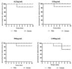
Survival curve of Tenebrio molitor after 10 days of exposure to the metabolite at different concentrations produced by Streptomyces sp. isolated from soil. Source: Author’s own work (2023).
3.4. Biofilm assays
For the biofilm assays, only the Candida species with lower MIC results were tested, as the extract showed toxicity at a concentration of 1,000 µg/mL. All tested samples were found to be biofilm-forming.
3.4.1. Effect of the extract on biofilm formation
Using the applied method, the Streptomyces metabolite reduced biofilm formation in all Candida species at the MIC concentration, with the strongest effects against C. krusei ATCC 6285 (67%) and C. albicans OCI37 (75%). At ½ and ¼ MIC, reductions were observed only for C. albicans OCI37 and C. glabrata VCI124. No inhibitory effect was detected for C. krusei ATCC 6285, C. albicans OCI40, or C. parapsilosis ATCC 22019 at sub-MIC concentrations (Table 3).
3.4.2. Effect of the extract on pre-formed biofilm
The metabolite was also capable of reducing pre-formed biofilm across all tested samples at higher concentrations. Once again, C. krusei ATCC 6285 and C. albicans OCI 37 showed the greatest reduction in biofilm, 68% and 83%, respectively. At ½ and ¼ of the MIC, it was only possible to reduce biofilm for C. albicans OCI 37 and C. glabrata VCI 124. For the species C. krusei ATCC 6285 and C. parapsilosis ATCC 22019, the extract did not show interference at any concentration beyond the MIC (Table 4).
3.5. Gas Chromatography coupled to Mass Spectrometry (GC-MS – HPSE)
Chromatographic analysis of the Streptomyces sp. extract revealed a profile dominated by volatile terpenes, fatty acids, and aromatic nitrogenous compounds, including chemotaxonomic markers geosmin and 2-methylisoborneol. Notably, compounds with reported antimicrobial activity, such as Hexadecanoic Acid, Octadecanoic Acid, Hedycaryol, and 4-(3-methyl-2-butenyl)-1H-indole, were also detected (Figure 4).

4. Discusion
Actinobacteria are a key group of microorganisms known for producing bioactive compounds. Their biotechnological importance has led to extensive studies, revealing their presence in soils, aquatic ecosystems, plants, and animals. In these environments, they contribute to nutrient cycling and pathogen defense. Their adaptation to diverse habitats has driven the evolution of varied biosynthetic pathways, allowing the production of a wide range of secondary metabolites (Jose et al., 2021).
Studies such as that of Lavan et al. (2016) report that impacted soils, such as landfills and sanitary landfills, harbor more genera of actinobacteria compared to other bacterial genera, indicating their proliferation in such soils. In works like Sonia et al. (2011), soil amendment with municipal solid waste compost was conducted, followed by comparison of microbial growth in treated and untreated soil, revealing a significant increase in actinobacteria presence in compost-amended soil, possibly due to increased availability of carbon and nitrogen.
The actinobacterium used in the study was isolated from a soil sample collected from an area impacted by urban solid waste. The isolated microorganism exhibited slow growth with formation of aerial mycelium, and notably, it excreted a burnt yellow pigment in the medium, suggesting a broad spectrum of biological activities, such as antimicrobial properties (Fernandes et al., 2021). Microscopic examination revealed structures characteristic of the genus Streptomyces sp., which is commonly isolated from soil. This observation is supported by studies like Oliveira (2018), where 30 soil samples were isolated, yielding 219 actinobacteria, with Streptomyces genus comprising 48.85% of the isolates.
The genus Streptomyces is well known for its ability to produce enzymes such as proteases and lipases (Al Dhabi et al., 2020), antiparasitic compounds effective against Toxoplasma gondii and Plasmodium falciparum (Pagmadulam et al., 2020), as well as antimicrobial compounds active against Gram-positive and Gram-negative bacteria (Tomaseto et al., 2020) and fungi (Ferreira et al., 2016).
In the present study, the extract from the isolated Streptomyces exhibited antifungal activity against all tested Candida species in the Agar Diffusion test. The largest inhibition zone observed was 17.3 ± 1.15 mm for Candida albicans ATCC 90028, and the Minimum Inhibitory Concentration (MIC) ranged as low as 62.5 μg/mL for Candida parapsilosis ATCC 22019. In a study by Oliveira (2018), the antimicrobial potential of secondary metabolites produced by Streptomyces isolated from Amazonian soil samples was analyzed. Among 153 ethyl acetate metabolite extracts tested, twelve exhibited antimicrobial activity, with five showing activity against Candida albicans ATCC 24433, with an MIC of 312.5 μg/mL and the largest inhibition zone of 12.0 ± 0.0 mm in the Agar Diffusion test.
In a study conducted in Mexico, actinobacterial strains isolated from soil in the state of Tabasco were cultured to produce secondary metabolites for testing their anti-Candida activity. Among the findings, the extract from Streptomyces parvisporogenes showed the lowest MIC (0.5 mg/mL), and the highest activity against Candida was exhibited by the extract produced by S. manipurensis (Réndiz, 2020).
In recent years, science has been seeking ethical and legal alternatives to the use of animals in laboratories, based on the 3Rs principle (replacement, reduction, and refinement), proposed by Russel and Burch in 1959. Brazilian legislation has also progressed in this area. Enacted in 2008, the Arouca Law (No. 11,794/08) established guidelines for the scientific use of animals in Brazil, aiming to ensure their protection and well-being by promoting the replacement with alternative methods whenever possible (Braz and Negri, 2025).
The alternative model using Tenebrio molitor larvae has been widely employed in research due to its ease of handling, low cost, robust immune system, and physiological similarities to vertebrates. T. molitor larvae have been used to investigate drug toxicity and to evaluate pharmacological activity (Cordeiro, 2023).
Systemic candidiasis is reported as a major cause of death in hospital-acquired and opportunistic fungal infections due to its ability to form biofilms that reduce fungal cell susceptibility to treatment (Atriwal et al., 2021). In the tests conducted in this study regarding biofilms, the extract demonstrated a prominent ability to reduce biofilm formation of the tested Candida species, as well as to disrupt pre-formed biofilms. Similar biofilm eradication effects have been observed in other studies, such as the work of Córdova-Dávalos et al. (2018), where an extract from a Streptomyces sp. isolated from soil reduced Candida albicans biofilm formation by approximately 10-30%.
Srivastava and Dubey (2016) characterized the anti-Candida property of the ADP4 strain of Streptomyces chrestomyceticu, isolated from desert soil. Several Candida species were used in the study, including C. albicans, C. krusei, C. tropicalis and C. parapsilosi, all ATCC. A 90% reduction in biofilm was observed, in the case of the 2-hour preformed biofilm, the concentration of the ADP4 metabolite was 8-16 µg/ml.
Among the main species described in bloodstream infections capable of forming biofilms, C. albicans stands out as primary. However, the emergence of non-albicans species is increasingly reported. Biofilms facilitate microbial adherence to medical devices such as catheters, artificial heart valves, and various tissues, enhancing their virulence and complicating treatment, thereby contributing to therapeutic failure (Lazzarotto et al., 2020). Despite evidence of increasing incidence of infections caused by non-albicans species, studies on the effect of Streptomyces extract in reducing biofilms in non-albicans Candida species remain scarce
Volatile organic compounds (VOCs) are low molecular weight substances (generally <300 Da) with low polarity but high vapor pressure. Antimicrobial VOCs are produced by bacteria such as Streptomyces spp (Huang et al., 2012)
The main compounds present in the studied extract, as indicated by some higher peaks, were identified as: 1- Hexadecanoic acid (Palmitic acid), with a retention time of ~21-22 min; 2- 1H-Indole, 4-(3-methyl-2-butenyl)- (Prenylated indole) with a retention time of ~15-16 min; 3- 4aH-Cycloprop[e]azulenol (Globulol analogs) with a retention time of ~17.5-18 min.
Compounds such as 2-methylisoborneol (MIB) and trans-1,10-dimethyl-trans-9-decalol (Geosmin) are responsible for the characteristic earthy smell of moist soils, being the main compounds produced by Streptomyces sp. (Cuervo et al., 2023). The isolate showed a significant production of such compounds, evidencing its contribution to the characteristic chemical profile of the genus.
Chemical characterization of the sample via GC-MS revealed the presence of secondary metabolites belonging to classes with antimicrobial bioactivity. The potential anti-Candida activity observed can be attributed to the distinct, yet complementary, mechanisms exerted by these constituents.
The presence of long-chain fatty acids, such as hexadecanoic (palmitic) acid and octadecanoic (stearic) acid, predominantly suggests a direct action in destabilizing the fungal plasma membrane. The main proposed mechanism of action for antifungal fatty acids states that they insert themselves into the lipid bilayers of fungal membranes, compromising membrane integrity and resulting in an uncontrolled release of intracellular electrolytes and proteins, eventually leading to cytoplasmic disintegration of fungal cells (Guimarães and Venâncio, 2022). Prasath et al. (2020) studied the effect of palmitic acid on C. tropicalis, reporting that the acid reduces C. tropicalis biofilm by decreasing surface hydrophobicity, reduces ergosterol biosynthesis, and has protease/lipase activity. While Guimarães and Venânico (2022) reported activity of Octadecinoic Acid against Candida krusei,
Studies such as that of Pereira et al. (2017) suggest that Sesquiterpenes may play an important role as antifungal agents. Literature points to a permeation capacity through the cell wall.
Tetradecane is a saturated aliphatic alkane hydrocarbon compound that has an oily nature and possesses broad antibacterial and antifungal properties. Experimental reports show antimicrobial activity attributed to isolated Tetradecane; however many publications indicate that effects observed in hexane extracts may result from synergy between n-alkanes and other lipophilic metabolites; therefore, direct mechanistic data in fungi are still scarce (Nasr et al., 2022).
Another component that suggests a synergistic mechanism is Isopropyl Palmitate. Being an ester of hexadecanoic acid, this compound presents a strategic advantage due to its high lipophilicity, which favors interaction with the membrane. Kaewbanjong et al. (2017), in order to define a complex system and potentiate the effect of the imidazole-based compound clotrimazole, studied the antifungal activity of fluid and gel microemulsions formulated with Tween 80 and Span 80 (1:1) 50%, isopropanol 20%, and isopropyl palmitate 30%. Ex vivo release studies, using chorioallantoic membranes that mimic the oral mucosa, emphasized fluid microemulsions as immediate-release systems and favored a broad area of inhibition of C. albicans.
The compound 1H-Indole, 4-(3-methyl-2-butenyl)- is a prenylated indole that appears in microbial and plant extracts active against fungi. Although few studies test this isolated compound against Candida, the literature on prenylated indole demonstrates that this class shows antifungal activity through multiple mechanisms — including membrane interaction, biofilm inhibition, and in some cases direct enzyme inhibition — suggesting that prenyl-indole may contribute to the activity observed in extracts (Li, 2010; Graziani et al., 2021; Makangara et al., 2004).
Acknowledgements
The authors thank Ceuma University (UNICEUMA), the Postgraduate Program in Biodiversity and Biotechnology (BIONORTE Network), and the Electrochemistry and Biotechnology Laboratory for providing infrastructure and technical support essential for conducting this study. The authors also acknowledge financial support from Maranhão State Research Foundation (FAPEMA), (Edital 38/2022; Process ACT-FAPEMA 02510/2023) and Coordination for the Improvement of Higher Education Personnel (CAPES). This research was funded by the Maranhão State Research Foundation (FAPEMA), under Edital FAPEMA 38/2022, Process ACT-FAPEMA 02510/2023.
Data Availability Statement
The dataset analyzed or produced in this study can be requested from the corresponding author.
References
-
ABDELMOHSEN, U.R., GRKOVIC, T., BALASUBRAMANIAN, S., KAMEL, M.S., QUINN, R.J. and HENTSCHEL, U., 2015. Elicitation of secondary metabolism in actinomycetes. Biotechnology Advances, vol. 33, no. 6, pp. 798-811. https://doi.org/10.1016/j.biotechadv.2015.06.003 PMid:26087412.
» https://doi.org/10.1016/j.biotechadv.2015.06.003 -
AL-DHABI, N.A., ESMAIL, G.A., GHILAN, A.M. and ARASU, M.V., 2020. Isolation and screening of Streptomyces sp. Al-Dhabi-49 from the environment of Saudi Arabia with concomitant production of lipase and protease in submerged fermentation. Saudi Journal of Biological Sciences, vol. 27, no. 1, pp. 474-479. https://doi.org/10.1016/j.sjbs.2019.11.011 PMid:31889873.
» https://doi.org/10.1016/j.sjbs.2019.11.011 -
AMORIM, E.A.F., CASTRO, E.J.M., DA SOUZA, S.V., ALVES, M.S., DIAS, L.R.L., MELO, M.H.F., DA SILVA, I.M.A., VILLIS, P.C.M., BONFIM, M.R.Q., FALCAI, A., SILVA, M.R.C., MONTEIRO-NETO, V., ALIANÇA, A., DA SILVA, L.C.N. and DE MIRANDA, R.C.M., 2020. Antimicrobial potential of Streptomyces ansochromogenes (PB3) isolated from a plant native to the Amazon against Pseudomonas aeruginosa. Frontiers in Microbiology, vol. 11, pp. 574693. https://doi.org/10.3389/fmicb.2020.574693 PMid:33162956.
» https://doi.org/10.3389/fmicb.2020.574693 -
ATRIWAL, T., AZEEM, K., HUSAIN, F.M., HUSSAIN, A., KHAN, M.N., ALAJMI, M.F. and ABID, M., 2021. Mechanistic understanding of Candida albicans biofilm formation and approaches for its inhibition. Frontiers in Microbiology, vol. 12, pp. 638609. https://doi.org/10.3389/fmicb.2021.638609 PMid:33995297.
» https://doi.org/10.3389/fmicb.2021.638609 -
BAUER, A.W., KIRBY, W.M., SHERRIS, J.C. and TURCK, M., 1966. Antibiotic susceptibility testing by a standardized single disk method. American Journal of Clinical Pathology, vol. 45, no. 4, pp. 493-496. https://doi.org/10.1093/ajcp/45.4_ts.493 PMid:5325707.
» https://doi.org/10.1093/ajcp/45.4_ts.493 - BRAZ, N.M. and NEGRI, M., 2025. Protocol optimization for LD50 assessment in Tenebrio molitor larvae using commercial antifungals as reference for screening new compounds São José dos Pinhais: Impacto Científico Publishing, pp. 1623-1651.
-
CLARK, F.E., 1965. Agar‐plate method for total microbial count. In: A.C. PAGE, eds. Methods of soil analysis: Part 2. Chemical and microbiological properties Madison: American Society of Agronomy, pp. 1460-1466. https://doi.org/10.2134/agronmonogr9.2.c48
» https://doi.org/10.2134/agronmonogr9.2.c48 - CLINICAL AND LABORATORY STANDARDS INSTITUTE – CLSI, 2008. M27-A3: reference method for broth dilution antifungal susceptibility testing of yeasts: approved standard Wayne: CLSI, vol. 28.
- CLINICAL AND LABORATORY STANDARDS INSTITUTE – CLSI, 2015. M100-S25: Performance standards for antimicrobial susceptibility testing: twenty-fifth informational supplement Wayne: CLSI.
- CORDEIRO, M.L.S., 2023. Extracts of Talisia esculenta contain bioactive compounds with antioxidant activity and anti-inflammatory potential in in vitro and in vivo models Natal: Universidade Federal do Rio Grande do Norte. Tese de Doutorado em Bioquímica e Biologia Molecular.
-
CÓRDOVA-DÁVALOS, L.E., ESCOBEDO-CHÁVEZ, K.G. and EVANGELISTA-MARTÍNEZ, Z., 2018. Inhibition of Candida albicans cell growth and biofilm formation by a bioactive extract produced by soil Streptomyces strain GCAL-25. Archives of Biological Sciences, vol. 70, no. 2, pp. 387-396. https://doi.org/10.2298/ABS170908057C
» https://doi.org/10.2298/ABS170908057C - CORRÊA, G.G., 2014. Potencial biotecnológico de actinobactérias da rizosfera de Caesalpinia pyramidalis Tul. do bioma Caatinga Recife: Universidade Federal de Pernambuco. Dissertação de Mestrado em Biotecnologia.
-
CUERVO, L., ÁLVAREZ-GARCÍA, S., SALAS, J.A., MÉNDEZ, C., OLANO, C. and MALMIERCA, M.G., 2023. The volatile organic compounds of Streptomyces spp.: an in-depth analysis of their antifungal properties. Microorganisms, vol. 11, no. 7, pp. 1820. https://doi.org/10.3390/microorganisms11071820 PMid:37512992.
» https://doi.org/10.3390/microorganisms11071820 -
FERNANDES, C.J., DODDAVARAPU, B., HARRY, A., DILIP, S.P.S. and RAVI, L., 2021. Isolation and identification of pigment producing actinomycete Saccharomonospora azurea SJCJABS01. Biomedicine and Pharmacotherapy Journal, vol. 14, no. 4, pp. 2261-2269. https://doi.org/10.13005/bpj/2326
» https://doi.org/10.13005/bpj/2326 -
FERREIRA, H.K.L., MACHADO, S.E.F., SANTANA, R.C.F., ALBUQUERQUE, L.E.F., SILVA, I.D.E.G., SILVA-LACERDA, G.R., ARAÚJO, J.M. and LIMA, G.M.S., 2016. Avaliação in vitro do potencial antimicrobiano de Streptomyces sp. G-27 contra microrganismos de interesse clínico. Revista Brasileira de Gestão Ambiental e Sustentabilidade, vol. 3, no. 6, pp. 367-373. https://doi.org/10.21438/rbgas.030610
» https://doi.org/10.21438/rbgas.030610 -
FERRO, T.A.F., COSTA MORAES, F., MENESES DA SILVA, A., PORCY, C., AMORIM SOARES, L., ANDRADE MONTEIRO, C., MELO LOBÃO, N.T., ASSIS DE MELLO, F.A., MONTEIRO-NETO, V. and FIGUEIRÊDO, M.S., 2012. Caracterização de fatores de virulência em cepas de Escherichia coli enteroagregativas e enteropatogênicas atípicas isoladas de crianças com diarreia. Advances in Infectious Diseases, vol. 2, no. 4, pp. 128-133. https://doi.org/10.4236/aid.2012.24022
» https://doi.org/10.4236/aid.2012.24022 - FRITSCH, L.N., DIAS, A.L.T., SILVA, N.C., FERNANDES, G.J.M. and RIBEIRO, F.B.A.O., 2021. Comparative analysis of biofilm formation by Candida albicans and Candida krusei in different types of contact lenses. Arquivos Brasileiros de Oftalmologia, vol. 85, no. 3, pp. 235-239. PMid:34586234.
-
GOULART, L.S., SOUZA, W.W.R., VIEIRA, C.A., LIMA, J.S., OLINDA, R.A. and ARAÚJO, C., 2018. Colonização oral por espécies de Candida em pacientes HIV positivo: estudo de associação e suscetibilidade antifúngica. Einstein, vol. 16, no. 3, pp. 1-6. https://doi.org/10.1590/s1679-45082018ao4224 PMid:30088546.
» https://doi.org/10.1590/s1679-45082018ao4224 -
GRAZIANI, F.S., POTENZA, N., D’ABROSCA, B., TROIANI, T., NAPOLITANO, S., FIORENTINO, A. and SCOGNAMIGLIO, M., 2021. NMR profiling of ononis diffusa identifies cytotoxic compounds against cetuximab-resistant colon cancer cell lines. Molecules, vol. 26, no. 11, pp. 3266. https://doi.org/10.3390/molecules26113266
» https://doi.org/10.3390/molecules26113266 -
GUIMARÃES, A. and VENÂNCIO, A., 2022. The potential of fatty acids and their derivatives as antifungal agents: a review. Toxins, vol. 14, no. 3, pp. 188. https://doi.org/10.3390/toxins14030188 PMid:35324685.
» https://doi.org/10.3390/toxins14030188 -
HUANG, R., CHE, H.J., ZHANG, J., YANG, L., JIANG, D.H. and LI, G.Q., 2012. Evaluation of Sporidiobolus pararoseus strain YCXT3 as biocontrol agent of Botrytis cinerea on post-harvest strawberry fruits. Biological Control, vol. 62, no. 1, pp. 53-63. https://doi.org/10.1016/j.biocontrol.2012.02.010
» https://doi.org/10.1016/j.biocontrol.2012.02.010 -
INSTITUTO BRASILEIRO DE GEOGRAFIA E ESTATÍSTICA – IBGE, 2021 [viewed 13 July 2026]. Cidades e Estados: Pinheiro (MA) [online]. Rio de Janeiro: IBGE. Available from: https://www.ibge.gov.br/cidades-e-estados/ma/pinheiro.html
» https://www.ibge.gov.br/cidades-e-estados/ma/pinheiro.html -
JOSE, P.A., MAHARSHI, A. and JHA, B., 2021. Actinobacteria in natural products research: progress and prospects. Microbiological Research, vol. 246, pp. 126708. https://doi.org/10.1016/j.micres.2021.126708 PMid:33529791.
» https://doi.org/10.1016/j.micres.2021.126708 -
KAEWBANJONG, J., WAN SIA HENG, P. and BOONME, P., 2017. Prapaporn Boonme, Clotrimazole microemulsion and microemulsion-based gel: evaluation of buccal drug delivery and irritancy using chick chorioallantoic membrane as the model. The Journal of Pharmacy and Pharmacology, vol. 69, no. 12, pp. 1716-1723. https://doi.org/10.1111/jphp.12809 PMid:28836273.
» https://doi.org/10.1111/jphp.12809 -
KAMJAM, M., SIVALINGAM, P., DENG, Z. and HONG, K., 2017. Deep sea actinomycetes and their secondary metabolites. Frontiers in Microbiology, vol. 8, pp. 760. https://doi.org/10.3389/fmicb.2017.00760 PMid:28507537.
» https://doi.org/10.3389/fmicb.2017.00760 - LAVAN, S., KAVITHA, D. and ILAKKIYA, S., 2016. Diversity of cellulose degrading actinomycetes in waste landfill ecosystem. Asian Journal of Microbiology, Biotechnology & Environmental Sciences : AJMBES, vol. 18, pp. 967-973.
-
LAZZAROTTO, E.S., DE MELO VASCO, J.F., LOPES, C.M., KRELLING, A., VILLA STANGLER AREND, L.N. and RODRIGUES, L.S., 2020. Identificação de leveduras do gênero Candida isoladas de hemoculturas de pacientes oncológicos e pesquisa de biofilme. Revista UNINOVE, vol. 57, no. 3, pp. 85-94. https://doi.org/10.46311/2318-0579.57.eUJ3502
» https://doi.org/10.46311/2318-0579.57.eUJ3502 -
LI, S.M., 2010. Prenylated indole derivatives from fungi: structure diversity, biological activities, biosynthesis and chemoenzymatic synthesis. Natural Product Reports, vol. 27, no. 1, pp. 57-78. https://doi.org/10.1039/B909987P PMid:20024094.
» https://doi.org/10.1039/B909987P - LIMA JUNIOR, A.A., 2020. Potencial biotecnológico de actinobactéria isolada da rizosfera de Aniba parviflora Syn Fragans (Macacaporanga) da Amazônia Recife: Universidade Federal de Pernambuco. Tese de Doutorado em Biotecnologia.
-
MAKANGARA, J.J., HENRY, L., JONKER, S.A. and NKUNYA, M.H., 2004. The caulindoles: dimeric prenylindoles from Isolona cauliflora. Phytochemistry, vol. 65, no. 2, pp. 227-232. https://doi.org/10.1016/j.phytochem.2003.10.010 PMid:14732283.
» https://doi.org/10.1016/j.phytochem.2003.10.010 - MARTINEZ-ROSSI, N.M., BITENCOURT, T.A., PERES, N.T.A., LANG, E.A.S., GOMES, E.V., QUARESEMIN, N.R., MARTINS, M.P., LOPES, L. and ROSSI, A., 2018. Resistência de dermatófitos a antifúngicos: mecanismos e prospecto. Frontiers in Microbiology, vol. 9, no. 108.
- MILAGRE, L.P., 2017. Caracterização de metabólitos secundários produzidos por isolados do gênero Streptomyces com atividade contra fungos fitopatogênicos Porto Alegre: Universidade Federal do Rio Grande do Sul.
- NASR, Z.S., EL-SHERSHABY, H., SALLAM, K., ABED, N., EL GHANY, I.A., SIDKEY, N.M., 2022. Evaluation of antimicrobial potential of tetradecane extracted from Pediococcus acidilactici DSM: 20284-CM isolated from curd milk. Egyptian Journal of Chemistry, vol. 65, no. 3, pp. 705-713.
- OLIVEIRA, R.C., 2018. Potencial antimicrobiano de actinomicetos de solos amazônicos Rio Branco: Universidade Federaldo do Acre.
-
PAGMADULAM, B., TSERENDULAM, D., RENTSENKHAND, T., IGARASHI, M., SAWA, R., NIHEI, C.I. and NISHIKAWA, Y., 2020. Isolation and characterization of antiprotozoal compound-producing Streptomyces species from Mongolian soils. Parasitology International, vol. 74, pp. 101961. https://doi.org/10.1016/j.parint.2019.101961 PMid:31437553.
» https://doi.org/10.1016/j.parint.2019.101961 -
PEREIRA, F.G., MARQUETE, R., DOMINGOS, L.T., ROCHA, M.E.N., FERREIRA-PEREIRA, A., MANSUR, E. and MOREIRA, D.L., 2017. Antifungal activities of the essential oil and its fractions rich in sesquiterpenes from leaves of Casearia sylvestris Sw. Anais da Academia Brasileira de Ciências, vol. 89, no. 4, pp. 2817-2824. https://doi.org/10.1590/0001-3765201720170339 PMid:29236852.
» https://doi.org/10.1590/0001-3765201720170339 -
PRASATH, K.G., THARANI, H., KUMAR, M.S. and PANDIAN, S.K., 2020. Palmitic acid inhibits the virulence factors of candida tropicalis: biofilms, cell surface hydrophobicity, ergosterol biosynthesis, and enzymatic activity. Frontiers in Microbiology, vol. 11, pp. 864. https://doi.org/10.3389/fmicb.2020.00864 PMid:32457728.
» https://doi.org/10.3389/fmicb.2020.00864 -
PUTRANTI, A., ASMARAWATI, T.P., RACHMAN, B.E., HADI, U. and NASRONUDIN., 2018. Oral candidiasis as clinical manifestation of HIV/AIDS infection in Airlangga University Hospital patients. IOP Conference Series. Earth and Environmental Science, vol. 125, no. 1, pp. 012063. https://doi.org/10.1088/1755-1315/125/1/012063
» https://doi.org/10.1088/1755-1315/125/1/012063 -
RAMOS, A.D., MARTINS, P.S.O., FIAUX, S.B. and LEITE, S.G.F., 2009. Solid-phase microextraction of 6-pentyl-α-pyrone produced by solid-state fermentation. Food Science and Technology, vol. 29, no. 3, pp. 523-528. https://doi.org/10.1590/S0101-20612009000300011
» https://doi.org/10.1590/S0101-20612009000300011 - RÉNDIZ, D.Y.E., 2020. Identificacoón molecular y evaluacoón anti-candida de cepas de Streptomyces aisladas de suelos del trópico mexicanos, cultivados en diferentes fuentes de carbono y nitrógeno Mérida, México: Centro de Investigación Científica de Yucatán. Tese de Doutorado.
-
SAYGIN, H., AY, H., GUVEN, K., CETIN, D. and SAHIN, N., 2020. Streptomyces cahuitamycinicus sp. nov., isolated from desert soil and reclassification of Streptomyces galilaeus as a later heterotypic synonym of Streptomyces bobili. International Journal of Systematic and Evolutionary Microbiology, vol. 70, no. 4, pp. 2750-2759. https://doi.org/10.1099/ijsem.0.004103 PMid:32176603.
» https://doi.org/10.1099/ijsem.0.004103 -
SHIN, J.H., KEE, S.J., SHIN, M.G., KIM, S.H., SHIN, D.H., LEE, S.K., SUH, S.P. and RYANG, D.W., 2002. Biofilm production by isolates of Candida species recovered from nonneutropenic patients: comparison of bloodstream isolates with isolates from other sources. Journal of Clinical Microbiology, vol. 40, no. 4, pp. 1244-1248. https://doi.org/10.1128/JCM.40.4.1244-1248.2002 PMid:11923339.
» https://doi.org/10.1128/JCM.40.4.1244-1248.2002 -
SHIRLING, E.B. and GOTTLIEB, D., 1966. Methods for characterization of Streptomyces species. International Journal of Systematic Bacteriology, vol. 16, no. 3, pp. 313-340. https://doi.org/10.1099/00207713-16-3-313
» https://doi.org/10.1099/00207713-16-3-313 -
SHIVLATA, L. and SATYANARAYANA, T., 2015. Thermophilic and alkaliphilic actinobacteria: biology and potential applications. Frontiers in Microbiology, vol. 6, no. 1014., pp. 1014. https://doi.org/10.3389/fmicb.2015.01014 PMid:26441937.
» https://doi.org/10.3389/fmicb.2015.01014 -
SONIA, M.T., NACEUR, J. and ABDENNACEUR, H., 2011. Studies on the ecology of actinomycetes in an agricultural soil amended with organic residues: I. Identification of the dominant groups of Actinomycetales. World Journal of Microbiology & Biotechnology, vol. 27, no. 10, pp. 2239-2241. https://doi.org/10.1007/s11274-011-0687-5
» https://doi.org/10.1007/s11274-011-0687-5 -
SPINLER, J.K., HAIDACHER, S.J., HOCH, K.M., LUNA, R.A. and HAAG, A.M., 2019. Discerning strain-specific β-lactam drug resistance by clonal isolates of multi-drug resistant Pseudomonas aeruginosa using selected reaction monitoring. International Journal of Mass Spectrometry, vol. 438, pp. 36-43. https://doi.org/10.1016/j.ijms.2018.12.013
» https://doi.org/10.1016/j.ijms.2018.12.013 -
SRIVASTAVA, V. and DUBEY, A.K., 2016. Anti-biofilm activity of the metabolites of Streptomyces chrestomyceticus strain ADP4 against Candida albicans. Journal of Bioscience and Bioengineering, vol. 122, no. 4, pp. 434-440. https://doi.org/10.1016/j.jbiosc.2016.03.013 PMid:27117484.
» https://doi.org/10.1016/j.jbiosc.2016.03.013 -
TODD, R.T., SOISANGWAN, N., PETERS, S., KEMP, B., CROOKS, T., GERSTEIN, A. and SELMECKI, A., 2023. Antifungal drug concentration impacts the spectrum of adaptive mutations in Candida albicans. Molecular Biology and Evolution, vol. 40, no. 1, pp. msad009. https://doi.org/10.1093/molbev/msad009 PMid:36649220.
» https://doi.org/10.1093/molbev/msad009 -
TOMASETO, A.A., ALPISTE, M.C., NASSAR, A.F.D.C. and DESTÉFANO, S.A.L., 2020. Atividade antimicrobiana de Streptomyces fitopatogênicas contra bactérias associadas a doenças de importância clínica. Arquivos do Instituto Biológico, vol. 87, pp. e0142020. https://doi.org/10.1590/1808-1657000142020
» https://doi.org/10.1590/1808-1657000142020 -
TRISUWAN, K., RUKACHAISIRIKUL, V., SUKPONDMA, Y., PREEDANON, S., PHONGPAICHIT, S., RUNGJINDAMAI, N. and SAKAYAROJ, J., 2008. Epoxydons and a pyrone from the marine-derived fungus Nigrospora sp. PSU-F5. Journal of Natural Products, vol. 71, no. 8, pp. 1323-1326. https://doi.org/10.1021/np8002595 PMid:18646829.
» https://doi.org/10.1021/np8002595 -
TSANG, P.W.K., BANDARA, H.M.H.N. and FONG, W.P., 2012. Purpurin suppresses Candida albicans biofilm formation and hyphal development. PLoS One, vol. 7, no. 11, pp. e50866. https://doi.org/10.1371/journal.pone.0050866 PMid:23226409.
» https://doi.org/10.1371/journal.pone.0050866
Edited by
-
Editor:
Takako Matsumura Tundisi


