
 Genomic prediction of diabetic retinopathy via VEGF-A variants: insights from meta-analysis, machine learning and molecular docking approaches
Abstract
Diabetic retinopathy (DR) is a complex ocular microvascular neurodegenerative condition resulting from chronic hyperglycemia and hypoxia. Genetic variants, particularly in the vascular endothelial growth factor A (VEGF-A) gene, play a key role in promoting pathological angiogenesis. This study conducted a systematic review and meta-analysis to identify key VEGF-A variants associated with DR development. Observational studies were retrieved from PubMed/Medline, Web of Science, Virtual Health Library, Embase, and Scopus, without time restrictions. Moreover, machine learning (ML) models were applied to enhance genetic risk prediction for DR. Additionally, molecular docking was performed to assess the impact of the −460T>C single-nucleotide variant (SNV) on the interaction between the VEGF-A promoter and the HIF1 transcription factor. Of the 20 studies included in the systematic review, 14 were eligible for meta-analysis. A significant association was found between the C allele of the rs833061 (−460T>C) variant and DR risk (OR = 1.49; 95% CI = 1.17–1.90; p = 0.0014), as well as in the dominant genotypic model (TT vs. TC + CC; OR = 1.76; 95% CI = 1.13–2.74; p = 0.0130). Although the −2549 I/D variant was not significant in the meta-analysis, ML models indicated its predictive value. Molecular docking revealed enhanced binding affinity of the −460C allele to HIF1, suggesting functional relevance. In conclusion, these findings support the potential of VEGF-A variants, particularly rs833061, as biomarkers for DR susceptibility and highlight the utility of integrating ML and molecular docking for predictive medicine.
Keywords:
VEGF-A; angiogenesis; diabetic retinopathy; docking molecular; machine learning
Resumo
A retinopatia diabética (RD) é uma complicação microvascular ocular complexa, resultante da hiperglicemia crônica e da hipóxia. Variantes genéticas, especialmente no gene do fator de crescimento endotelial vascular A (VEGF-A), desempenham papel fundamental na promoção da angiogênese patológica. Este estudo realizou uma revisão sistemática e meta-análise para identificar variantes-chave do gene VEGF-A associadas ao desenvolvimento da RD. Foram incluídos estudos observacionais das bases PubMed/Medline, Web of Science, Biblioteca Virtual em Saúde, Embase e Scopus, sem restrições de período. Modelos de aprendizado de máquina (ML) foram aplicados para aprimorar a predição de risco genético para RD. Além disso, foi realizada análise de docking molecular para avaliar o impacto da variante −460T>C (rs833061) na interação entre o promotor do gene VEGF-A e o fator de transcrição HIF1. Dos 20 estudos incluídos na revisão sistemática, 14 foram elegíveis para a meta-análise. Observou-se associação significativa entre o alelo C da variante rs833061 e o risco de RD (OR = 1,49; IC 95% = 1,17–1,90; p = 0,0014), bem como no modelo genotípico dominante (OR = 1,76; IC 95% = 1,13–2,74; p = 0,0130). Embora a variante −2549 I/D não tenha sido significativa na meta-análise, os modelos de ML indicaram valor preditivo. O docking molecular revelou maior afinidade de ligação do alelo −460C ao HIF1, sugerindo relevância funcional. Esses achados reforçam o potencial das variantes do VEGF-A como biomarcadores de suscetibilidade à RD e destacam a integração de ML e docking molecular na medicina preditiva.
Palavras-chave:
VEGF-A; angiogênese; retinopatia diabética; docking molecular; aprendizado de máquina
1. Introduction
Diabetic retinopathy (DR) is a major microvascular complication of diabetes mellitus and the leading cause of vision loss (Lin et al., 2021). It progresses from a non‑proliferative stage (NPDR), characterized by increased vascular permeability and retinal ischemia, to a proliferative stage (PDR), marked by pathological neovascularization and vitreous hemorrhage (Crasto et al., 2021; Chaudhary et al., 2021). In DR, chronic hyperglycemia, hypoxia (Fung et al., 2022), and inflammation activate biochemical pathways that lead to capillary damage (Moriya and Ferrara, 2015), pericyte apoptosis, and vasoconstriction (Wang and Lo, 2018), promoting increased secretion of angiogenic factors and pathological retinal neovascularization (Invernizzi et al., 2023). These processes upregulate transcription factors and pro-angiogenic growth factors, leading to PDR and diabetic macular edema (DME) (Tsai et al., 2018).
This microcomplication is a complex, multifactorial condition influenced by poor glycemic control, disease duration (Tarasewicz et al., 2023), hypertension (Hainsworth et al., 2019), and genetic factors (Sienkiewicz-Szłapka et al., 2023). Among these, VEGF-A is highlighted as a key gene, located on chromosome 6 (6p21.1) (NCBI, 2024), encoding multiple isoforms that regulate angiogenesis and vascular homeostasis (Peach et al., 2018).
VEGF-A maintains retinal vascular homeostasis and regulates angiogenesis (Apte et al., 2019). Under normal conditions, its expression is low, promoting vascular repair. However, hyperglycemia markedly upregulates VEGF-A, increasing vascular permeability and promoting neovascularization (Bucolo et al., 2021; Simmons et al., 2018), which contributes to DR progression (Arrigo et al., 2022).
In diabetes, hyperglycemia and dyslipidemia induce hypoxia, causing VEGF-A overexpression via hypoxia-inducible factor 1α (HIF-1α) activation (Yin et al., 2021). HIF binds to hypoxia response elements (HRE) in the VEGF-A promoter, improving transcription and disrupting vascular homeostasis (Alves et al., 2021; Youngblood et al., 2019; Semenza, 2017). VEGF-A contributes to DR pathogenesis, with elevated vitreous levels associated with increased vascular permeability and neovascularization (Bucolo et al., 2021; Singh et al., 2019). This review aimed to identify VEGF‑A variants associated with DR and to clarify their role in disease susceptibility.
2. Materials and Methods
2.1. Registration and search strategy
A systematic review was performed to identify VEGF-A polymorphisms associated with DR susceptibility. To avoid duplication, the study protocol was registered on the International Prospective Registry of Systematic Reviews Platform (PROSPERO) on March 19, 2023 (number CRD42023406487) and reported according to the Preferred Reporting Items for Systematic Reviews and Meta-Analyses (PRISMA) guidelines.
The literature search was conducted from March to July 2023 in PubMed/Medline, Web of Science, Virtual Health Library (VHL), Embase, and Scopus. The strategy (Table S1, Supplementary Material) followed the PEO acronym: "P" for diabetic with patients with DR, "E" for SNVs in the VEGF-A gene, and "O" for their association with DR. “Genetic Polymorphism”, “VEGF‑A gene”, and “diabetic retinopathy” were adapted to each database
2.2. Inclusion and exclusion criteria
Observational studies in humans investigating the association between VEGF‑A gene variants and DR were included. Animal studies, reviews, and studies not related to DR or VEGF‑A were excluded. No temporal restriction was applied.
2.3. Selection of articles and evaluation of methodological quality
The selected studies were exported to Rayyan platform and evaluated by two reviewers (SSNLC and CCPC) in two phases: titles and abstracts, and full text, following pre-established inclusion and exclusion criteria. Methodological risk was bias assessed using the Joanna Briggs Institute (JBI) Critical Assessment tool, with studies with at least 70% positive responses considered low risk. Disagreements were resolved by consensus among the reviewers.
2.4. Data extraction
The data extraction from the articles selected in phase II was performed by 2 independent reviewers (SSNLC and CCPC). Data were inserted into a standardized form, containing the following information: (1) authors and year of publication, (2) type of study, (3) population, (4) ethnicity, (5) average age, (6) gene, (7) analyzed polymorphism and (8) genotypic and allele frequencies.
2.5. Statistical analysis
The association between genetic variants and DR was estimated by odds ratios (ORs) with 95% confidence interval (CI), using the dominant genetic model. Heterogeneity was assessed by the Higgins inconsistency test (I2), defining the model as fixed (I2< 25%) or random (I2>25%). Publication bias was assessed by Egger’s test, considering a p-value < 0,05 and all analyses were performed in RStudio®, version 4.0.4.
To analyze the association of SNVs with disease, supervised machine learning (ML) was applied using five models: support vector machine (SVM), logistic regression (LR), decision tree (DT), random forest (RF), and Bayesian models, based on predictive performance.
2.6. In silico analysis
The transcription factor for docking with the VEGF-A gene promoter was selected based on scientific literature. The HIF1a (Q16665) and ARNT (P27540) subunits were obtained from Uniprot, and the 3D model of HIF1 was generated via SWISS-MODEL, following standard comparative modeling steps.
The FASTA sequences of the HIF1A and ARNT subunits were used as input for modeling. The generated models were validated using the Saves 3.4 tool, which includes PROCHECK (Ramachandran angle analysis) and ERRAT (structural evaluation by statistical comparison). Models with more than 90% of the amino acids in favorable regions were considered high quality.
2.6.1. Modeling of the HRE-like region in the promoter region of the VEGF-A gene in relation to the -460T>C polymorphism
The 3D DNA structures were made from the 5′GTGTT′3 sequence present in the VEGF-A promoter region at positions −459 and −463 (reference position chr6:43769749 (GRCh 38.p14), containing the T alleles or C of the −460T>C variation site (rs833061), along with 14 flanking nucleotides (5′ TGTGGGGTTGAGGGT/CGTTGGAGCGGGGAG ′3, referring to nucleotides −461 to −457), using Discovery Studio 5 software. The choice of the selected sequence was made according to the presence of the polymorphism and its similarity with the HRE sequence 5′GCGTG′3, where HIF1 binding occurs.
2.6.2. Molecular docking
HADDOCK v.2.4 software was used to perform protein-DNA docking between HIF1 and the promoter region of the VEGFA gene containing the -460T or -460C alleles (Van Zundert et al., 2016; Dominguez et al., 2003). Amino acids S22, R23, A26, A27, R29, and R30 were selected as active residues for the α subunit (HIF1a), and H94, E98, R101, and R102 for the β subunit (ARNT), due to their high conservation among HIF1 family proteins (Wu et al., 2015; Wu et al., 2016). For the DNA, the nucleotides −459G, −460T/C, −461G, −462T, and −463T were chosen, corresponding to the HRE-like sequence and the polymorphism analyzed. The server automatically defined the passive residues.
Docking was performed using default parameters, with specific adjustments to temperature and sampling settings, as previously described (Honorato et al., 2019; Van Zundert et al., 2016). Protein–DNA interactions were subsequently analyzed using the Protein–Ligand Interaction Profiler (PLIP) server (Adasme et al., 2021).
3. Results
3.1. Individual study results and meta-analysis
After the initial search, 422 articles were identified. In Phase I, 175 duplicates and 186 articles that were not considered eligible were excluded, leaving 62 articles for full‑text review. In Phase II, 31 articles were excluded after detailed assessment (Figure 1), resulting in 20 studies (13 case-control and 7 cohort) included in the systematic review.
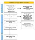
All selected articles were published in English. Sample sizes varied across studies: 33 to 200 cases and 35 to 493 controls (Frame S1). The meta-analysis was conducted using 14 articles selected from the systematic review, performed with five genetic variants. The association between genetic variants in the VEGF-A and the risk of DR was evaluated using the dominant (Wild vs. Heterozygous + mutant) and allelic models.
This systematic review meta-analyzed the variants: -152A>G (rs13207351), -2578C>A (rs699947), -634G>C (rs2010963), and -460T>C (rs833061) and variant of insertion/deletion -2549 (I/D) with risk being estimated by OR and 95% CI. No association was found for A-152G (OR= 0,19; CI 95%=0,05-0,71; p=0,0129), C-2578A (OR= 0,47; CI 95%=0,04-5,79; p=0,5572) and G-634C (OR=0,72; CI 95%=0,44-1,17; p=0,1869) in genotypic and allelic comparisons (Figure S1, Supplementary Material). However, T-460C (rs833061) showed a significant association for the mutant allele, with 1.49-fold increased risk of developing DR (95% CI= 1,17-1,90; p=0,0014) and genotypic comparison (OR=1,76; CI 95% =1,13-2,74; p=0,0130) (Figure 2).

Forest plot for genotypic and allelic comparisons of SNV T-460C (rs833061). A) allele frequency; B) genotype frequency.
Additionally, the I/D -2549 variant showed no significant association between the D allele and the risk of DR (OR=0,84; CI 95%=0,65-1,09; p=0,1971), nor in the genotypic analysis under the dominant model (II vs. ID + DD) (OR=0,84; CI 95%=0,65-1,09; p=0,84) (Figure 3).

Forest plot for genotypic and allelic comparisons of the I-2549D Insertion/Deletion variant. A) allele frequency and B) genotypic frequency.
In the publication bias analysis, the funnel plot did not identify a significant risk for any of the SNVs (Figures S2 to S4, Supplementary Material), while the Egger test for allelic (A-152G: p=0.16; G-634C: p=0.09), and genotypic (A-152G: p=0.10; G-634C: p=0.15) comparisons also was found no risk of publication bias. For SNV C-2578A, the genotypic frequency analysis showed no evidence of publication bias (p=0.56); however, the allelic frequency test could not be performed. Similarly, bias analysis for the T‑460C and I‑2549D SNVs could not be performed due to the limited number of included studies.
3.2. Supervised machine learning approach
The adequacy of the ML models was evaluated by three parameters: accuracy, precision, and recall. The RF model was selected based on the parameter adjustment (Table S2, Supplementary Material). The results showed that the rs833061 and I/D -2549 variants were the main factors observed to predict DR in the selected studies.
Figure 4 shows the results of the evaluative parameters of the RF model applied to the training and test data for the rs833061 (Figure 4A) and I/D -2549 (Figure 4B) variants. The metrics used to evaluate the performance of each study represent estimates that range from 0 to 1, indicating the accuracy of the analyzed dataset. Both variants presented results similar to 1 for the evaluated parameters, indicating a positive prediction. The other variants did not present a good prediction, considering the parameters analyzed (Figure S5, Supplementary Material).

Parameters analyzed by machine learning of the SNVs: A) T-460C (rs833061). B) Insertion/deletion variant -2549.
3.3. In silico analysis and molecular docking
Four homology models were generated, showing 82.97% identity with the HIF-2α:ARNT complex bound to HRE DNA (PDB ID: 4zpk). Structural validation using SAVES (Ramachandran and ERRAT) indicated good stereochemical quality (Table S3, Supplementary Material). The HIF1 model from Swiss-Model had 83.9% residues in favorable regions, supporting its suitability for docking.
Protein-DNA docking was performed in HADDOCK v2.4 using HIF1 and DNA sequences with −460T or −460C alleles. For HIF1-460T, 293 structures were grouped into 28 clusters (73%), and for HIF1-460C, 291 structures into 24 clusters (72%). Clusters were ranked by HADDOCK score, and the best poses (clusters 28 and 21) were selected as final complexes (Table S4, Supplementary Material).
Intermolecular interactions were analyzed with PLIP and visualized in PyMol. The HIF1-460T complex formed five hydrogen bonds, while HIF1-460C formed 13, including seven with the HRE region. Key residues involved included H94, E98, R102 (ARNT) and R30 (HIF1α), with E98 binding −460C and −459rC. The −460C allele increased hydrogen bonding and altered the binding pattern, enhancing molecular interaction.
4. Discussion
This systematic review and meta-analysis summarized 14 studies, highlighting SNVs: rs13207351, rs699947, rs2010963, and rs833061. and the insertion/deletion variant -2549 (I/D). The meta-analysis found no significant association between rs13207351, rs699947, rs2010963, and the −2549 I/D variant with DR risk under the dominant model; however, previous literature reports remain inconsistent (Churchill et al., 2008; Nakamura et al., 2008; Awata et al., 2002; Singh et al., 2021; Xie et al., 2017).
Several genetic epidemiological studies have reported an association between the rs13207351 variant and DR susceptibility and severity. (Churchill et al., 2008; Wagih et al., 2022; Yang et al., 2011). Wagih et al. (2022) also revealed elevated VEGF-A levels and a strong association between PDR and rs13207351 combined with 14 VEGF-A SNVs (OR=18.26; p=0.00622). Conversely, Yang et al. (2011) and Wagih et al. (2022) described that the wild-type genotype (AA) increased the DR risk progression. On the other hand, in this present meta-analysis, no significant association was observed for this variant. These conflicting results may reflect ethnic differences or other risk factors requiring further investigation.
The SNV rs699947 has been associated with increased DR risk in Asian populations (Nakamura et al., 2008; Singh et al., 2021; Wijaya et al., 2021). For instance, Singh et al. (2021) demonstrated a 1.03-fold risk (p=0.004), while Yang et al. (2011) identified a 4-fold increase for the AA mutant genotype (OR=3.98; 95% CI=1.38–11.42; p=0.031). Similarly, Nakamura et al. (2008) found a strong association with the AA genotype in Japanese patients (OR= 7.5; p=0.002). However, our findings did not confirm this association, showing no significant effect of rs699947 on DR risk.
The SNV rs2010963 described no significant association with DR risk in Chinese individuals, according to Yuan et al. (2014), which corroborates our meta-analysis findings. In contrast, studies by Awata et al. (2002) and Yang et al. (2010) reported a significant association of the CC genotype with a 2.16 and 2.60-fold risk DR, respectively. Additionally, Awata et al. (2005) further suggested that this variant may increase VEGF-A transcriptional activity and serum levels, contributing to disease progression.
In a study involving Indian patients, individuals with DR exhibited a higher frequency of heterozygous genotypes, and the mutant C allele was identified as risk factor. (Suganthalakshmi et al., 2006). Consistently, Awata et al. (2005) observed in Japanese patients that the heterozygous genotype of variant 634G>C was associated with an increased DR risk and DME, regardless of disease severity. It has been suggested that the G>C substitution may enhance VEGF‑A expression in the retina, thereby contributing to the development of DR and DME. Differences among studies may be attributed to population‑specific genetic backgrounds, sample size, methodological approaches, and the influence of additional risk factors, suggesting the need for additional investigation.
Interestingly, this systematic review and meta-analysis identified only one insertion/deletion variant (-2549 I/D) in the VEGF gene. Singh et al. (2021) described that the DD genotype confers a 2.27-fold increased risk of DR, with the (D) allele associated with enhanced VEGF-A transcription. Despite this, in our pooled analysis, no significant association was observed for allele frequencies or genotypic distributions. Although evidence remains limited, Buraczynska et al. (2007) suggest that the (D) allele may contribute to DR susceptibility through higher transcriptional activity compared to the (I) allele, potentially leading to elevated VEGF-A levels. These findings support the need for large scale, well‑designed studies to clarify the role of this variant in DR pathogenesis.
Among the analyzed variants, the −460T/C SNV (rs833061) is of particular interest because it is located in the promoter region of the VEGF gene, which is critical for transcriptional regulation. This polymorphism showed a significant association with increased risk of DR in both allelic and genotypic comparisons. The TC + CC genotypes and the C allele were more frequent among diabetic patients, suggesting an increased predisposition to DR. This association may reflect differences in VEGF-A expression levels among individuals, contributing to pathological processes such as DR (Xie et al., 2017). Consistent with our findings, previous meta‑analyses (Xie et al., 2017; Gong and Sun, 2013) have indicated an increased risk associated with the C allele in relation to DR susceptibility.
Gong and Sun (2013) demonstrated increased DR risk for CT + CC versus TT (OR=1.78; p=0.04) and CC versus TT + TC (OR = 1.76; p=0.02), which aligns with our findings for the dominant model (OR=1.49; p=0.0014). In total, these results reinforce the hypothesis that the C allele of SNV rs833061 may contribute to DR susceptibility, possibly through increased promoter activity and VEGF-A expression. This interpretation is further supported by our molecular docking analysis, which revealed that the -460C allele enhances HIF1 binding to the VEGF-A promoter, potentially amplifying transcriptional activation and promoting pathological angiogenesis in the retina. Similar structural studies have shown that nucleotide changes in HRE-like regions can modulate HIF1-DNA binding affinity and transcriptional regulation (Alves et al., 2021; Wu et al., 2015), providing a mechanistic basis for these observations.
Our molecular docking analysis demonstrated that the −460C allele (rs833061) significantly altered the binding pattern of the HIF1 transcription factor to the VEGF‑A promoter region. The mutant allele formed a greater number of hydrogen bonds compared to the wild-type allele, indicating a more stable interaction. Notably, the E98 residue, a conserved site within the bHLH-PAS family, was involved in the interaction only in the mutant complex, reinforcing the hypothesis of increased binding specificity.
These structural insights suggest that the -460C allele enhances the stability and specificity of the HIF1–VEGF-A complex, potentially leading to increased transcriptional activity. This mechanistic evidence supports the observed association between the rs833061 variant and elevated VEGF-A expression, which may contribute to pathological angiogenesis and increased vascular permeability in DR.
Additionally, the results of the meta-analysis for these SNVs, rs13207351, rs699947, rs2010963, and the insertion/deletion variant -2549 (I/D) variant revealed no association with DR risk but should be interpreted with caution due to high heterogeneity. Subgroup analyses were not possible due to the small number of studies included. According to the Cochrane Collaboration, this type of analysis is uncommon in systematic reviews because separate participant data are rarely published (Higgins et al., 2023).
Our findings were subsequently used for a complementary analysis employing supervised ML (MacEachern and Forkert, 2021). Among the models tested, random forest was selected due to its strong performance in prediction tasks and its growing application in precision medicine (Myszczynska et al., 2020; Greener et al., 2022; Islam et al., 2023). Of the five variants analyzed, only rs833061 showed statistical significance in the meta-analysis. Interestingly, in the ML approach, the I/D -2549 variant, previously non-significant in the meta-analysis, demonstrated positive predictive value for DR risk. This finding indicates that ML may complement traditional meta‑analytic approaches by capturing complex, non‑linear interactions between genetic variants and disease susceptibility.
The SNV rs833061 (−460T/C) has been reported to influence promoter activity and VEGF‑A gene expression, potentially contributing to dysregulated angiogenesis and increased retinal vascular permeability (Yang et al., 2011;). In our analysis, the C allele was significantly associated with DR risk (OR=1.49; 95%CI=1.17–1.90; p=0.0014), a finding also observed in the dominant genotypic model (OR=1.76; 95%CI=1.13–2.74; p=0.0130), suggesting that this variant may contribute to DR susceptibility.
To our knowledge, this is the first study to apply ML approaches to the development and evaluation of predictive models assessing susceptibility to DR associated with VEGF‑A gene variants. None of the articles included in the systematic review and meta-analysis employed this approach (Islam et al., 2023; Philip et al., 2007; Gulshan et al., 2016; Ting et al., 2017; Gargeya and Leng, 2017). While previous studies have demonstrated promising results using ML for DR prediction, these models were primarily based on imaging data. In contrast, the present study utilized genotyping data from patients with and without DR, focusing on genetic variants identified through systematic screening and meta-analysis. This strategy provides a complementary perspective and highlights the potential of integrating genomic information into predictive models for DR risk.
In conclusion, this study demonstrated a significant association between the -460T>C variant (rs833061) in the VEGF-A gene and DR, indicating the predictive potential of rs833061 and I/D -2549 through ML analysis, and revealed via molecular docking that the -460C allele enhances HIF1 binding to the VEGF-A promoter. These findings suggest a possible mechanism for increased transcriptional activation and angiogenesis, underscoring the need for functional validation and integration with clinical data to advance risk stratification and personalized therapies.
5. Limitations
Although this meta-analysis provided a comprehensive evaluation of the association between VEGF-A gene variants and the risk of DR, several limitations should be acknowledged. First, DR is a complex and multifactorial condition, and the number of studies included was limited due to the relatively scarce literature on these specific genetic variants.
Another constraint was the lack of diversity among study populations, as few investigations assessed the association of VEGF-A variants with DR across different ethnic groups. Additionally, some studies were excluded from meta-analysis because they examined distinct SNVs, making data harmonization challenging. The ML models employed were also limited by the small dataset, which restricted the sample size and potentially affected the robustness of the predictive outcomes.
Supplementary Material
Supplementary material accompanies this paper.
Table S1
Table S2
Table S3
Table S4
Frame S1
Figure S1
Figure S2
Figure S3
Figure S4
Figure S5
This material is available as part of the online article from https://doi.org/10.1590/1519-6984.303374
Acknowledgements
We thank CAPES (Coordenação de Aperfeiçoamento de Pessoal de Nível Superior) for supporting this project through master’s scholarships awarded to S.S.N.L.d.C., and D.G.S., as well as a doctoral scholarship awarded to C.C.P.C. and K.F.S. CNPq (Conselho Nacional de Desenvolvimento Científico e Tecnológico) for financial support to A.A.S.R. (Process No. 409166/2025‑1).
Data Availability Statement
Research data is available upon request.
References
-
ADASME, M.F., LINNEMANN, K.L., BOLZ, S.N., KAISER, F., SALENTIN, S., HAUPT, V.J. and SCHROEDER, M., 2021. PLIP 2021: expanding the scope of the protein–ligand interaction profiler to DNA and RNA. Nucleic Acids Research, vol. 49, no. W1, pp. W530-W534. https://doi.org/10.1093/nar/gkab294 PMid:33950214.
» https://doi.org/10.1093/nar/gkab294 -
ALVES, C.C., DONADI, E.A. and GIULIATTI, S., 2021. Structural characterization of the interaction of hypoxia inducible factor-1 with its hypoxia responsive element at the− 964g> a variation site of the hla-g promoter region. International Journal of Molecular Sciences, vol. 22, no. 23, pp. 13046. https://doi.org/10.3390/ijms222313046 PMid:34884849.
» https://doi.org/10.3390/ijms222313046 -
APTE, R.S., CHEN, D.S. and FERRARA, N., 2019. VEGF in signaling and disease: beyond discovery and development. Cell, vol. 176, no. 6, pp. 1248-1264. https://doi.org/10.1016/j.cell.2019.01.021 PMid:30849371.
» https://doi.org/10.1016/j.cell.2019.01.021 -
ARRIGO, A., ARAGONA, E. and BANDELLO, F., 2022. VEGF-targeting drugs for the treatment of retinal neovascularization in diabetic retinopathy. Annals of Medicine, vol. 54, no. 1, pp. 1089-1111. https://doi.org/10.1080/07853890.2022.2064541 PMid:35451900.
» https://doi.org/10.1080/07853890.2022.2064541 -
AWATA, T., INOUE, K., KURIHARA, S., OHKUBO, T., WATANABE, M., INUKAI, K., INOUE, I. and KATAYAMA, S., 2002. A common polymorphism in the 5′-untranslated region of the VEGF gene is associated with diabetic retinopathy in type 2 diabetes. Diabetes, vol. 51, no. 5, pp. 1635-1639. https://doi.org/10.2337/diabetes.51.5.1635 PMid:11978667.
» https://doi.org/10.2337/diabetes.51.5.1635 -
AWATA, T., KURIHARA, S., TAKATA, N., NEDA, T., IIZUKA, H., OHKUBO, T., OSAKI, M., WATANABE, M., NAKASHIMA, Y., INUKAI, K., INOUE, I., KAWASAKI, I., MORI, K., YONEYA, S. and KATAYAMA, S., 2005. Functional VEGF C-634G polymorphism is associated with development of diabetic macular edema and correlated with macular retinal thickness in type 2 diabetes. Biochemical and Biophysical Research Communications, vol. 333, no. 3, pp. 679-685. https://doi.org/10.1016/j.bbrc.2005.05.167 PMid:15963467.
» https://doi.org/10.1016/j.bbrc.2005.05.167 -
BUCOLO, C., BARBIERI, A., VIGANÒ, I., MARCHESI, N., BANDELLO, F., DRAGO, F., GOVONI, S., ZERBINI, G. and PASCALE, A., 2021. Short-and long-term expression of VEGF: a temporal regulation of a key factor in diabetic retinopathy. Frontiers in Pharmacology, vol. 12, pp. 707909. https://doi.org/10.3389/fphar.2021.707909 PMid:34489701.
» https://doi.org/10.3389/fphar.2021.707909 -
BURACZYNSKA, M., KSIAZEK, P., BARANOWICZ-GASZCZYK, I. and JOZWIAK, L., 2007. Association of the VEGF gene polymorphism with diabetic retinopathy in type 2 diabetes patients. Nephrology, Dialysis, Transplantation : Official Publication of the European Dialysis and Transplant Association - European Renal Association, vol. 22, no. 3, pp. 827-832. https://doi.org/10.1093/ndt/gfl641 PMid:17121786.
» https://doi.org/10.1093/ndt/gfl641 -
CHAUDHARY, S., ZAVERI, J. and BECKER, N., 2021. Proliferative diabetic retinopathy (PDR). Disease-a-Month : DM, vol. 67, no. 5, pp. 101140. https://doi.org/10.1016/j.disamonth.2021.101140 PMid:33546872.
» https://doi.org/10.1016/j.disamonth.2021.101140 -
CHURCHILL, A.J., CARTER, J.G., RAMSDEN, C., TURNER, S.J., YEUNG, A., BRENCHLEY, P.E. and RAY, D.W., 2008. VEGF polymorphisms are associated with severity of diabetic retinopathy. Investigative Ophthalmology & Visual Science, vol. 49, no. 8, pp. 3611-3616. https://doi.org/10.1167/iovs.07-1383 PMid:18441306.
» https://doi.org/10.1167/iovs.07-1383 -
CRASTO, W., PATEL, V., DAVIES, M.J. and KHUNTI, K., 2021. Prevention of microvascular complications of diabetes. Endocrinology and Metabolism Clinics of North America, vol. 50, no. 3, pp. 431-455. https://doi.org/10.1016/j.ecl.2021.05.005 PMid:34399955.
» https://doi.org/10.1016/j.ecl.2021.05.005 -
DOMINGUEZ, C., BOELENS, R. and BONVIN, A.M.J.J., 2003. HADDOCK: A protein-protein docking approach based on biochemical or biophysical information. Journal of the American Chemical Society, vol. 125, no. 7, pp. 1731-1737. https://doi.org/10.1021/ja026939x PMid:12580598.
» https://doi.org/10.1021/ja026939x -
FUNG, T.H., PATEL, B., WILMOT, E.G. and AMOAKU, W.M., 2022. Diabetic retinopathy for the non-ophthalmologist. Clinical Medicine, vol. 22, no. 2, pp. 112-116. https://doi.org/10.7861/clinmed.2021-0792 PMid:35304370.
» https://doi.org/10.7861/clinmed.2021-0792 -
GARGEYA, R. and LENG, T., 2017. Automated identification of diabetic retinopathy using deep learning. Ophthalmology, vol. 124, no. 7, pp. 962-969. https://doi.org/10.1016/j.ophtha.2017.02.008 PMid:28359545.
» https://doi.org/10.1016/j.ophtha.2017.02.008 -
GONG, J.Y. and SUN, Y.H., 2013. Association of VEGF gene polymorphisms with diabetic retinopathy: A meta-analysis. PLoS One, vol. 8, no. 12, pp. e84069. https://doi.org/10.1371/journal.pone.0084069 PMid:24376787.
» https://doi.org/10.1371/journal.pone.0084069 -
GREENER, J.G., KANDATHIL, S.M., MOFFAT, L. and JONES, D.T., 2022. A guide to ML for biologists. Nature Reviews. Molecular Cell Biology, vol. 23, no. 1, pp. 40-55. https://doi.org/10.1038/s41580-021-00407-0 PMid:34518686.
» https://doi.org/10.1038/s41580-021-00407-0 -
ISLAM, M.M., RAHMAN, M.J., RABBY, M.S., ALAM, M.J., POLLOB, S.M.A.I., AHMED, N.A.M.F., TAWABUNNAHAR, M., ROY, D.C., SHIN, J. and MANIRUZZAMAN, M., 2023. Predicting the risk of diabetic retinopathy using explainable machine learning algorithms. Diabetes & Metabolic Syndrome, vol. 17, no. 12, pp. 102919. https://doi.org/10.1016/j.dsx.2023.102919 PMid:38091881.
» https://doi.org/10.1016/j.dsx.2023.102919 -
INVERNIZZI, A., CHHABLANI, J., VIOLA, F., GABRIELLE, P.H., ZARRANZ-VENTURA, J. and STAURENGHI, G., 2023. Diabetic retinopathy in the pediatric population: Pathophysiology, screening, current and future treatments. Pharmacological Research, vol. 188, pp. 106670. https://doi.org/10.1016/j.phrs.2023.106670 PMid:36681366.
» https://doi.org/10.1016/j.phrs.2023.106670 -
GULSHAN, V., PENG, L., CORAM, M., STUMPE, M.C., WU, D., NARAYANASWAMY, A., VENUGOPALAN, S., WIDNER, K., MADAMS, T., CUADROS, J., KIM, R., RAMAN, R., NELSON, P.C., MEGA, J.L. and WEBSTER, D.R., 2016. Development and validation of a deep learning algorithm for detection of diabetic retinopathy in retinal fundus photographs. Journal of the American Medical Association, vol. 316, no. 22, pp. 2402-2410. https://doi.org/10.1001/jama.2016.17216 PMid:27898976.
» https://doi.org/10.1001/jama.2016.17216 -
HAINSWORTH, D., BEBU, I., AIELLO, L.P., SIVITZ, W., GUBITOSI-KLUG, R., MALONE, J., WHITE, N.H., DANIS, R., WALLIA, A., GAO, X., BARKMEIER, A.J., DAS, A., PATEL, S., GARDNER, T.W. and LACHIN, J.M., 2019. Risk factors for retinopathy in type 1 diabetes: the DCCT/EDIC study. Diabetes Care, vol. 42, no. 5, pp. 875-882. https://doi.org/10.2337/dc18-2308 PMid:30833368.
» https://doi.org/10.2337/dc18-2308 -
HIGGINS, J.P.T., THOMAS, J., CHANDLER, J., CUMPSTON, M., LI, T., PAGE, M.J., WELCH, V. and FLEMYNG, E., 2023 [viewed 18 September 2024]. Cochrane Handbook for Systematic Reviews of Interventions version 6.4 (updated August 2023) [online]. Available from: www.training.cochrane.org/handbook
» www.training.cochrane.org/handbook -
HONORATO, R.V., ROEL-TOURIS, J. and BONVIN, A.M., 2019. MARTINI-based protein-DNA coarse-grained HADDOCKing. Frontiers in Molecular Biosciences, vol. 6, pp. 102. https://doi.org/10.3389/fmolb.2019.00102 PMid:31632986.
» https://doi.org/10.3389/fmolb.2019.00102 -
LIN, K.Y., HSIH, W.H., LIN, Y.B., WEN, C.Y. and CHANG, T.J., 2021. Update in the epidemiology, risk factors, screening, and treatment of diabetic retinopathy. Journal of Diabetes Investigation, vol. 12, no. 8, pp. 1322-1325. https://doi.org/10.1111/jdi.13480 PMid:33316144.
» https://doi.org/10.1111/jdi.13480 -
MACEACHERN, S.J. and FORKERT, N.D., 2021. Machine learning for precision medicine. Genome, vol. 64, no. 4, pp. 416-425. https://doi.org/10.1139/gen-2020-0131 PMid:33091314.
» https://doi.org/10.1139/gen-2020-0131 -
MORIYA, J. and FERRARA, N., 2015. Inhibition of protein kinase C enhances angiogenesis induced by platelet-derived growth factor C in hyperglycemic endothelial cells. Cardiovascular Diabetology, vol. 14, no. 1, pp. 19. https://doi.org/10.1186/s12933-015-0180-9 PMid:25849290.
» https://doi.org/10.1186/s12933-015-0180-9 -
MYSZCZYNSKA, M.A., OJAMIES, P.N., LACOSTE, A.M.B., NEIL, D., SAFFARI, A., MEAD, R., HAUTBERGUE, G.M., HOLBROOK, J.D. and FERRAIUOLO, L., 2020. Applications of machine learning to diagnosis and treatment of neurodegenerative diseases. Nature Reviews. Neurology, vol. 16, no. 8, pp. 440-456. https://doi.org/10.1038/s41582-020-0377-8 PMid:32669685.
» https://doi.org/10.1038/s41582-020-0377-8 -
NAKAMURA, S., IWASAKI, N., FUNATSU, H., KITANO, S. and IWAMOTO, Y., 2008. Impact of variants in the VEGF gene on progression of proliferative diabetic retinopathy. Graefe’s Archive for Clinical and Experimental Ophthalmology = Albrecht Von Graefes Archiv für Klinische und Experimentelle Ophthalmologie, vol. 247, no. 1, pp. 21-26. https://doi.org/10.1007/s00417-008-0915-3 PMid:18709380.
» https://doi.org/10.1007/s00417-008-0915-3 -
NATIONAL CENTER FOR BIOTECHNOLOGY INFORMATION – NCBI, 2024 [viewed 18 September 2024]. Homo sapiens vascular endothelial growth factor A (VEGFA), RefSeqGene – Nucleotide [base de dados] [1988] [online]. Bethesda: U.S. National Library of Medicine. Available from: https://ncbi.nlm.nih.gov/nuccore/NG_008732.1
» https://ncbi.nlm.nih.gov/nuccore/NG_008732.1 - PAINE, S.K., BASU, A., MONDAL, L.K., SEN, A., CHOUDHURI, S., CHOWDHURY, I.H., SAHA, A., BHADHURI, G., MUKHERJEE, A. and BHATTACHARYA, B., 2012. Association of vascular endothelial growth factor, transforming growth factor beta, and interferon gamma gene polymorphisms with proliferative diabetic retinopathy in patients with type 2 diabetes. Molecular Vision, vol. 18, pp. 2749-2757. PMid:23213275.
-
PEACH, C.J., MIGNONE, V.W., ARRUDA, M.A., ALCOBIA, D.C., HILL, S.J., KILPATRICK, L.E. and WOOLARD, J., 2018. Molecular pharmacology of VEGF-A isoforms: binding and signalling at VEGFR2. International Journal of Molecular Sciences, vol. 19, no. 4, pp. 1264. https://doi.org/10.3390/ijms19041264 PMid:29690653.
» https://doi.org/10.3390/ijms19041264 -
PHILIP, S., FLEMING, A.D., GOATMAN, K.A., FONSECA, S., MCNAMEE, P., SCOTLAND, G.S., PRESCOTT, G.J., SHARP, P.F. and OLSON, J.A., 2007. The efficacy of automated “disease/no disease” grading for diabetic retinopathy in a systematic screening programme. The British Journal of Ophthalmology, vol. 91, no. 11, pp. 1512-1517. https://doi.org/10.1136/bjo.2007.119453 PMid:17504851.
» https://doi.org/10.1136/bjo.2007.119453 -
RAY, D., MISHRA, M., RALPH, S., READ, I., DAVIES, R. and BRENCHLEY, P., 2004. Association of the VEGF gene with proliferative diabetic retinopathy but not proteinuria in diabetes. Diabetes, vol. 53, no. 3, pp. 861-864. https://doi.org/10.2337/diabetes.53.3.861 PMid:14988276.
» https://doi.org/10.2337/diabetes.53.3.861 -
SEMENZA, G.L., 2017. A compendium of proteins that interact with HIF-1α. Experimental Cell Research, vol. 356, no. 2, pp. 128-135. https://doi.org/10.1016/j.yexcr.2017.03.041 PMid:28336293.
» https://doi.org/10.1016/j.yexcr.2017.03.041 -
SIENKIEWICZ-SZŁAPKA, E., FIEDOROWICZ, E., KRÓL-GRZYMAŁA, A., KORDULEWSKA, N., ROZMUS, D., CIEŚLIŃSKA, A. and GRZYBOWSKI, A., 2023. The role of genetic polymorphisms in diabetic retinopathy: narrative review. International Journal of Molecular Sciences, vol. 24, no. 21, pp. 15865. https://doi.org/10.3390/ijms242115865 PMid:37958858.
» https://doi.org/10.3390/ijms242115865 -
SIMMONS, A.B., BRETZ, C.A., WANG, H., KUNZ, E., HAJJ, K., KENNEDY, C., YANG, Z., SUWANMANEE, T., KAFRI, T. and HARTNETT, M.E., 2018. Gene therapy knockdown of VEGFR2 in retinal endothelial cells to treat retinopathy. Angiogenesis, vol. 21, no. 4, pp. 751-764. https://doi.org/10.1007/s10456-018-9618-5 PMid:29730824.
» https://doi.org/10.1007/s10456-018-9618-5 -
SINGH, R.P., ELMAN, M.J., SINGH, S.K., FUNG, A.E. and STOILOV, I., 2019. Advances in the treatment of diabetic retinopathy. Journal of Diabetes and Its Complications, vol. 33, no. 12, pp. 107417. https://doi.org/10.1016/j.jdiacomp.2019.107417 PMid:31669065.
» https://doi.org/10.1016/j.jdiacomp.2019.107417 -
SINGH, R.A., GUPTA, R., SHUKLA, M., JAIN, A. and SHUKLA, D., 2021. Association of VEGFA promoter polymorphisms rs699947 and rs35569394 with diabetic retinopathy among North-Central Indian subjects: a caso-Controle study. Ophthalmic Genetics, vol. 43, no. 1, pp. 80-87. https://doi.org/10.1080/13816810.2021.1992786 PMid:34693874.
» https://doi.org/10.1080/13816810.2021.1992786 - SUGANTHALAKSHMI, B., ANAND, R., KIM, R., MAHALAKSHMI, R., KARTHIKPRAKASH, S., NAMPERUMALSAMY, P. and SUNDARESAN, P., 2006. Association of VEGF and eNOS gene polymorphisms in type 2 diabetic retinopathy. Molecular Vision, vol. 12, no. 1, pp. 336-341. PMid:16636650.
-
TARASEWICZ, D., CONELL, C., GILLIAM, L.K. and MELLES, R.B., 2023. Quantification of risk factors for diabetic retinopathy progression. Acta Diabetologica, vol. 60, no. 3, pp. 363-369. https://doi.org/10.1007/s00592-022-02007-6 PMid:36527502.
» https://doi.org/10.1007/s00592-022-02007-6 -
TING, D.S.W., CHEUNG, C.Y., LIM, G., TAN, G.S.W., QUANG, N.D., GAN, A., HAMZAH, H., GARCIA-FRANCO, R., SAN YEO, I.Y., LEE, S.Y., WONG, E.Y.M., SABANAYAGAM, C., BASKARAN, M., IBRAHIM, F., TAN, N.C., FINKELSTEIN, E.A., LAMOUREUX, E.L., WONG, I.Y., BRESSLER, N.M., SIVAPRASAD, S., VARMA, R., JONAS, J.B., HE, M.G., CHENG, C.Y., CHEUNG, G.C.M., AUNG, T., HSU, W., LEE, M.L. and WONG, T.Y., 2017. Development and validation of a deep learning system for diabetic retinopathy and related eye diseases using retinal images from multiethnic populations with diabetes. Journal of the American Medical Association, vol. 318, no. 22, pp. 2211-2223. https://doi.org/10.1001/jama.2017.18152 PMid:29234807.
» https://doi.org/10.1001/jama.2017.18152 -
TSAI, T., KUEHN, S., TSIAMPALIS, N., VU, M.K., KAKKASSERY, V., STUTE, G., DICK, H.B. and JOACHIM, S.C., 2018. Anti-inflammatory cytokine and angiogenic factors levels in vitreous samples of diabetic retinopathy patients. PLoS One, vol. 13, no. 3, pp. e0194603. https://doi.org/10.1371/journal.pone.0194603 PMid:29584759.
» https://doi.org/10.1371/journal.pone.0194603 -
VAN ZUNDERT, G.C.P., RODRIGUES, J.P.G.L.M., TRELLET, M., SCHMITZ, C., KASTRITIS, P.L., KARACA, E., MELQUIOND, A.S.J., VAN DIJK, M., DE VRIES, S.J. and BONVIN, A.M.J.J., 2016. The HADDOCK2.2 web server: user-friendly integrative modeling of biomolecular complexes. Journal of Molecular Biology, vol. 428, no. 4, pp. 720-725. https://doi.org/10.1016/j.jmb.2015.09.014 PMid:26410586.
» https://doi.org/10.1016/j.jmb.2015.09.014 -
WAGIH, S., HUSSEIN, M.M., RIZK, K.A., ABDEL AZEEM, A.A. and EL-HABIT, O.H., 2022. A study of the genotyping and vascular endothelial growth factor polymorphism differences in diabetic and diabetic retinopathy patients. The Egyptian Journal of Medical Human Genetics, vol. 23, no. 1, pp. 58. https://doi.org/10.1186/s43042-022-00277-x
» https://doi.org/10.1186/s43042-022-00277-x -
WANG, W. and LO, A.C., 2018. Diabetic retinopathy: pathophysiology and treatments. International Journal of Molecular Sciences, vol. 19, no. 6, pp. 1816. https://doi.org/10.3390/ijms19061816 PMid:29925789.
» https://doi.org/10.3390/ijms19061816 -
WIJAYA, A.R., SURUDARMA, I.W., WIHANDANI, D.M. and PUTRA, I.W.A.S., 2021. Polymorphisms of vascular endothelial growth factor-2578C/A rs699947 are risk factors for diabetic retinopathy in type-2 diabetes mellitus patients in Bali, Indonesia. Biomedicine, vol. 11, no. 2, pp. 11-17. https://doi.org/10.37796/2211-8039.1170 PMid:35223399.
» https://doi.org/10.37796/2211-8039.1170 -
WU, D., POTLURI, N., LU, J., KIM, Y. and RASTINEJAD, F., 2015. Structural integration in hypoxia-inducible factors. Nature, vol. 524, no. 7565, pp. 303-308. https://doi.org/10.1038/nature14883 PMid:26245371.
» https://doi.org/10.1038/nature14883 -
WU, D., SU, X., POTLURI, N., KIM, Y. and RASTINEJAD, F., 2016. NPAS1-ARNT and NPAS3-ARNT crystal structures implicate the bHLH-PAS family as multi-ligand binding transcription factors. eLife, vol. 5, pp. e18790. https://doi.org/10.7554/eLife.18790 PMid:27782878.
» https://doi.org/10.7554/eLife.18790 -
XIE, X.J., YANG, Y.M., JIANG, J.K. and LU, Y.Q., 2017. Association between the vascular endothelial growth factor single nucleotide polymorphisms and diabetic retinopathy risk: A meta-analysis. Journal of Diabetes, vol. 9, no. 8, pp. 738-753. https://doi.org/10.1111/1753-0407.12480 PMid:27613596.
» https://doi.org/10.1111/1753-0407.12480 - YANG, X., DENG, Y., GU, H., LIM, A., ALTANKHUYAG, A., JIA, W., MA, K., XU, J., ZOU, Y., SNELLINGEN, T., LIU, X., WANG, N. and LIU, N., 2011. Polymorphisms in the vascular endothelial growth factor gene and the risk of diabetic retinopathy in Chinese patients with type 2 diabetes. Molecular Vision, vol. 17, pp. 3088-3096. PMid:22162628.
-
YANG, Y., ANDRESEN, B.T., YANG, K., ZHANG, Y., LI, X., LI, X. and WANG, H., 2010. Association of vascular endothelial growth factor -634C/G polymorphism and diabetic retinopathy in type 2 diabetic Han Chinese. Experimental Biology and Medicine, vol. 235, no. 10, pp. 1204-1211. https://doi.org/10.1258/ebm.2010.010102 PMid:20801867.
» https://doi.org/10.1258/ebm.2010.010102 -
YIN, Z., TAN, R., YUAN, T., CHEN, S., QUAN, Y., HAO, Q., ZENG, J., ZHAO, J. and LI, L., 2021. Berberine prevents diabetic retinopathy through inhibiting HIF-1α/VEGF/ NF-κB pathway in db/db mice. Die Pharmazie, vol. 76, no. 4, pp. 165-171. https://doi.org/10.31083/ph.2021.01012 PMid:33849702.
» https://doi.org/10.31083/ph.2021.01012 -
YOUNGBLOOD, H., ROBINSON, R., SHARMA, A. and SHARMA, S., 2019. Proteomic biomarkers of retinal inflammation in diabetic retinopathy. International Journal of Molecular Sciences, vol. 20, no. 19, pp. 4755. https://doi.org/10.3390/ijms20194755 PMid:31557880.
» https://doi.org/10.3390/ijms20194755 -
YUAN, Y., WEN, Z., GUAN, Y., SUN, Y., YANG, J., FAN, X., YANG, X. and LIU, R., 2014. The relationships between type 2 diabetic retinopathy and VEGF-634G/C and VEGF-460C/T polymorphisms in Han Chinese subjects. Journal of Diabetes and Its Complications, vol. 28, no. 6, pp. 785-790. https://doi.org/10.1016/j.jdiacomp.2014.08.003 PMid:25217793.
» https://doi.org/10.1016/j.jdiacomp.2014.08.003
Edited by
-
Editor:
Marcelo A.M. Esquisatto


