A promising oral anticancer of hexadecanoic acid on genotoxicity evaluation of micronuclei and apoptosis induction
Abstract
Micronuclei serve as a biomarker of cancer cell due to genetic mutations and an indicator of deoxyribonucleic acid (DNA) damage. The aim of this study to examine the effect of hexadecanoic acid in Musa paradisiaca (MP) as anticancer by reducing the frequency of micronuclei and inducing apoptosis.. Twenty male rats were divided into five groups. Negative control was given aquades orally (K-) and positive control was induced with 0.5% DMBA (K+). In addition, the treatment groups were induced with 0.5% DMBA and given ethanol extract of MP (EEMP) at doses of 1 mg/kgBW/day (EEMP1), 2 mg/kgBW/day (EEMP2), and 4 mg/kgBW/day (EEMP3), respectively. DMBA was applied to the buccal mucosa for 14 weeks, followed by the administration of EEMP orally after nodules appeared for 10 days. MN examination was carried out using the papanicolaou method, while apoptosis was assessed using the TUNEL assay. Oral administration of EEMP at a dose of 4 mg/kg BW for 10 days in rats showed a lower frequency of MN compared to other groups with significant difference at p=0.000 (p<0.05). In addition, EEMP dose of 4 mg/kgBW increased apoptosis of epithelial cell that transformed towards malignancy, as showed by green-stained epithelial cell (TdT) with significant different at p=0.000 (p<0.05), and the green-stained cell exhibited a linear increase with the increasing dose. The administration of EEMP could reduce the frequency of MN in DMBA-induced precancerous lesions of the buccal mucosa. The decrease in MN was caused by EEMP through the enhancement of apoptosis, which prevented oral cancer.
Keywords:
hexadecanoic acid; oral cancer; DNA repair failure; micronuclei; apoptosis
Resumo
Os micronúcleos servem como um biomarcador de células cancerígenas devido a mutações genéticas e um indicador de danos ao ácido desoxirribonucleico (DNA). O objetivo deste estudo é examinar o efeito do ácido hexadecanoico presernte em Musa paradisiaca (MP) como anticancerígeno, reduzindo a frequência de micronúcleos e induzindo apoptose. Vinte ratos machos foram divididos em cinco grupos. O controle negativo recebeu água destilada por via oral (K-) e o controle positivo foi induzido com 0,5% de DMBA (K+). Além disso, os grupos de tratamento foram induzidos com 0,5% de DMBA e receberam extrato etanólico de MP (EEMP) em doses de 1 mg/kg de peso corporal/dia (EEMP1), 2 mg/kgde peso corporal/dia (EEMP2) e 4 mg/kgde peso corporal/dia (EEMP3), respectivamente. O DMBA foi aplicado na mucosa bucal por 14 semanas, seguido pela administração de EEMP oralmente após o aparecimento de nódulos por 10 dias. O exame de micronúleos foi realizado usando o método de papanicolau, enquanto a apoptose foi avaliada usando o ensaio TUNEL. A administração oral de EEMP na dose de 4 mg/kg de peso corporal por 10 dias em ratos mostrou uma frequência menor de micronúleos em comparação com outros grupos, com diferença significativa em p = 0,000 (p < 0,05). Além disso, a dose de 4 mg/kg de EEMP aumentou a apoptose de células epiteliais que se transformaram em malignas, como mostrado pelas células epiteliais coradas de verde (TdT) com diferença significativa em p = 0,000 (p < 0,05), sendo que as células coradas de verde exibiram um aumento linear com o aumento da dose. A administração de EEMP pode reduzir a frequência de micronúcleos em lesões pré-cancerígenas induzidas por DMBA da mucosa bucal. A diminuição dos micronúcleos foi causada pelo EEMP através do aumento da apoptose, o que preveniu o câncer oral.
Palavras-chave:
ácido hexadecanoico; câncer oral; falha na reparação do DNA; micronúcleos; apoptose
1. Introduction
Micronuclei (MN) are small structures that serve as markers for the transition of normal cell toward cancerous cell (Krupina et al., 2021; de Souza et al., 2022). These structures typically manifest as additional nuclei characterized by small size and a rounded to oval shape, consisting of cytoplasmic chromatin located near the main nucleus (Fenech et al., 2020). In addition, the formation of MN often occurs due to exposure to genotoxic compounds, such as 7,12-dimethylbenz(a)anthracene (DMBA) or benzopyrene, which cause chromosomes and DNA damage (Fenech et al., 2020; Silva Junior et al., 2021; Hakura et al., 2022).
Several studies have also shown that MN serve as a biomarker for the growth of cancer cell due to genetic mutations, leading to their use as an early indicator of deoxyribonucleic acid (DNA) damage for preventive purposes (Meschini et al., 2015; Fenech et al., 2016). In pathological conditions, such as epithelial mucosa oral cancer, abnormal cell show aberrant chromatid replication and chromosomal fragmentations, leading to the failure of proper segregation towards cell poles. This defective segregation typically leads to the formation of nucleoplasmic bridges (NPB) and MN (Khlifi et al., 2013; Kumari et al., 2022). NPB originate from dicentric chromosomes, which are typically formed due to errors in DNA damage repair, telomere end fusion, and failures in decatenation.
Optimal treatment for patients with oral cancer necessitates a comprehensive method, comprising surgery, chemotherapy, and radiation therapy (Marta et al., 2015; Mendenhall et al., 2021;). Radiation and chemotherapy have also been reported to play an essential role in the management of the condition. In response to these challenges, several studies have been carried out to develop new drugs as well as effective and specific therapeutic methods targeting cancer cell proteins. A potential candidate under investigation as an anticancer agent is sap from Musa paradisiaca var. sapientum (L) Kunz (MP). This plant, along with its sap, has showed healing properties by promoting wound healing through enhanced fibroblast proliferation and angiogenesis (Budi and Astuti, 2019; Budi et al., 2022a).
In addition, chemical analysis of the ethanol extract of Musa paradisiaca (EEMP) has showed the presence of several compounds with potential anticancer properties through caspase-3 activation (Budi et al., 2022b). These compounds include hexadecanoic acid, methyl ester, n-hexadecanoic acid, pentadecanoic acid, 14-methyl-, methyl ester, heptadecanoic acid, 16-methyl-, methyl ester, and 9,12-octadecadienoic acid, all belonging to the palmitic acid derivates. Hexadecanoic acid has been reported to have the highest composition in EEMP and exhibits cytotoxicity in oral cancer cell cultures with an inhibitory concentration (IC50) of 15 μg/mL (Budi et al., 2022b). Palmitic acid compounds in MP also play a role in suppressing cancers, such as colon, lung, and melanoma (Zhou et al., 2023). The anticancer mechanisms of these compounds are associated with cytotoxicity, cell cycle arrest, apoptosis, antioxidant effects, and anti-inflammatory effects. Therefore, this study aims to determine the effect of EEMP on reducing the frequency of MN formation in the buccal mucosa of rats induced with DMBA and apoptosis induction.
2. Materials and Methods
2.1. Ethical clearance
This study was approved by the Ethical Clearance of Health Experiment Committee Faculty of Dental Medicine, Universitas Airlangga, number: 477/HRECC.FODM/VII/2022, using rats as a model for oral cancer. Furthermore, the rats were acclimatized in line with the principles of international regulations for the management of study animals. The study comprising animals adhered to the three Rs principle (3R) and Five freedom (5F) principles. A total of 20 male rats were divided into five groups. Negative control was given aquades (K-) and positive control was induced with 0.5% DMBA (K+), while treatment groups were induced with 0.5% DMBA and administered ethanol extract of MP (EEMP) at doses of 1 mg/kgBW/day (EEMP1), 2 mg/kgBW/day (EEMP2), and 4 mg/kgBW/day (EEMP3), respectively.
2.2. Ethanol extract of Musa paradisiaca var. sapientum (L) Kunz preparation
The Ambonese banana tree used in the extract preparation was obtained from the Plant Conservation Center. Furthermore, the sample was 12-13 months old, with a height of 2.5 – 3 meters and a stem diameter ranging from 12.3 – 18.9 cm. The MP stems were cleaned and cut into small pieces measuring 0.5 – 1 cm. The banana stem pieces were then collected and dried in an oven at 50 °C for 3 hours. After drying, the stems were processed into powder, and maceration was performed using 1 kg with 2 liters of 96% ethanol. The maceration product obtained was filtered using double-layered Whatman No. 41 filter paper, followed by the separation of the extract and solvent using a heidolph evaporator at 50 °C with a speed of 200 rpm for 2 hours. This process led to the production of a concentrated extract, which was stored in a light-protected, sealed container (Budi et al., 2022a).
2.3. DMBA induction in rat buccal mucosa and administration of EEMP
A 0.5% DMBA solution was prepared by dissolving DMBA powder in 1 ml of corn oil and stirring until homogeneous using a stirrer for 25 minutes. Topical induction of the DMBA solution was then applied on the buccal mucosa of the experimental animals using a micro brush three times a week for 14 weeks, leading to the formation of nodules. Furthermore, the administration of EEMP began 1 day after the nodules were observed on the buccal mucosa during clinical examination. EEMP1, EEMP2, and EEMP3 were then administered orally using a feeding tube for 10 days (Nagini and Kowshik, 2016).
2.4. Examination of micronuclei and nucleoplasmic bridge in rat buccal mucosal epithelial cell
The epithelial cell of the buccal mucosa in rats were obtained by swabbing with a cytobrush. Swabbing was performed under ketamine anesthesia on day 11 to facilitate sample collection. Furthermore, the swabbing targeted the swollen area of the buccal mucosa due to the carcinogenesis process following DMBA induction. The obtained samples were then placed on a clean slide, and 1 drop of 0.09% sodium chloride solution was added. Fixation was carried out using methanol-acetic acid (3:1). The slides were immersed in paraformaldehyde at room temperature for 15 minutes, followed by washing with distilled water for 10-15 minutes and staining using the papanicolaou method. Histopathological examination of the prepared slides for the observation of MN and NPB was conducted using a light microscope at a magnification of 400x over 5 fields of view (Fenech et al., 2011).
2.5. Apoptosis analysis by immunofluorescence staining using TdT-mediated dUTP nick end labeling (TUNEL) assay
Fresh fixation of the buccal mucosa tissue of rats in the lesion area was carried out using 3.7% (w/v) formaldehyde, followed by preparations by embedding them in paraffin blocks. The tissue was cut to a thickness of 4 µm and placed on a glass slide for TUNEL assay examination. Furthermore, apoptosis detection procedure was based on the instructions on the ApopTage kit (Merck, S7110), and observation was carried out through fluorescence microscopy using appropriate excitation and emission filters (Grimm et al., 2014).
2.6. Statistical analysis
The data obtained were tabulated and analyzed using the Statistical Package for the Social Sciences (SPSS) with a one-way ANOVA test at a 95% confidence level. The results were presented as mean ± standard deviation (X ± SD). Furthermore, differences between groups were analyzed using the Post-Hoc Tukey Honest Significant Difference (HSD) test at a significance level of p-value (p) < 0.05).
3. Results
3.1. The administration of EEMP showed a reduction in MN and NPB formation
The induction of DMBA into the oral mucosa demonstrated that normal epithelial cells experienced a metamorphosis that led to the development of malignancy. This change was defined by an increase in the frequency of MN between the non-induced group (Figure 1A) and as presented in the positive control (Figure 1B). Furthermore, the administration of EEMP could reduce the frequency of the formation of MN and non-nucleated epithelial cell showing apoptosis (Figure 1C). The frequency of MN represented the ratio of the number of epithelial cells containing MN to every 500 cells.
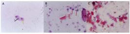
Rat buccal mucosa epithelial cells stained with Papanicolaou stain under a light microscope at 400x magnification. (A) Normal epithelial cells; (B) Precancerous epithelial cell transformation; (C) Buccal mucosa epithelial cells in rats treated with EEMP. Cell nucleus (black arrow), Cytoplasm (yellow arrow), MN (red arrow), and NPB (green arrow).
The results showed that there was a significant difference in the MN frequency between groups with p=0.000, as shown in Table 1. The administration of EEMP at a dose of 4 mg/kg BW for 10 days orally in rats showed a lower MN frequency compared to the positive control group, as well as EEMP doses of 1 mg/kg BW, and 2 mg/kg BW. The mean MN frequency between the negative control group and the EEMP dose of 4 mg/kg BW group showed the absence of significant differences, as shown in Figure 2.

Post hoc analysis using tukey honest significant difference (HSD) showed a significant effect between groups. nsnot significant; *statistically significant at p<0.05; **statistically significant at p<0.01; ***statistically significant at p<0.001.
3.2. Oral administration of EEMP induces apoptosis of the buccal mucosal epithelial cells by immunofluorescence
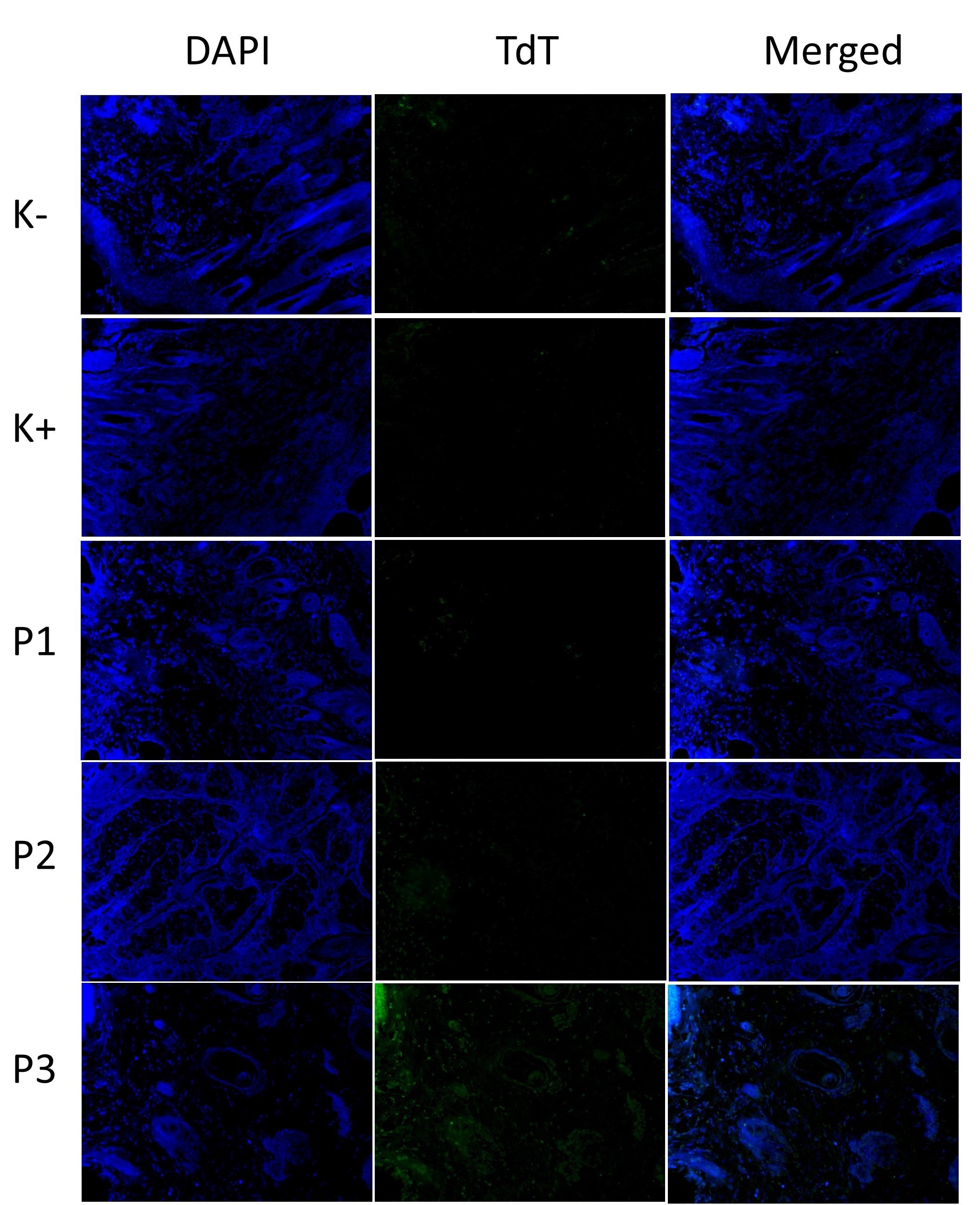
Oral administration of EEMP to DMBA-induced rats showed the presence of apoptosis through TUNEL examination. At a dose of EEMP 4 mg/kgBW, it could enhance apoptosis in epithelial cells that had transformed towards malignancy, as showed by green-stained epithelial cells. Furthermore, the number of green-stained cells increased linearly with the dose increment, as shown in Figure 3.

Analysis of apoptosis using TUNEL assay. Nuclei are stained with the terminal deoxynucleotidyl transferase (TdT) kit in the buccal mucosa epithelial cells induced by DMBA and treated with EEMP. TdT and DAPI (4',6-diamidino-2-phenylindole) images are merged to produce overlay images. After EEMP administration, positively apoptotic cells are green-stained. Negative control: K-; Positive control: K+; P1: EEMP1 (1 mg/kgBW); P2: EEMP2 (2 mg/kgBW); and P3: EEMP3 (4 mg/kgBW). The scale bar of images is 50 μm.
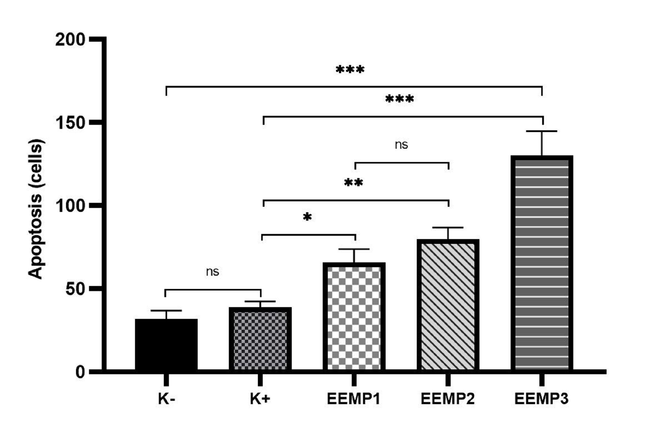
The apoptosis cells in the negative control (without DMBA-induced) and positive control (DMBA-induced) were lower compared to EEMP1 (P1), EEMP2 (P2), and EEMP3 (P3) groups, as shown in Table 2. There was a significant difference in the apoptosis cell between groups with p=0.000 (p<0.05), but there was no significant between negative and positive groups, as shown in Figure 4.

Post hoc analysis using Tukey Honest Significant Difference (HSD) showed a significant effect between groups on apoptosis of epithelial buccal cells induced by DMBA. nsnot significant; *statistically significant at p<0.05; **statistically significant at p<0.01; ***statistically significant at p<0.001.
4. Discussion
In our study, the development of OSCC in the buccal mucosa of male Wistar rats by topically applying a solution containing 0.5% DMBA. The carcinogenic and mutagenic effects of DMBA depended on metabolic activation by endogenous enzymes. Furthermore, the breakdown product of DMBA, namely DMBA-3,4-diol-1,2-epoxides (DMBADEs), had been reported to have the ability to bind to amino groups of deoxyguanosine and deoxyadenosine, leading to the formation of additional DNA adducts. Previous reports showed that depurination could result in the formation of apurinic sites (AP sites) in DNA. Damage to DNA could cause strand breaks, leading to the formation of MN and NPB. Ethanol extract of MP (Musa paradisiaca) reduced the formation of MN and NPB. The results showed that the administration of a dose of 4 mg/kgBW in rats caused a significant decrease compared to those induced with DMBA and positive control (K+). The reduction in the frequency of MN formation showed that hexadecanoic in EEMP had an effect in preventing the transformation of normal cells towards malignancy, such as in the induction of the buccal mucosa with DMBA for the occurrence of oral cancer. NPB as a result of decatenation failure, misrepair of DNA breaks, telomere end fusions, or incorrect separation of sister chromatids during anaphase was found at groups induced DMBA.
MN are small compartments delimited by a membrane containing DNA wrapped in a nuclear envelope and are spatially separated from the primary nucleus. Several studies had shown that genome fragmentation, mutagenesis, and MN had long been associated with their presence (Poetsch, 2020; Krupina et al., 2021). Furthermore, cancer, aging, and the effects of genotoxic stress had been identified as common symptoms. In various human and non-human models, the accumulation of MN had been used as a biological marker for genotoxic stress and genetic instability (Hartwig et al., 2020).
GC-MS analysis of the ethanol extract and ethyl acetate extract of MP stems showed the presence of several bioactive chemical compounds, including hexadecanoic acid (5.40%), heptadecanoic acid, 16-methyl; and 9,12-octadecadienoic acid (11.47%), which was a compound belonging to the palmitic acid and stearic acid groups (Budi et al., 2022a). Hexadecanoic acid was a possible source of anti-inflammatory agents because it selectively induced G2/M arrest and apoptosis in MCF-7 cells. This effect was mediated by the upregulation of p53 and the Bax/Bcl-2 ratio (Ghate et al., 2016). According to previous studies, the compound also exhibited cytotoxicity against human leukemia cells as well as inhibited phagocytic activity and production of nitric oxide in specific cells (Achakzai et al., 2019; Mellado et al., 2019). Reactive oxygen species (ROS) were attenuated by antioxidants in the body, such as SOD, in normal cells experiencing oxidative stress. DNA damage could be triggered by cell damage that was allowed to continue by high ROS and low SOD activity. Damaged DNA typically triggered the repair process in cells by inducing apoptosis pathway. The formation of MN stopped in epithelial cells that had undergone the process of apoptosis (Boice and Bouchier-Hayes, 2020).
The terminal deoxyribonucleotidyl transferase dUTP nick end labeling (TUNEL) method could be used to detect apoptosis by examining DNA fragmentation. This method provided an overview of apoptosis process at the single-cell level, hence it was more specific and had high accuracy. The administration of EEMP showed that there was apoptosis process through a positive reaction to TdT staining (green fluorescence). Furthermore, increasing the EEMP dose from 1 mg/kgBW to 4 mg/kgBW led to the production of more green fluorescent cells. This showed that hexadecanoic acid content in EEMP was able to prevent the transformation of normal cells into malignancy through apoptosis process. An anticancer agent may be developed when the administration of a drug candidate induces apoptosis in malignant cells, such as oral cancer.
Hexadecanoic acid, or palmitic acid compounds, could be well absorbed in the human intestinal tract. This was because palmitic acid was a fatty acid that was highly soluble in fat, easily penetrated cell membranes, and was found in the subcellular environment in mitochondria. Based on the results obtained, banana stem extract made from ethanol and ethyl acetate had the potential to be administered orally. When the ethanol extract of MP stems was applied, there was an increase in caspase-3 levels, showing that cancer cells were more active in apoptosis process. The high amount of caspase-3 showed that hexadecanoic acid could suppress the proliferation of oral cancer cells (OSCC).
The role of inflammation in the initiation and progression of cancer was well understood, and the underlying molecular mechanisms had been studied extensively. Therefore, in recent decades, targeting inflammatory pathways for cancer prevention and therapy had become possible. DNA repair proteins, caspases, lipid peroxidation, mutations, and NF-κB activation had been reported to contribute to various diseases, including cancer (Xiao et al., 2024). Caspases were a type of proapoptotic protease that cleaved key sites for apoptosis execution. According to previous studies, caspase-3 was an important member of this family that caused apoptosis by inducing nuclear alterations (Budi et al., 2022b). Caspase-3 is a key factor in the process of programmed cell death, known as apoptosis, which is triggered when cells are exposed to cytotoxic medicines, radiotherapy, or immunotherapy. It is frequently employed as an indicator of the effectiveness of cancer treatment (Kozubek et al., 2023).
5. Conclusion
The administration of EEMP could reduce the frequency of MN in DMBA-induced precancerous lesions of the buccal mucosa. The reduction of MN is a consequence of hexadecanoic acid in EEMP, which is believed to occur through the apoptotic pathway in order to prevent oral cancer.
Acknowledgements
We appreciate Universitas Airlangga and the Minister of Education, Culture, Research, and Technology of Indonesia (DRTPM) for support the research.
References
-
ACHAKZAI, J.K., ANWAR PANEZAI, M., KAKAR, M.A., KAKAR, A.M., KAKAR, S., KHAN, J., KHAN, N.Y., KHILJI, I. and TAREEN, A.K., 2019. In vitro anticancer MCF-7, anti-inflammatory, and Brine Shrimp Lethality Assay (BSLA) and GC-MS analysis of Whole Plant Butanol Fraction of Rheum ribes (WBFRR). BioMed Research International, vol. 2019, no. 1, pp. 3264846. http://doi.org/10.1155/2019/3264846
» http://doi.org/10.1155/2019/3264846 -
BOICE, A. and BOUCHIER-HAYES, L., 2020. Targeting apoptotic caspases in cancer. Biochimica et Biophysica Acta. Molecular Cell Research, vol. 1867, no. 6, pp. 118688. http://doi.org/10.1016/j.bbamcr.2020.118688
» http://doi.org/10.1016/j.bbamcr.2020.118688 - BUDI, H.S. and ASTUTI, E.R., 2019. The MMP-2, MMP-9 expression and collagen density of the ambonese banana stem sap administration on wound healing. Journal of International Dental and Medical Research, vol. 12, no. 2, pp. 492-497.
-
BUDI, H.S., ANITASARI, S., ULFA, N.M., JULIASTUTI, W.S., ALJUNAID, M., RAMADAN, D.E., MUZARI, K. and SHEN, Y.K., 2022a. Topical medicine potency of Musa paradisiaca var. sapientum (L.) kuntze as oral gel for wound healing: an in vitro, in vivo study. European Journal of Dentistry, vol. 16, no. 4, pp. 848-855. http://doi.org/10.1055/s-0041-1740226 PMid:35181871.
» http://doi.org/10.1055/s-0041-1740226 -
BUDI, H.S., ANITASARI, S., ULFA, N.M., SETIABUDI, M.A., RAMASAMY, R., WU, C.Z. and SHEN, Y.K., 2022b. Palmitic acid of Musa Paradisiaca induces apoptosis through caspase-3 in human oral squamous cell carcinoma. European Review for Medical and Pharmacological Sciences, vol. 26, no. 19, pp. 7099-7114. http://doi.org/10.26355/eurrev_202210_29895 PMid:36263558.
» http://doi.org/10.26355/eurrev_202210_29895 -
FENECH, M., KIRSCH-VOLDERS, M., NATARAJAN, A.T., SURRALLES, J., CROTT, J.W., PARRY, J., NORPPA, H., EASTMOND, D.A., TUCKER, J.D. and THOMAS, P., 2011. Molecular mechanisms of micronucleus, nucleoplasmic bridge and nuclear bud formation in mammalian and human cells. Mutagenesis, vol. 26, no. 1, pp. 125-132. http://doi.org/10.1093/mutage/geq052 PMid:21164193.
» http://doi.org/10.1093/mutage/geq052 - FENECH, M., KNASMUELLER, S., BOLOGNESI, C., BONASSI, S., HOLLAND, N., MIGLIORE, L., PALITTI, F., NATARAJAN, A.T. and KIRSCH-VOLDERS, M., 2016. Molecular mechanisms by which in vivo exposure to exogenous chemical genotoxic agents can lead to micronucleus formation in lymphocytes in vivo and ex vivo in humans. Mutation Research/Reviews in Mutation Research, vol. 770, no. Pt A, pp. 12-25. PMid:27894682.
- FENECH, M., KNASMUELLER, S., BOLOGNESI, C., HOLLAND, N., BONASSI, S. and KIRSCH-VOLDERS, M., 2020. Micronuclei as biomarkers of DNA damage, aneuploidy, inducers of chromosomal hypermutation and as sources of pro-inflammatory DNA in humans. Mutation Research/Reviews in Mutation Research, vol. 786, pp. 108342. PMid:33339572.
-
GHATE, N.B., DAS, A., CHAUDHURI, D., PANJA, S. and MANDAL, N., 2016. Sundew plant, a potential source of anti-inflammatory agents, selectively induces G2/M arrest and apoptosis in MCF-7 cells through upregulation of p53 and Bax/Bcl-2 ratio. Cell Death Discovery, vol. 2, no. 1, pp. 15062. http://doi.org/10.1038/cddiscovery.2015.62 PMid:27551490.
» http://doi.org/10.1038/cddiscovery.2015.62 -
GRIMM, M., CETINDIS, M., LEHMANN, M., BIEGNER, T., MUNZ, A., TERIETE, P., KRAUT, W. and REINERT, S., 2014. Association of cancer metabolism-related proteins with oral carcinogenesis - indications for chemoprevention and metabolic sensitizing of oral squamous cell carcinoma? Journal of Translational Medicine, vol. 12, no. 1, pp. 208. http://doi.org/10.1186/1479-5876-12-208 PMid:25048361.
» http://doi.org/10.1186/1479-5876-12-208 -
HARTWIG, A., ARAND, M., EPE, B., GUTH, S., JAHNKE, G., LAMPEN, A., MARTUS, H.J., MONIEN, B., RIETJENS, I.M.C.M., SCHMITZ-SPANKE, S., SCHRIEVER-SCHWEMMER, G., STEINBERG, P. and EISENBRAND, G., 2020. Mode of action-based risk assessment of genotoxic carcinogens. Archives of Toxicology, vol. 94, no. 6, pp. 1787-1877. http://doi.org/10.1007/s00204-020-02733-2 PMid:32542409.
» http://doi.org/10.1007/s00204-020-02733-2 -
HAKURA, A., KOYAMA, N., SEKI, Y., SONODA, J. and ASAKURA, S., 2022. O-Aminoazotoluene, 7,12-dimethylbenz[a]anthracene, and N-ethyl-N-nitrosourea, which are mutagenic but not carcinogenic in the colon, rapidly induce colonic tumors in mice with dextran sulfate sodium-induced colitis. Genes and Environment : the Official Journal of the Japanese Environmental Mutagen Society, vol. 44, no. 1, pp. 11. http://doi.org/10.1186/s41021-022-00240-7 PMid:35351212.
» http://doi.org/10.1186/s41021-022-00240-7 -
KHLIFI, R., TRABELSI-KSIBI, F., CHAKROUN, A., REBAI, A. and HAMZA-CHAFFAI, A., 2013. Cytogenetic abnormality in exfoliated cells of buccal mucosa in head and neck cancer patients in the Tunisian population: impact of different exposure sources. BioMed Research International, vol. 2013, pp. 905252. http://doi.org/10.1155/2013/905252 PMid:23957010.
» http://doi.org/10.1155/2013/905252 -
KOZUBEK, P., WOŁOSZCZAK, J. and GOMUŁKA, K., 2023. Immune reactions in major types of oncological treatment. International Journal of Molecular Sciences, vol. 24, no. 14, pp. 11257. http://doi.org/10.3390/ijms241411257 PMid:37511017.
» http://doi.org/10.3390/ijms241411257 - KRUPINA, K., GOGINASHVILI, A. and CLEVELAND, D.W., 2021. Causes and consequences of micronuclei. Current Opinion in Cell Biology, vol. 70, pp. 91-99. PMid:33610905.
- KUMARI, P., DEBTA, P. and DIXIT, A., 2022. Oral potentially malignant disorders: etiology, pathogenesis, and transformation into oral cancer. Frontiers in Pharmacology, vol. 13, pp. 825266. PMid:35517828.
-
MARTA, G.N., RIERA, R., BOSSI, P., ZHONG, L.P., LICITRA, L., MACEDO, C.R., CASTRO JUNIOR, G., CARVALHO, A.L., WILLIAM JUNIOR, W.N. and KOWALSKI, L.P., 2015. Induction chemotherapy prior to surgery with or without postoperative radiotherapy for oral cavity cancer patients: systematic review and meta-analysis. European Journal of Cancer, vol. 51, no. 17, pp. 2596-2603. http://doi.org/10.1016/j.ejca.2015.08.007 PMid:26318725.
» http://doi.org/10.1016/j.ejca.2015.08.007 - MELLADO, M., SOTO, M., MADRID, A., MONTENEGRO, I., JARA-GUTIÉRREZ, C., VILLENA, J., WERNER, E., GODOY, P. and AGUILAR, L.F., 2019. In vitro antioxidant and antiproliferative effect of the extracts of Ephedra chilensis K Presl aerial parts. BMC Complementary and Alternative Medicine, vol. 19, no. 1, pp. 53. PMid:30832627.
- MENDENHALL, W.M., HOLTZMAN, A.L., DAGAN, R., BRYANT, C.M., HITCHCOCK, K.E., AMDUR, R.J. and FERNANDES, R.P., 2021. Current role of radiotherapy in the management of oral cavity squamous cell carcinoma. Craniomaxillofacial Trauma & Reconstruction, vol. 14, no. 1, pp. 79-83. PMid:33613841.
- MESCHINI, R., BERNI, A., FILIPPI, S., PEPE, G., GROSSI, M.R., NATARAJAN, A.T. and PALITTI, F., 2015. The micronucleus assay in mammalian cells in vitro to assess health benefits of various phytochemicals. Mutation Research/Genetic Toxicology and Environmental Mutagenesis, vol. 793, pp. 79-85. PMid:26520376.
-
NAGINI, S. and KOWSHIK, J., 2016. The hamster buccal pouch model of oral carcinogenesis. Methods in Molecular Biology, vol. 1422, pp. 341-350. http://doi.org/10.1007/978-1-4939-3603-8_29 PMid:27246045.
» http://doi.org/10.1007/978-1-4939-3603-8_29 - POETSCH, A.R., 2020. The genomics of oxidative DNA damage, repair, and resulting mutagenesis. Computational and Structural Biotechnology Journal, vol. 18, pp. 207-219. PMid:31993111.
-
SILVA JUNIOR, F.C., FELIPE, M.B.M.C., CASTRO, D.E.F., ARAÚJO, S.C.D.S., SISENANDO, H.C.N. and BATISTUZZO DE MEDEIROS, S.R., 2021. A look beyond the priority: a systematic review of the genotoxic, mutagenic, and carcinogenic endpoints of non-priority. Environmental Pollution, vol. 278, pp. 116838. http://doi.org/10.1016/j.envpol.2021.116838 PMid:33714059.
» http://doi.org/10.1016/j.envpol.2021.116838 -
SOUZA, D.V., ANJOS ROSARIO, B., TAKESHITA, W.M., BARROS VIANA, M., NAGAOKA, M.R., SANTOS, J.N. and RIBEIRO, D.A., 2022. Is micronucleus assay in oral exfoliated cells a suitable biomarker for predicting cancer risk in individuals with oral potentially malignant disorders? A systematic review with meta-analysis. Pathology, Research and Practice, vol. 232, pp. 153828. http://doi.org/10.1016/j.prp.2022.153828 PMid:35279481.
» http://doi.org/10.1016/j.prp.2022.153828 -
XIAO, L., XIAN, M., ZHANG, C., GUO, Q. and YI, Q., 2024. Lipid peroxidation of immune cells in cancer. Frontiers in Immunology, vol. 14, pp. 1322746. http://doi.org/10.3389/fimmu.2023.1322746 PMid:38259464.
» http://doi.org/10.3389/fimmu.2023.1322746 - ZHOU, B., HAO, Q., LIANG, Y. and KONG, E., 2023. Protein palmitoylation in cancer: molecular functions and therapeutic potential. Molecular Oncology, vol. 17, no. 1, pp. 3-26. PMid:36018061.