Evaluation of tissue changes induced by hyaluronic acid in the interdental papilla of rats
ABSTRACT
Objective The present study aimed to evaluate whether the application of hyaluronic acid (HA) in the interdental papilla (IP) of rats promotes an exacerbation of leukocyte infiltration and hemorrhage (PURPOSE-GAP).
Material and Methods A total of 51 rats were used in a split-mouth experimental model, randomly distributed through group randomization, and all animals received the first application of HA (Experimental Group – EG) and saline solution (SS) (Control Group – CG) at the same time. The groups were divided so that some received only one application, while others received a second application after 7 days, with euthanasia performed at experimental time points of 12 hours, 72 hours, and 7 days. In the comparison between EG1 and EG6, the intensity of leukocyte infiltration decreased significantly (p<0.005).
Results EG1 showed a higher intensity of inflammatory infiltration than CG1 (12h) (p<0.005). In the EG, there was a significant presence of mild and moderate hemorrhage compared to the CG (p<0.005). It was concluded that the application of HA in the IP of rats increased the intensity of leukocyte infiltration, which progressively regressed until the 14th day. The hemorrhage caused by HA application was mild to moderate in the EG; moreover, there was a significant presence of hemorrhage in groups that received only one application of HA.
Conclusion The results suggest that hyaluronic acid has promising potential in the regeneration of the interdental papilla, providing support for future research and possible applications in the field of periodontal aesthetics and regenerative dentistry.
KEYWORDS:
Hemorrhage; Histology; Hyaluronic acid; Interdental papilla; Leukocytic infiltrate
RESUMO
Objetivo O presente estudo objetivou avaliar se a aplicação de ácido hialurônico (HA) na papila interdentária (PI) de ratos promove exacerbação do infiltrado leucocitário e hemorragia (PROPÓSITO-GAP).
Material e Métodos Utilizou-se 51 ratos no modelo experimental boca dividida distribuídos aleatoriamente através da randomização dos grupos e todos os animais receberam a primeira aplicação de HA (Grupo experimental – GE) e solução salina (SS) (Grupo controle – GC) ao mesmo tempo. Os grupos foram divididos de modo que houve grupos com apenas uma aplicação e outros contaram com nova aplicação após 7 dias, sendo eutanasiados nos tempos experimentais de 12 horas, 72 horas e 7 dias.
Resultados Na comparação entre os GE1 com GE6[CS1], a intensidade do infiltrado leucocitário diminui consideravelmente (p<0,005). O GE1 apresentou maior intensidade de infiltrado inflamatório do que e GC1 [jcr2] (12h) (p<0,005). No GE houve significativa presença de hemorragia leve e moderada quando comparado ao GC (p<0,005). Concluiu-se que a aplicação de HA na PI de ratos promoveu aumento da intensidade do infiltrado leucocitário e regride progressivamente até o 14° dia. A hemorragia causada pela aplicação de HA apresentou-se mais leve e moderada no GE, além disso, houve significativa presença de hemorragia nos grupos que receberam apenas uma aplicação de HA.
Conclusão The results suggest that hyaluronic acid has promising potential in the regeneration of the interdental papilla, providing support for future research and possible applications in the field of periodontal aesthetics and regenerative dentistry.
PALAVRAS-CHAVE:
Hemorragia; Histologia; Ácido hialurônico; Papila interdental; Inflamatório leucocitário
INTRODUCTION
The interdental papilla (IP), present in both anterior and posterior teeth, plays an important role in smile aesthetics, as well as serving as a barrier and protective defense for periodontal tissues. The loss of this region, especially in the anterior maxilla, can lead to "black triangles," open gingival embrasures, phonetic problems, and lateral impaction of food debris. This results in negative effects on oral health quality and the patient’s self-esteem [1]. Research indicates that the formation of a "black space" is the third most common aesthetic complaint among patients, behind only cavities and exposed crown margins. Consequently, this issue is a significant concern for dentists, especially due to the difficulty in treating it [2].
Various treatments can be adopted for the reconstruction of the IP; however, it remains a complex aesthetic procedure with low predictability of success [1], given that the IP is a delicate, small area with limited blood supply, making its reconstruction a significant challenge for periodontics [3]. Thus, to meet the need for less invasive procedures, new non-surgical techniques have emerged, and the use of hyaluronic acid (HA) has proven effective.
HA possesses unique hygroscopic, rheological, and viscoelastic properties, and it can influence cellular behavior by affecting the macro and microenvironment surrounding the cells. This acid has gained prominence in dentistry for treating inflammations, oral lesions, aesthetics, periodontal treatment, and IP augmentation [3-6]. Precisely because HA plays a role with various functions in the healing process, similar to what occurs in periodontal tissues [7].
A study conducted by Bertl et al. (2017) [8] involving the injection of HA into the interdental papilla around two implants to fill the missing space demonstrated that HA’s hygroscopic action, along with its high affinity for water, caused partial occlusion of blood vessels, thereby preventing infection or allergic reactions to this material.
The application of HA in the papillary region is a treatment modality that has been shown to be safe and minimally invasive for minor IP losses [6]. Literature has highlighted the promising potential of HA use [9-13]. However, the limited blood supply in this area has already led to reports of adverse reactions after HA injection in the oral mucosa [3,14]. In the study by Bertl et al. (2017) [3], adverse reactions were observed but disappeared after 7 days; these included swelling, significant sensitivity at the site, and a burning sensation near the injection area.
The initial tissue response to trauma or the presence of biomaterials involves fundamental events such as hemorrhage and inflammation, which determine the quality and predictability of healing. Hemorrhage reflects vascular rupture and activation of the inflammatory cascade; it is necessary for the onset of hemostasis and clot formation, which acts as a temporary matrix for cell migration [15,16]. Subsequently, the inflammatory process plays a central role in removing tissue debris, combating potential microorganisms, and recruiting reparative cells, making it an essential step for the transition to the proliferative phase [17,18].
However, the persistence or exacerbation of inflammation can have deleterious effects, inducing excessive fibrosis, chronic pain, or regeneration failures, especially when associated with biomaterials [19,20]. Furthermore, prolonged inflammatory responses can compromise the remodeling of the extracellular matrix and favor the formation of exuberant scar tissue [21,22]. Recent studies reinforce that low-grade inflammatory microenvironments can evolve into chronic processes, reducing the functional and aesthetic quality of repaired tissues [23].
Thus, the objective of the present study was to evaluate whether the application of HA in the IP of rats promotes the exacerbation of leukocyte infiltration and hemorrhage.
MATERIAL AND METHODS
This research was approved by the Ethics Committee on Animal Use of the Federal University of Mato Grosso do Sul under protocol n. 887/2017. The sample consisted of 54 young adult male rats (Rattus norvegicus albinus wistar), weighing approximately 250g, from the vivarium of the Federal University of Mato Grosso do Sul (Campo Grande - MS). Of these animals, three died after intraperitoneal anesthesia. The animals were kept in unit cages of 25cm3, allowing enough space for movement and rest, under controlled ambient temperature, lighting and hygiene, fed with balanced feed (Nuvilab CR - 1® - Colombo - PR) and water throughout the experiment. The animals remained isolated to ensure that they were stable and healthy, maintaining contact only with the people involved in the research.
Prior to the procedures, the animals were weighed and the amounts of anesthetics were individualized. The anesthesia was composed by the association of ketamine hydrochloride (75 mg/kg) (CETAMIN® - Hortolândia - SP) and xylazine hydrochloride (10 mg/kg) (DOPASER® - Hortolândia - SP) and the application was intraperitoneal.
In this research 2 substances were applied in each rat: Hyaluronic acid 24mg/mL, lidocaine 0.3% (JUVÉDERM® ULTRA - Pringy - France); Experimental group – GE: The application of 0.02mL of HA into the IP of the rat upper and lower central incisors; Saline solution (SS) (Sodium chloride 0.9% - EQUIPLEX® - São Paulo - SP); Control group – CG: The application of 0.02 mL of SS into the interdental papilla of the upper and lower central incisors of the rats.
The substances were inserted into a 0.5 mL 15/64'' syringe with a 31G (6x0.25 mm) attached needle (UNIQMED® - São Paulo - SP). The needle insertion was parallel to the long axis of the tooth in the crown-apex direction and a volume of 0.02 mL of the materials was deposited 2-3 mm from the papilla apex in all animals. The application was conducted in a controlled manner by a single experienced and calibrated operator. For pain control, the analgesic butorphanol (Torbugesic® - Campinas - SP), active ingredient: butorphanol tartrate, was injected by intramuscular, with the volume of 0.1mL/300g, after completing the applications in each group.
Formation of the groups
Fifty-one rats were used in a split-mouth experimental model [14], that is, the same animal is EG and CG, they were randomly distributed through group randomization and all animals received application of the substances at the same time.
Specimens from groups EG1, EG2, EG3, and CG1, CG2, CG3, were euthanized at the predefined experimental times: (1) 12 hours, (2) 72 hours, and (3) 7 days, respectively (Table I).
Specimens from groups EG4, EG5, EG6, and CG4, CG5, CG6, received a new application 7 days after the first intervention and were euthanized at the predefined experimental times: (4) 12 hours, (5) 72 hours, and (6) 7 days respectively (Table I).
Euthanasia was performed at the predetermined times by anesthetic overdose (Ketamine + Xylazine) at 5 times the initial dose. The maxillae and mandibles were separated from the rest of the skull using a 15c scalpel blade (Solidor®- Diadema - SP) and fixed with 10% buffered formaldehyde at neutral pH (Bio-Aplica® - São Paulo - SP) for a period of 48 hours.
Histologycal analysis
After 48 hours, the specimens were washed in Phosphate-buffered saline - PBS (buffer) (Dinâmica® - Indaiatuba - SP), dehydrated with the alcohol series 70%, 80%, 95% and 100% (Santa Cruz® - Guarulhos - SP), diaphanized in xylene (Xileno® - São Paulo - SP) and demineralized with ethylenediaminetetraacetic acid (EDTA) at a concentration of 0.1 molar and pH 11 (Asfer® - São Caetano do Sul - SP). The demineralization process was completed when the pieces presented borrachoid consistency (cartilage) and embedded in paraffin (Isokad® - São Paulo - SP) at 60°C.
In the microtomy, the specimens were cut 5 µm thick in the longitudinal direction. The sections were stained with hematoxylin-eosin (HE) according to Lillie and Fullmer’s (1976) [24] guidelines for histomorphological evaluation.
The fragments in which the exogenous material was present were considered viable for histomorphological analysis. The slides belonging to the control group were considered viable when they were free of artifacts.
The analysis was performed by two previously calibrated and blinded examiners. The Kappa test (Kw) for intra-examiner and inter-examiner calibration was performed with the pre-analysis of 10 slides randomly selected in a time interval of 7 and 14 days from the first reading. The results obtained were Intraclass Correlation inter-examiner ICC = 0.80 and intra-examiner ICC = 0.88, which gave satisfactory agreement results for the subsequent analysis of the experimental and control slides.
The slides were analyzed with a light microscope (Olympus BX41 - Japan), at 100x and 400x magnification. The evaluated area was the portion of the IP between the connective tissue adjacent to the gingival epithelium (incisal portion) up to the height of an imaginary line perpendicular to the long axis of the central incisors and tangential to the most convex point of the amelogenic epithelium (cervical portion).
Within this pre-established area, the subarea with the highest intensity of the criteria to be evaluated was chosen. In the EG, the HA was observed as an amorphous, acellular, basophilic material. The intensity of the leukocyte infiltrate and of hemorrhage were evaluated. For leukocyte infiltrate intensity, the scores 0 - absent, 1 - mild, 2 - moderate, and 3 - intense were used. For hemorrhage, we used the scores 0 - absent, 1 - mild, and 2 - intense. The assignment of scores was done comparatively between the sections.
Statistical analysis
For statistical analysis the software Stata v.14 (College Station, TX, USA) was used. Initially the Kolmogorov-Smirnov test was performed to verify the normality of the outcome curves (hemorrhage, leukocyte infiltrate intensity). The results showed that the curves were not normal, and non-parametric tests were selected for the analyses. As each specimen generated an average of four slides and each slide contained three sections, each specimen generated 12 sections. For each outcome the central tendency measure mode (the value that appears most) was used to characterize the outcome of the given specimen. The mean and median, in this case, could not be applied because they would tend to give a measure that is not categorical and would have decimal values, not allowing a practical applicability of the category measured by each outcome. After that, absolute and relative frequency tests were processed, with the respective 95% confidence intervals.
Histogram charts were prepared to complement the descriptive analysis of the outcomes analyzed. After that, statistical tests of chi-square proportions were performed to verify statistical significance between the groups. The significance level adopted was 5% (p<0.05).
RESULTS
The animals remained healthy during the experimental times, and only 3 animals died due to complications after intraperitoneal general anesthesia. Therefore, 51 animals were included in the experiment according to the following distribution: CG1 (n=7), EG1 (n=7), CG2 (n=9), EG2 (n=9), CG3 (n=9), EG3 (n=9), CG4 (n=9), EG4 (n=9), CG5 (n=8), EG5 (n=8), CG6 (n=9), EG6 (n=9).
Histologycal evaluation
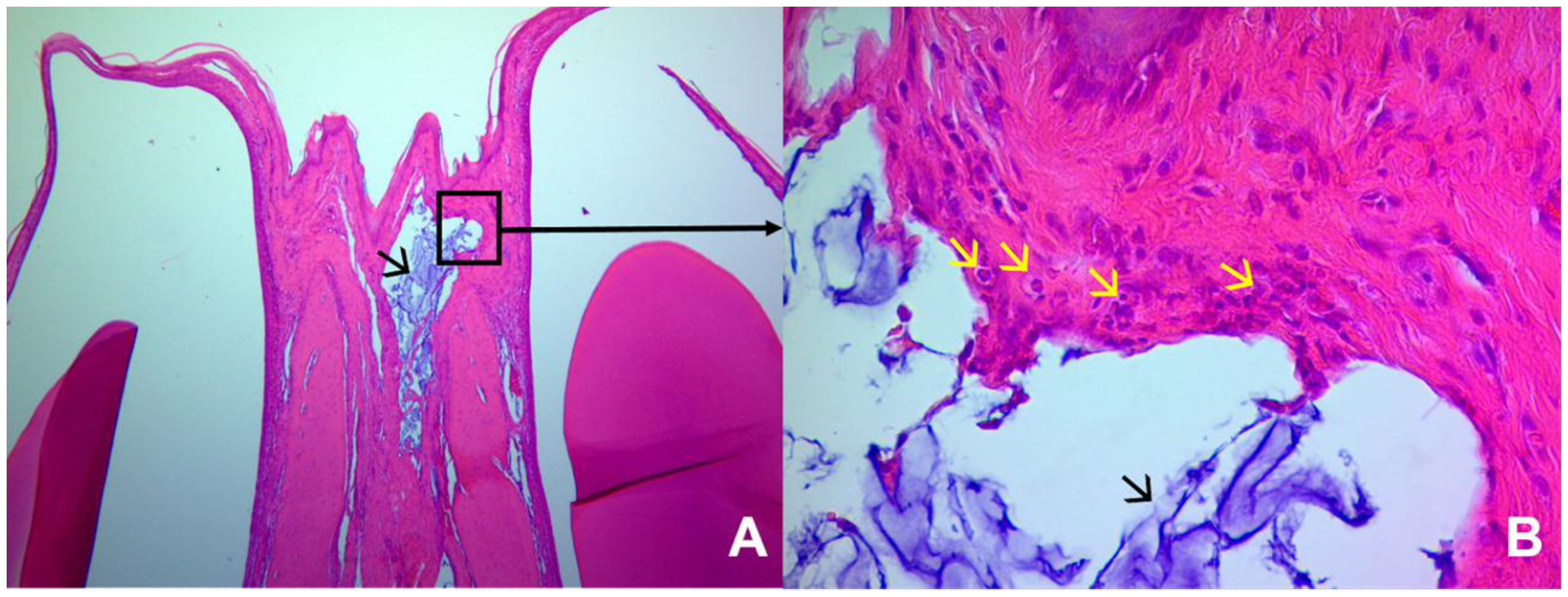
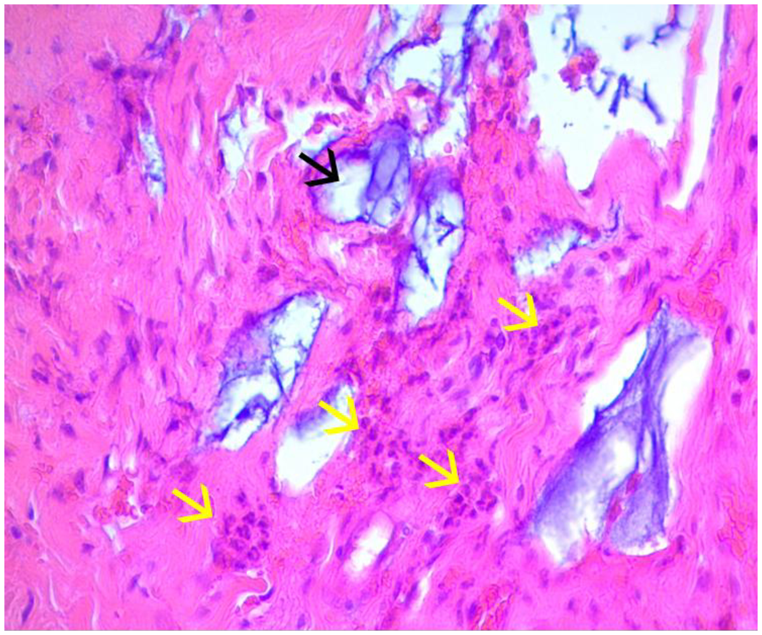
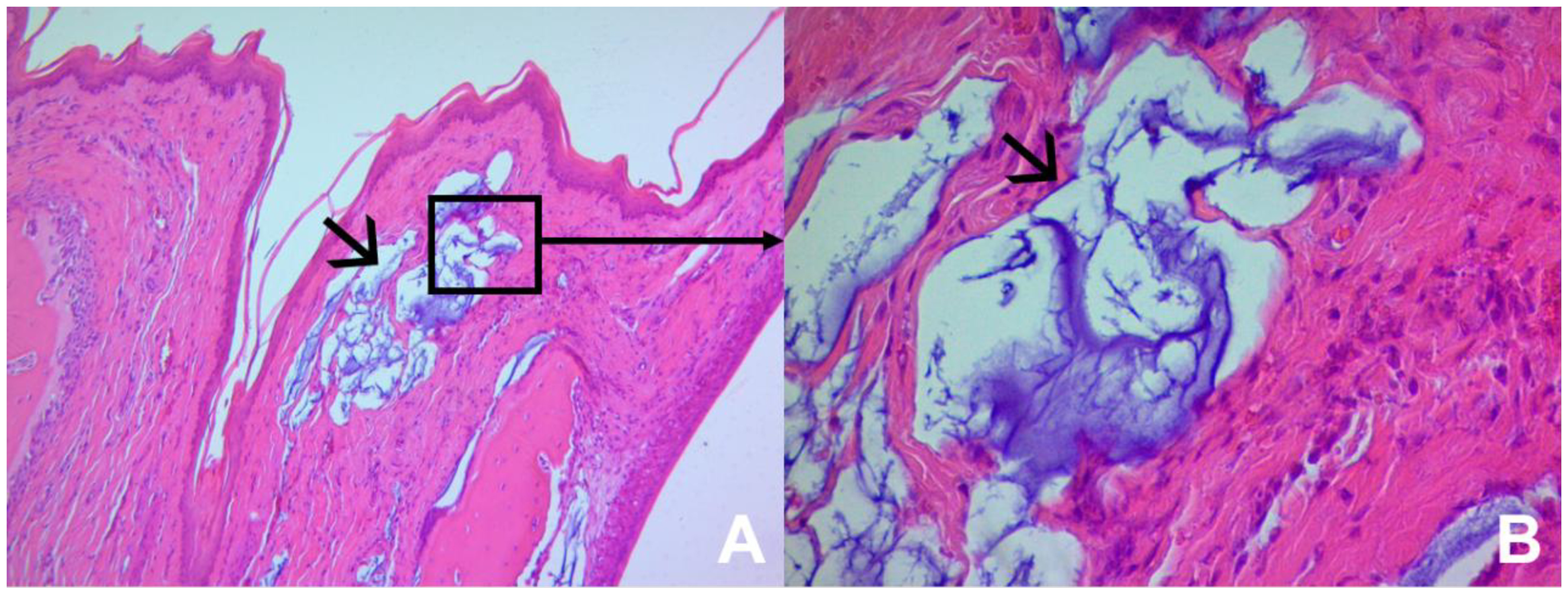
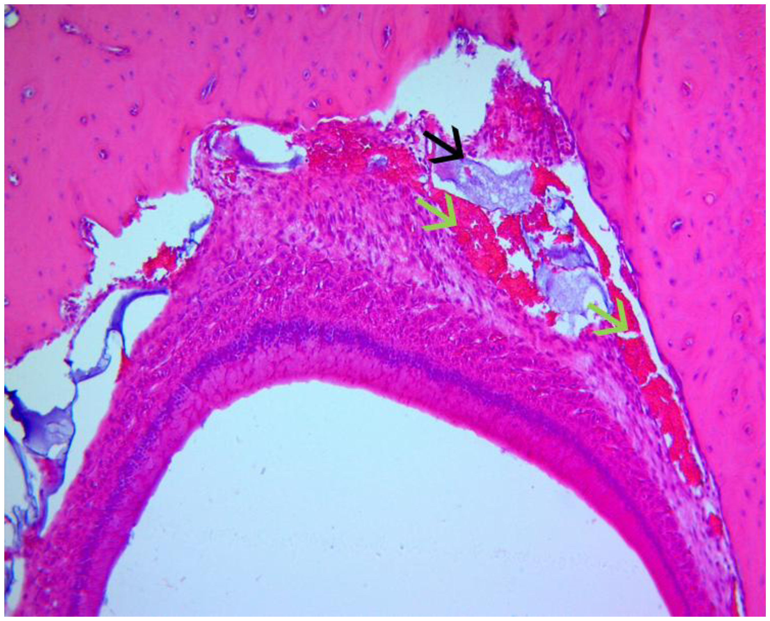
The slides were stained with HE and in the EG the HA was basophilic, whereas in the CG there were no changes in the staining profile. In the SLIDES analysis, some sections showed leukocyte infiltration in the IP after one or two applications of the HA (Figures 1, 2 and 3), and the presence of hemorrhage (Figure 4).
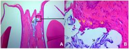
- A - Histological slide of EG1 (12h), showing the presence of HA within the connective tissue of the IP 40x; B - Higher magnification of Figure 1A showing presence of HA, presence of polymorphonuclear cells surrounding the HA 100x.
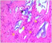
- Histological slide of EG1 (12h), showing the presence of HA within the connective tissue of the IP and polymorphonuclear inflammatory cells surrounding the HA 400x.
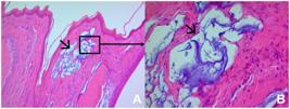
-A - Histological slide of EG1 (12h), showing the presence of HA within the connective tissue of the IP 100x; B - Higher magnification of Figure 3A showing presence of HA and absence of inflammatory cells surrounding the HA 400x.
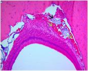
- Histological slide of EG1 (12h), showing the presence of HA within the connective tissue of the IP and presence of hemorrhage around the material in the gingival ligament 100x.
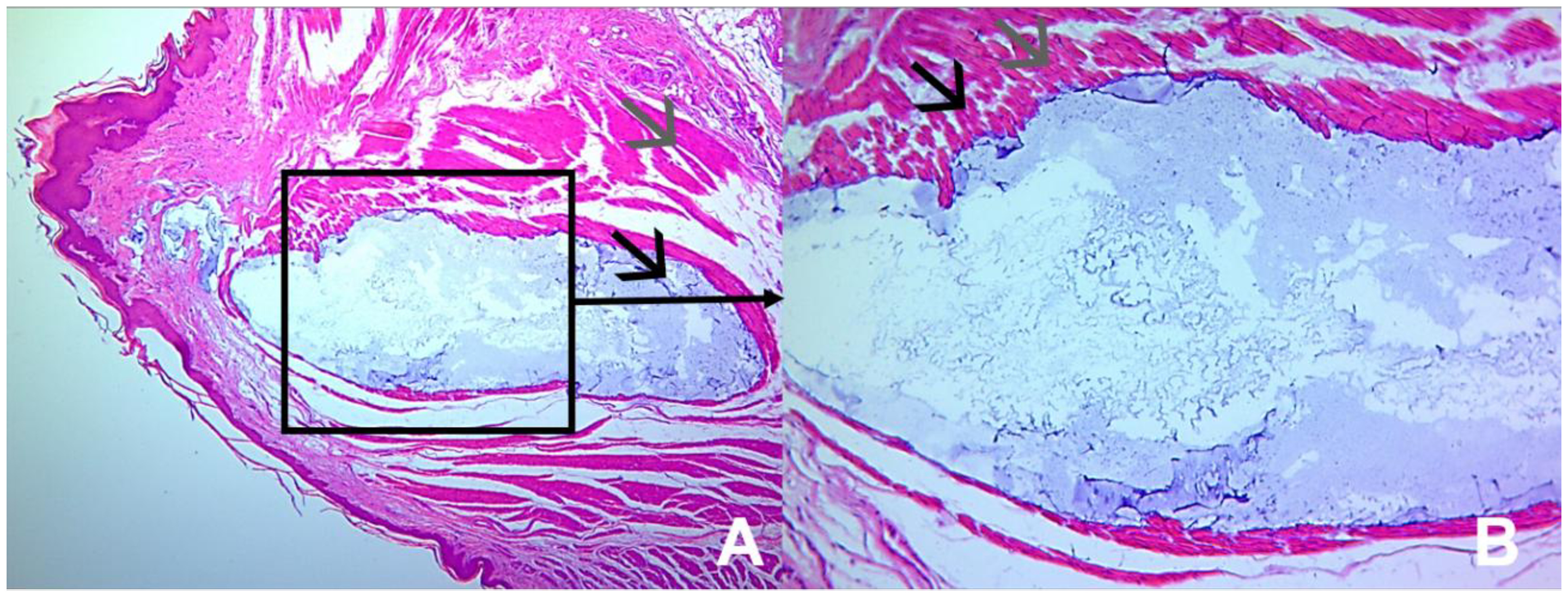
It was observed in all slides of the EG considerable deposition of HA in the muscle tissue and the absence of leukocyte infiltrate in this tissue (Figure 5).
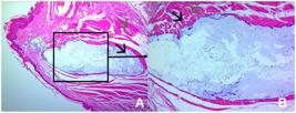
- A - Histological slide of EG1 (12), showing the presence of HA in the muscle tissue region 100x; B - Higher magnification of Figure 5A showing HA deposition in the muscle tissue, with absence of hemorrhage or leukocyte infiltrate 400x.
Comparing all the CG with the EG, there was a greater appearance of intense and moderate leukocyte infiltrate in the EG, and a prevalence of mild intensity infiltrate in the CG (p = 0.004). When comparing the EG1 with EG6, the intensity of the leukocyte infiltrate decreased considerably, being lighter at the end of the procedures (p = 0.0184). The leukocyte infiltrate between groups GE1 and GC1 showed a statistical difference (p = 0.0026). In other comparisons between groups with the same experimental times, the inflammatory reactions were considered similar (Table II).
- Description of the inflammatory infiltrate intensity values, expressed as percentages of times the score appeared on each histological section
Considering the hemorrhage patterns after the applications, most animals in the CG and EG did not bleed, however, in the EG there was significant presence of mild and moderate bleeding when compared to the CG (p = 0.0001). When comparing the CG and EG at the same experimental times, there was a statistical difference between the groups: G1C and G1E (p = 0.0217), G2C and G2E (p = 0.0042), and G3C and G3E (p = 0.0001). Between G4C and G4E, G5C and G5E, and G6C and G6E there was no statistical difference (p > 0.05) (Table III).
DISCUSSION
Hyaluronic acid (HA) has been increasingly used in the field of aesthetics, and its benefits have been widely disseminated in society. The present study sought, for the first time, to evaluate the presence of inflammatory infiltrate and the occurrence of hemorrhage after HA applications in the interdental papilla (IP) of rats. The results of this research pointed to the prevalence of mild inflammatory infiltrate across all experimental time points. Additionally, when analyzing the occurrence of hemorrhage, it was higher in the experimental group and significantly more intense in animals euthanized after the first application.
Previously published clinical trials mostly focus on HA application protocols in the IP [13]. However, with the high demand for this procedure, reports of adverse reactions have also been published [7]. Therefore, animal studies are necessary for histological evaluations to rule out potential side effects of HA application [14]. In the present study, a standardized dose of 0.2 ml of HA was initially used, and in half of the cases, a new application was performed after 7 days. The literature diverges regarding recommended dosages, but clinical trials have demonstrated the use of the dosage employed in this study [6,24-27].
Analyzing the histological sections concerning the intensity of leukocyte infiltrate, a prevalence of mild intensity was observed at all experimental time points. Thus, the body is able to quickly stabilize the inflammatory reaction caused by HA, despite the possibility of an exacerbated reaction at some point [4]. Despite the application of two distinct substances, the intensity remained mild in all groups, highlighting the biocompatibility of both products, given that the IP region frequently experiences inflammatory reactions due to constant aggression from chewing and habits [5].
When comparing EG1 with CG1 (euthanasia 12 hours after application), greater inflammatory infiltrate was observed in the experimental group. This finding indicates an acute reaction following the HA injection. The sections predominantly showed polymorphonuclear cells, reinforcing the argument of an acute inflammatory reaction in the IP at this experimental time point [28]. In subsequent comparisons, the intensity of the infiltrate remained similar between the groups and became progressively milder over time (14 days).
A literature review revealed that animal studies have not focused on investigating the occurrence of hemorrhage, which is why the present study analyzed this event following HA application. In this study, mild to moderate hemorrhage predominated in the EG, generally occurring in the periodontal ligament region and the central area of the lower IP, where a large-caliber vessel is present [14]. Based on this finding, the authors believe that the insertion of HA, being a cross-linked and dense substance, may promote tissue expansion, compression, and vessel injury, leading to blood extravasation. This event may result from the injected volume, application force, and speed [4].
When comparing EG with CG, animals euthanized before the second application showed significantly greater hemorrhage in the EG compared to the animals at the second experimental time point. It is believed that after the first application, HA integrated into the tissue, promoting tissue expansion. By the second application, the mucosal tissue was more prepared to receive the product, confirming the viscoelastic properties [29] of HA and the need for a second or third application as suggested by clinical studies [3,6,8,10,24-26].
Considering the occurrence of hemorrhage up to 7 days post-application, it is suggested that HA be gradually inserted into the IP. During the first application, a smaller quantity could be deposited compared to the second or third application to prepare the tissue for expansion.
Some limitations should be considered when analyzing the results of the present study. The rat IP is approximately ten times smaller than the human IP. Moreover, being an animal study, the results may not be directly translatable to humans. Additionally, the rats had no defects in their IP. Therefore, further publications are recommended to better investigate gingival defects for a more in-depth analysis.
The analysis of the observed patterns of hemorrhage and inflammation presents important clinical relevance, as these events constitute fundamental stages of the tissue response to trauma and the presence of biomaterials. The initial hemorrhage reflects vascular rupture and the activation of the inflammatory cascade, which are expected phenomena in early repair. Its progressive reduction is indicative of efficient hemostasis and conditions favorable for healing [15,16]. Similarly, the acute inflammatory infiltrate observed for up to 14 days is compatible with the physiological repair process, promoting cell recruitment and the removal of tissue debris [17,18]. However, the persistence or intensification of inflammation beyond this period could indicate an adverse reaction, with potential clinical repercussions such as chronic pain, excessive fibrosis, or impaired local regeneration [19,20].
Considering a time horizon of more than 14 days, it is possible that prolonged inflammatory responses compromise the proliferative and remodeling phases, favoring the formation of exuberant scar tissue, inadequate remodeling of the extracellular matrix, or even bone resorption, depending on the area involved [21,22]. Furthermore, the maintenance of hemorrhagic or inflammatory microfoci can evolve into a low-grade chronic process, reducing the functional and aesthetic quality of the repaired tissue [23]. Clinically, such changes could result in treatment instability, recurrence of the initial condition, or the need for new interventions. These aspects reinforce the importance of monitoring over longer periods to comprehensively understand the dynamics of repair and ensure greater therapeutic predictability.
It should be noted that the vascularization of the interdental papilla in rats differs from that found in humans. In rodents, the capillary network is less complex and proportionally reduced, which can influence the diffusion, absorption, and permanence of injected substances, such as hyaluronic acid. In humans, the greater density and calibration of blood vessels favor more dynamic conditions for tissue remodeling and repair.
CONCLUSION
Based on the results found in this study, it may be concluded that the application of hyaluronic acid into the interdental papilla of rats promotes an increase in the intensity of the leukocyte infiltrate, with a predominance of polymorphonuclear cells, 12 hours after its application, progressively regressing until day 14. The hemorrhage caused by the application of HA was histologically milder and more moderate when compared to saline solution; furthermore, there was greater presence of hemorrhage in the groups that received only one application of HA.
Acknowledgements
To Thalita Lorrayne, for her dedication and assistance in the experimental execution of this research.
-
How to cite:
Chaparro DM, Bocchi YG, Bomfim RA, Antunes DM, Ferrão JP Jr, Silva CO. Evaluation of tissue changes induced by hyaluronic acid in the interdental papilla of rats. Braz. Dent. Sci. 2026;29:e4627. https://doi.org/10.4322/bds.2026.e4627
-
Funding
The authors declare that no financial support was received.
-
Regulatory Statement
This study was conducted in accordance with all the provisions of the local animal subjects oversight committee guidelines and policies of: National Council for Animal Control and Experimentation (CONCEA). This study protocol was reviewed and approved by Ethics Committee on Animal Use at the Federal University of Mato Grosso do Sul (UFMS), approval number 887/2017.
Data availability
The data that support the findings of this study are available from the corresponding author upon reasonable request.
REFERENCES
-
1 Patel M, Guni A, Nibali L, Garcia-Sanchez R. Interdental papilla reconstruction: a systematic review. Clin Oral Investig. 2024;28(1):101. https://doi.org/10.1007/s00784-023-05409-0 PMid:38231354.
» https://doi.org/10.1007/s00784-023-05409-0 - 2 Cunliffe J, Pretty I. Patients’ ranking of interdental “black triangles” against other common aesthetic problems. Eur J Prosthodont Restor Dent. 2009;17(4):177-81. PMid:20158060.
-
3 Bertl K, Gotfredsen K, Jensen SS, Bruckmann C, Stavropoulos A. Can hyaluronan injections augment deficient papillae at implant-supported crowns in the anterior maxilla? A randomized controlled clinical trial with 6months follow-up. Clin Oral Implants Res. 2017;28(9):1054-61. https://doi.org/10.1111/clr.12917 PMid:27378556.
» https://doi.org/10.1111/clr.12917 - 4 Baisse P, Piantoni B. Action of hyaluronic acid on the wound healing process following extraction. Dent Inf. 2004;7(1):1-9.
-
5 Ballini A, Cantore S, Capodiferro S, Grassi FR. Esterified hyaluronic acid and autologous bone in the surgical correction of the infra-bone defects. Int J Med Sci. 2009;6(2):65-71. https://doi.org/10.7150/ijms.6.65 PMid:19277251.
» https://doi.org/10.7150/ijms.6.65 -
6 Becker W, Gabitov I, Stepanov M, Kois J, Smidt A, Becker BE. Minimally invasive treatment for papillae deficiencies in the esthetic zone: a pilot study. Clin Implant Dent Relat Res. 2010;12(1):1-8. https://doi.org/10.1111/j.1708-8208.2009.00247.x PMid:19843105.
» https://doi.org/10.1111/j.1708-8208.2009.00247.x - 7 Wulandari P, Satria D, Amalia M. Effectiveness freeze dried platelet rich plasma combination of hyaluronic acid after periodontal surgical healing: an experimental study on animal model:(In Vivo Studies). Braz Dent Sci. 2023;26(2):e3680.
-
8 Bertl K, Gotfredsen K, Jensen SS, Bruckmann C, Stavropoulos A. Adverse reaction after hyaluronan injection for minimally invasive papilla volume augmentation. A report on two cases. Clin Oral Implants Res. 2017;28(7):871-6. https://doi.org/10.1111/clr.12892 PMid:27252126.
» https://doi.org/10.1111/clr.12892 -
9 Awartani FA, Tatakis DN. Interdental papilla loss: treatment by hyaluronic acid gel injection: a case series. Clin Oral Investig. 2016;20(7):1775-80. https://doi.org/10.1007/s00784-015-1677-z PMid:26613740.
» https://doi.org/10.1007/s00784-015-1677-z - 10 Dall'Magro AK, Dalacort M, Santos R, Rocha RV, Del Conte Valcanaia T, Dall'Magro E. Neoformação de papila gengival com ácido hialurônico: relato de caso. Rev Faculdade Odontologia-UPF. 2016;21(1).
-
11 Lee WP, Kim HJ, Yu SJ, Kim BO. Six month clinical evaluation of interdental papilla reconstruction with injectable hyaluronic acid gel using an image analysis system. J Esthet Restor Dent. 2016;28(4):221-30. https://doi.org/10.1111/jerd.12216 PMid:27159838.
» https://doi.org/10.1111/jerd.12216 -
12 Alsharif SB, Aljahdali B. The use of hyaluronic acid injection for treatment of black triangle and reconstruction of lost interdental papilla in anterior teeth: a systematic review. Acta Odontol Scand. 2024;83:371-91. https://doi.org/10.2340/aos.v83.40864 PMid:38864684.
» https://doi.org/10.2340/aos.v83.40864 - 13 Do Sacramento IC, Pinto ACS, Lessa AMG, Lessa AG. O uso do ácido hialurônico para preenchimento de papilas interdentais: Uma Revisão de Literatura/The use of hyaluronic acid to fill interdental papillae: A Literature Review. Revista de Psicologia. 2019;13(45):415-25.
-
14 Bertl K, Bruckmann C, Isberg PE, Klinge B, Gotfredsen K, Stavropoulos A. Hyaluronan in non-surgical and surgical periodontal therapy: a systematic review. J Clin Periodontol. 2015;42(3):236-46. https://doi.org/10.1111/jcpe.12371 PMid:25640222.
» https://doi.org/10.1111/jcpe.12371 - 15 Clark RAF. Wound repair: overview and general considerations. In: Clark RAF, editor. The molecular and cellular biology of wound repair. 2nd ed. Boston: Springer, 1996. p. 3-50.
-
16 Eming SA, Krieg T, Davidson JM. Inflammation in wound repair: molecular and cellular mechanisms. J Invest Dermatol. 2007;127(3):514-25. https://doi.org/10.1038/sj.jid.5700701 PMid:17299434.
» https://doi.org/10.1038/sj.jid.5700701 -
17 Singer AJ, Clark RAF. Cutaneous wound healing. N Engl J Med. 1999;341(10):738-46. https://doi.org/10.1056/NEJM199909023411006 PMid:10471461.
» https://doi.org/10.1056/NEJM199909023411006 -
18 Gurtner GC, Werner S, Barrandon Y, Longaker MT. Wound repair and regeneration. Nature. 2008;453(7193):314-21. https://doi.org/10.1038/nature07039 PMid:18480812.
» https://doi.org/10.1038/nature07039 -
19 Anderson JM, Rodriguez A, Chang DT. Foreign body reaction to biomaterials. Semin Immunol. 2008;20(2):86-100. https://doi.org/10.1016/j.smim.2007.11.004 PMid:18162407.
» https://doi.org/10.1016/j.smim.2007.11.004 -
20 Landén NX, Li D, Ståhle M. Transition from inflammation to proliferation: a critical step during wound healing. Cell Mol Life Sci. 2016;73(20):3861-85. https://doi.org/10.1007/s00018-016-2268-0 PMid:27180275.
» https://doi.org/10.1007/s00018-016-2268-0 -
21 Wynn TA, Ramalingam TR. Mechanisms of fibrosis: therapeutic translation for fibrotic disease. Nat Med. 2012;18(7):1028-40. https://doi.org/10.1038/nm.2807 PMid:22772564.
» https://doi.org/10.1038/nm.2807 - 22 Rodero MP, Khosrotehrani K. Skin wound healing modulation by macrophages. Int J Clin Exp Pathol. 2010;3(7):643-53. PMid:20830235.
-
23 Krzyszczyk P, Schloss R, Palmer A, Berthiaume F. The role of macrophages in acute and chronic wound healing and interventions to promote pro-wound healing phenotypes. Front Physiol. 2018;9:419. https://doi.org/10.3389/fphys.2018.00419 PMid:29765329.
» https://doi.org/10.3389/fphys.2018.00419 - 24 Lillie RD, Fullmer HM. Histopathologic technique and practical histochemistry. 4th ed. New York: McGraw-Hill; 1976.
-
25 Pi S, Choi YJ, Hwang S, Lee DW, Yook JI, Kim KH, et al. Local injection of hyaluronic acid filler improves open gingival embrasure: validation through a rat model. J Periodontol. 2017;88(11):1221-30. https://doi.org/10.1902/jop.2017.170101 PMid:28671505.
» https://doi.org/10.1902/jop.2017.170101 -
26 Singh S, Vandana KL. Use of different concentrations of hyaluronic acid in interdental papillary deficiency treatment: A clinical study. J Indian Soc Periodontol. 2019;23(1):35-41. https://doi.org/10.4103/jisp.jisp_332_18 PMid:30692741.
» https://doi.org/10.4103/jisp.jisp_332_18 -
27 Tanwar J, Hungund SA. Hyaluronic acid: hope of light to black triangles. J Int Soc Prev Community Dent. 2016;6(5):497-500. https://doi.org/10.4103/2231-0762.192948 PMid:27891319.
» https://doi.org/10.4103/2231-0762.192948 -
28 Tran C, Carraux P, Micheels P, Kaya G, Salomon D. In vivo bio-integration of three hyaluronic acid fillers in human skin: a histological study. Dermatology. 2014;228(1):47-54. https://doi.org/10.1159/000354384 PMid:24503674.
» https://doi.org/10.1159/000354384 -
29 Eick S, Renatus A, Heinicke M, Pfister W, Stratul SI, Jentsch H. Hyaluronic Acid as an adjunct after scaling and root planing: a prospective randomized clinical trial. J Periodontol. 2013;84(7):941-9. https://doi.org/10.1902/jop.2012.120269 PMid:23088524.
» https://doi.org/10.1902/jop.2012.120269
Edited by
-
Editor-in-chief:
Sergio Eduardo de Paiva Gonçalves
-
Editor:
Maristela Maia Lobo