Developing solutions for complex oral rehabilitation: the potential of subtractive manufacturing
ABSTRACT
Background The use of digital technologies expands the possibilities for manufacturing complete dentures, allowing for greater standardization of procedures and a reduction in laboratory steps. Among these approaches, subtractive manufacturing enables the fabrication of dentures from pre-polymerized discs with conventional mechanical properties.
Objective To present a clinical case of oral rehabilitation with complete dentures manufactured using a digital workflow with subtractive production.
Case report A 70-year-old edentulous patient presented with aesthetic dissatisfaction, reduced masticatory efficiency, loss of vertical dimension of occlusion, and compromised mandibular retention associated with prolonged use of complete dentures. After obtaining study models and functional impressions, trial bases with wax rims were fabricated to determine the vertical dimension of occlusion and centric relation. The models were scanned with a benchtop scanner for digital planning. The prostheses were manufactured by milling from a monolithic pre-polymerized PMMA disc (Ivotion, Ivoclar, Schaan, Liechtenstein), which allows milling of the base and teeth in a single step.
Results After delivery, the prostheses showed adequate retention and stability. The patient reported improvements in comfort, masticatory function, and esthetic appearance.
Conclusion The digital workflow can represent a viable alternative for the fabrication of complete dentures, enabling efficient clinical procedures while providing satisfactory retention, stability, and patient comfort.
KEYWORDS:
CAD/CAM; Complete denture; Dental prosthesis design; Digital technology; Mouth rehabilitation
RESUMO
Background O uso de tecnologias digitais têm ampliado as possibilidades de fabricação de próteses totais, permitindo maior padronização dos procedimentos e redução de etapas laboratoriais. Entre essas abordagens, a manufatura subtrativa possibilita a confecção de próteses a partir de discos pré-polimerizados com propriedades mecânicas homogêneas.
Objetivo Apresentar um caso clínico de reabilitação oral com próteses totais confeccionadas por meio de fluxo digital utilizando manufatura subtrativa.
Relato de caso Uma paciente edêntula de 70 anos apresentou insatisfação estética, redução da eficiência mastigatória, perda da dimensão vertical de oclusão e comprometimento da retenção mandibular associados ao uso prolongado de próteses totais. Após obtenção de modelos de estudo e moldagem funcional, foram confeccionadas bases de prova com planos de cera para determinação da dimensão vertical de oclusão e relação cêntrica. Os modelos foram escaneados com scanner de bancada para planejamento digital. As próteses foram confeccionadas por fresagem a partir de um disco monolítico de PMMA pré-polimerizado (Ivotion, Ivoclar, Schaan, Liechtenstein), que permite a fresagem da base e dos dentes em uma única etapa.
Resultados Na instalação, as próteses apresentaram retenção e estabilidade satisfatórias. A paciente relatou melhora no conforto, na mastigação e na estética.
Conclusão O fluxo de trabalho digital pode representar uma alternativa viável para a fabricação de próteses totais, permitindo procedimentos clínicos eficientes e, ao mesmo tempo, proporcionando retenção, estabilidade e conforto satisfatórios para o paciente.
PALAVRAS-CHAVE:
CAD/CAM; Prótese total; Planejamento de prótese dentária; Tecnologia digital; Reabilitação bucal
INTRODUCTION
Oral rehabilitation with complete dentures aims to restore the overall health of the edentulous patient by reinstating functions such as mastication, phonetics, and aesthetics. In addition to restoring self-esteem, the complete denture aims to preserve alveolar ridges and integrate the patient psychologically and emotionally into society, promoting a better quality of life [1,2]. The fabrication process of complete dentures consists of a series of clinical and laboratory procedures, with each step being relevant to the treatment outcome. Conventional manufacturing methods involve multiple clinical appointments and several laboratory stages, which can lead to processing errors, inaccuracies, and increased time and cost [3].
Maeda et al. [1] in the 1990s introduced the concept of applying technology to the fabrication of complete dentures, which has since generated considerable interest [4]. The integration of technologies has simplified manufacturing steps and improved the results of this type of rehabilitation through the use of Computer-Aided Design and Computer-Aided Manufacturing (CAD/CAM) systems, which have enabled workflow agility compared to analog technologies [5,6]. This procedure involves digitizing the final impressions in plaster and a maxillomandibular record, designing the denture base, and arranging the artificial teeth using software, and then manufacturing the prosthesis through additive (3D printing) or subtractive (milling) techniques [7].
Digital workflows in the fabrication of complete dentures offer several advantages, such as reduced clinical time and increased efficiency. However, they also present certain clinical limitations compared to conventional techniques. These limitations primarily relate to retention, functional performance, and the need for additional adjustments. The digital workflow, despite its practicality and efficiency, still presents limitations in the scanning of soft tissues. According to Peroz et al. [8] in 2022, maxillary dentures fabricated entirely through digital scanning showed retention problems. This was attributed to overextended borders at the time of insertion, likely related to the difficulty in accurately capturing soft tissue morphology during the initial scanning process. Maxillary dentures fabricated using conventional impression techniques were able to reproduce the peripheral seal more effectively and, statistically, demonstrated greater retention [8].
Despite the limitations of the digital workflow in accurately capturing soft tissues, a hybrid approach combining conventional impressions using irreversible hydrocolloid (alginate) with subsequent model scanning for digital design and fabrication has been reported to provide improved retention without the need for adhesives, as well as better adaptation to the supporting tissues [8].
The milling process and fabrication of prostheses can be developed through various techniques: milling of discs in pink shades and in discs with tooth shades, and after milling, they are bonded together using resin cement. However, there are discs that facilitate the manufacturing process for dentists, such as those from Ivoclar (Ivotion), where the gum and tooth shades are all in a single disc. The digital process reduces the number of clinical appointments, treatment time, and, importantly, assists the dentist in treatment planning, allowing for more effective and clear communication with the patient, who can actively participate in the planning and also enables predictability of the final work. This manufacturing process is known for high precision and durability [9].
CAD/CAM can offer results similar or superior to conventional methods of complete denture fabrication, including better fit of surfaces, improved mechanical properties, and greater patient satisfaction, but the clinical success of digital dentures is still questioned [8,10]. Thus, the aim of this study is to present an oral rehabilitation with a complete denture fabricated through the digital workflow, involving scanning, digital case planning, and milling of the prosthetic work.
CLINICAL REPORT
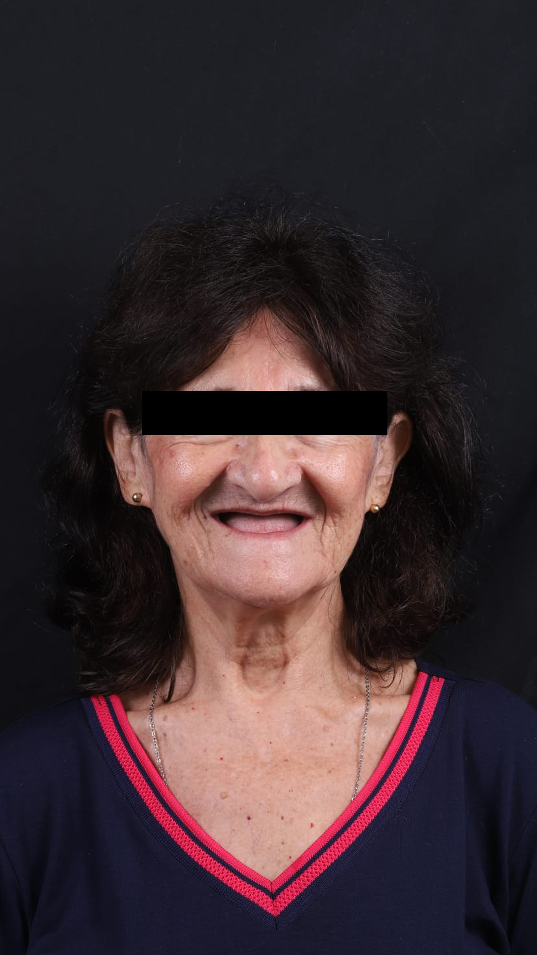
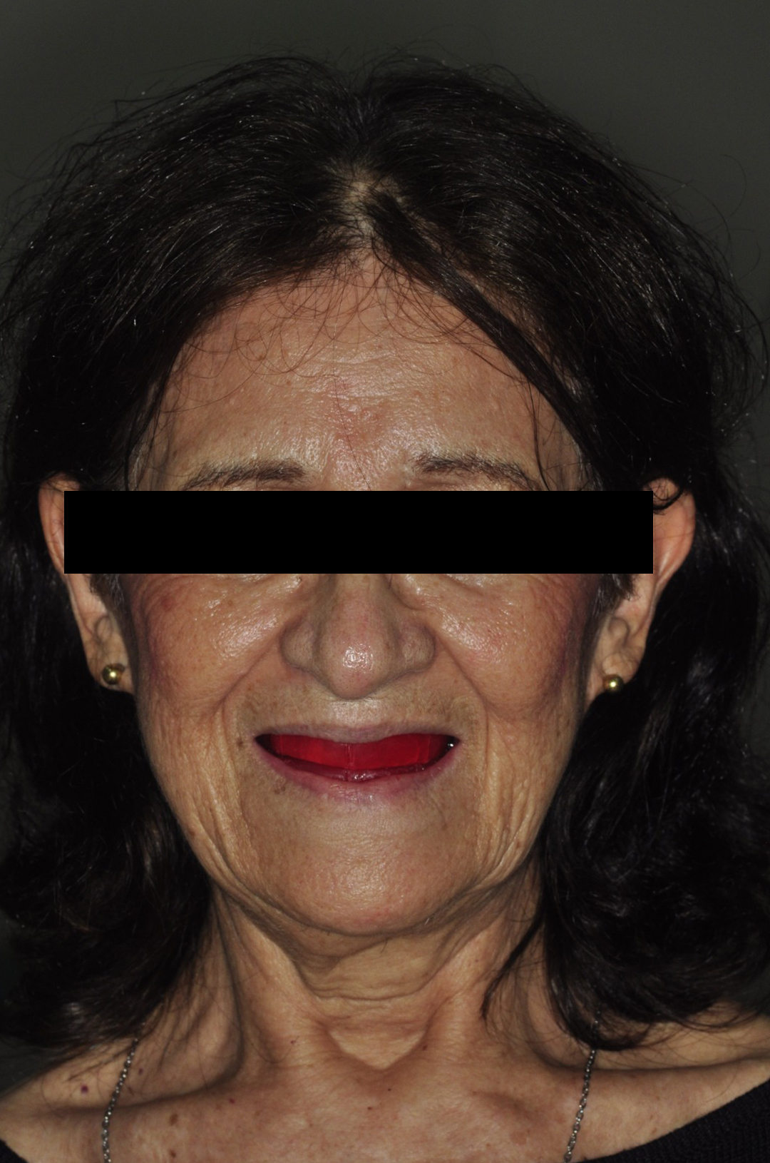
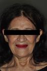
A female patient, healthy, 70 years old, sought treatment at the School of Dentistry of the São Paulo State University, in São José dos Campos (Figure 1 - initial). Her main complaint consisted of esthetic dissatisfaction and chewing difficulty regarding the previous complete denture (maxillary and mandibular).The patient presented with maxillary and mandibular complete dentures showing reduced retention, occlusal wear, and loss of vertical dimension of occlusion. aiming for the fabrication of new prostheses. Prior to the beginning of treatment, the patient received detailed explanations about the procedures that would be performed, and she consented to the execution of all stages.
Data collection involved clinical examination, no additional radiograph examination was required, detailed patient history, photographs (Figures 1 and 2), and conventional impressions with irreversible hydrocolloid (alginate) to obtain a study model, it was possible to observe a thick alveolar ridge, mainly in the mandibular region. Individual trays were fabricated and after this procedure, a functional impression was taken using additional silicone, and a working model was obtained. A try-in base was made in acrylic resin on the final model, with wax orientation planes for determination of occlusal vertical dimension, centric relation, and reference lines. After defining the orientation planes, compensation curve formation, and interocclusal registration, mounting was performed on an articulator.
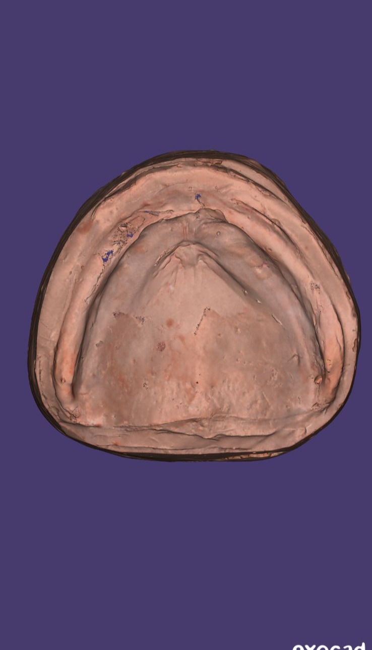
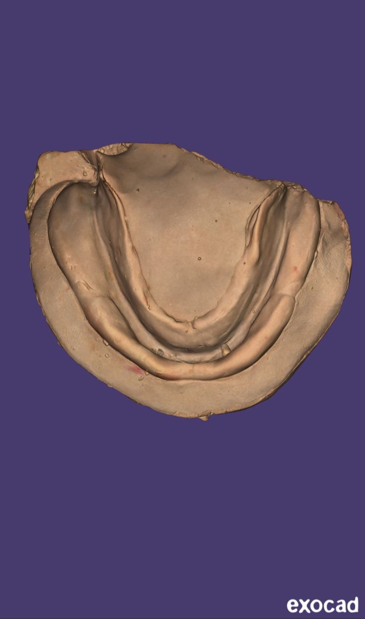
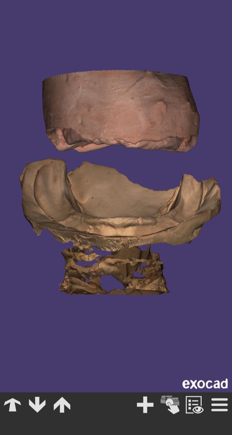
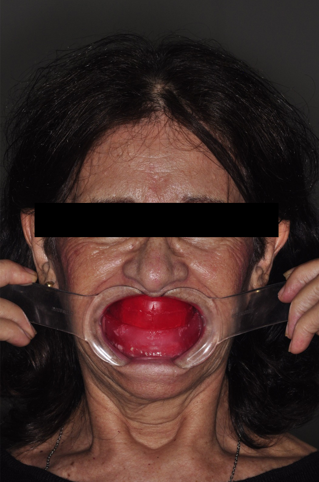
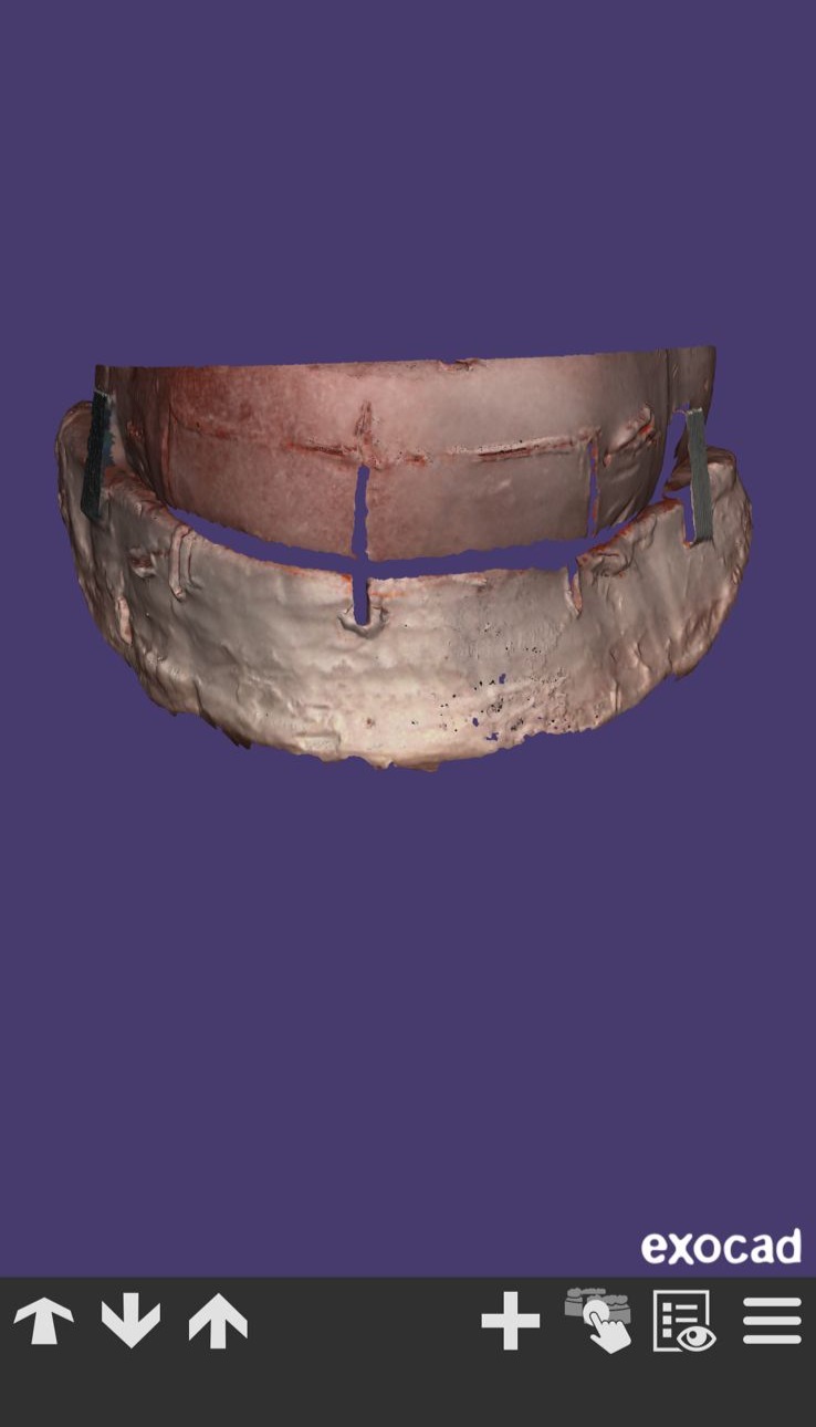
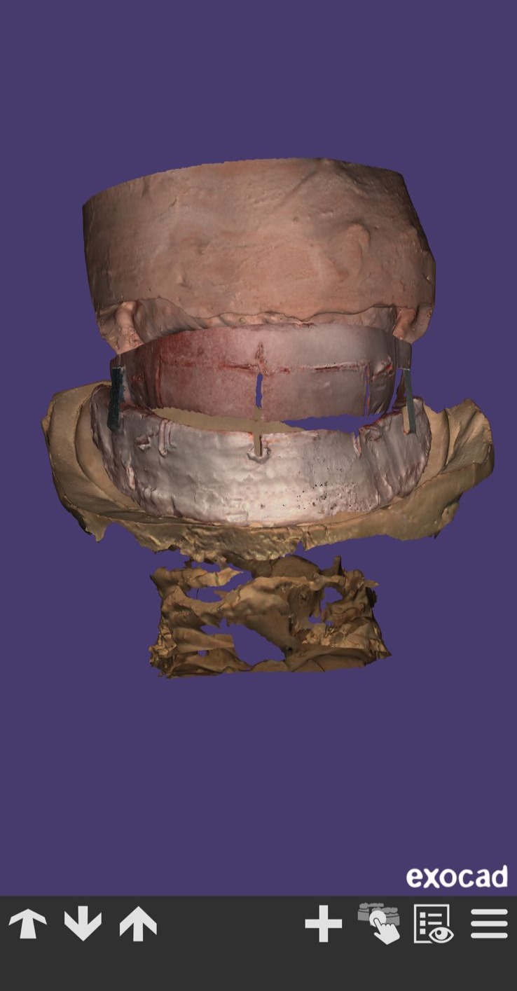
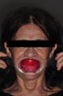
Model scanning was selected instead of benchtop scanning (PrograScan PS5, Ivoclar) (Figures 3, 4 and 5) to allow better capture of peripheral seal areas and data collection. The data were then imported into DentalCAD Rijeka 3.1 (Exocad), where esthetic and functional reference points were defined, including the upper smile line, pupillary line, and nasal wing to perform the wax rim mounting as a guide plane. After scanning the models with the guide plane, extraoral photographs were also taken with the guide plane in the mouth (Figures 6 and 7), allowing the prosthetic design to be forwarded for tooth fabrication. In addition to digitizing the models with the orientation plane, extraoral photographs were also taken with the orientation plane in the mouth, allowing for the creation of the compensation curves.
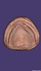
- The superior model in design software, obtained through functional impression and digitized with a benchtop scanner.
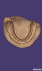
- The inferior model in design software, obtained through functional impression and digitized with a benchtop scanner.
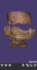
- Inferior and superior on occlusion model in design software, obtained through functional impression and digitized with a benchtop scanner.
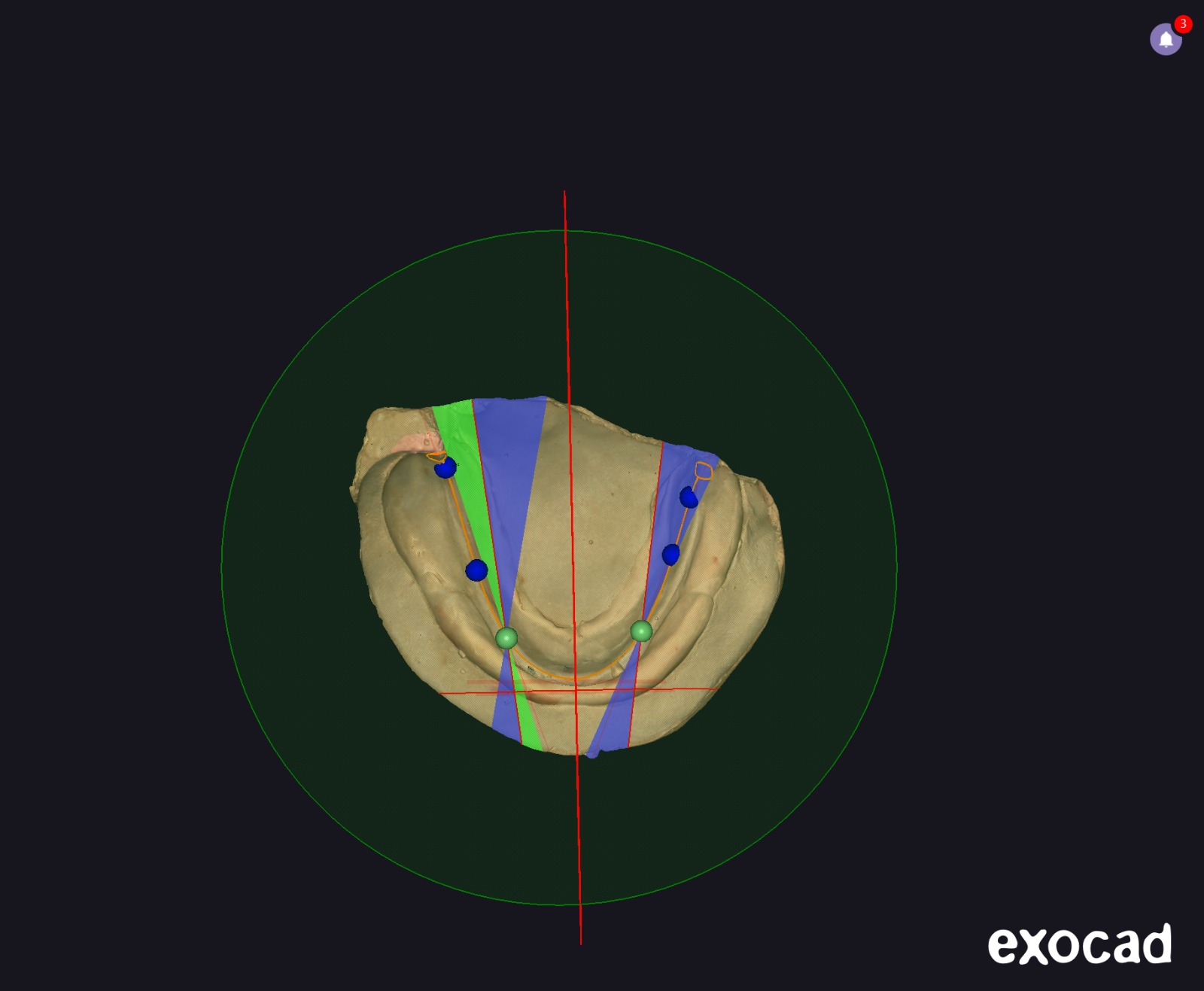
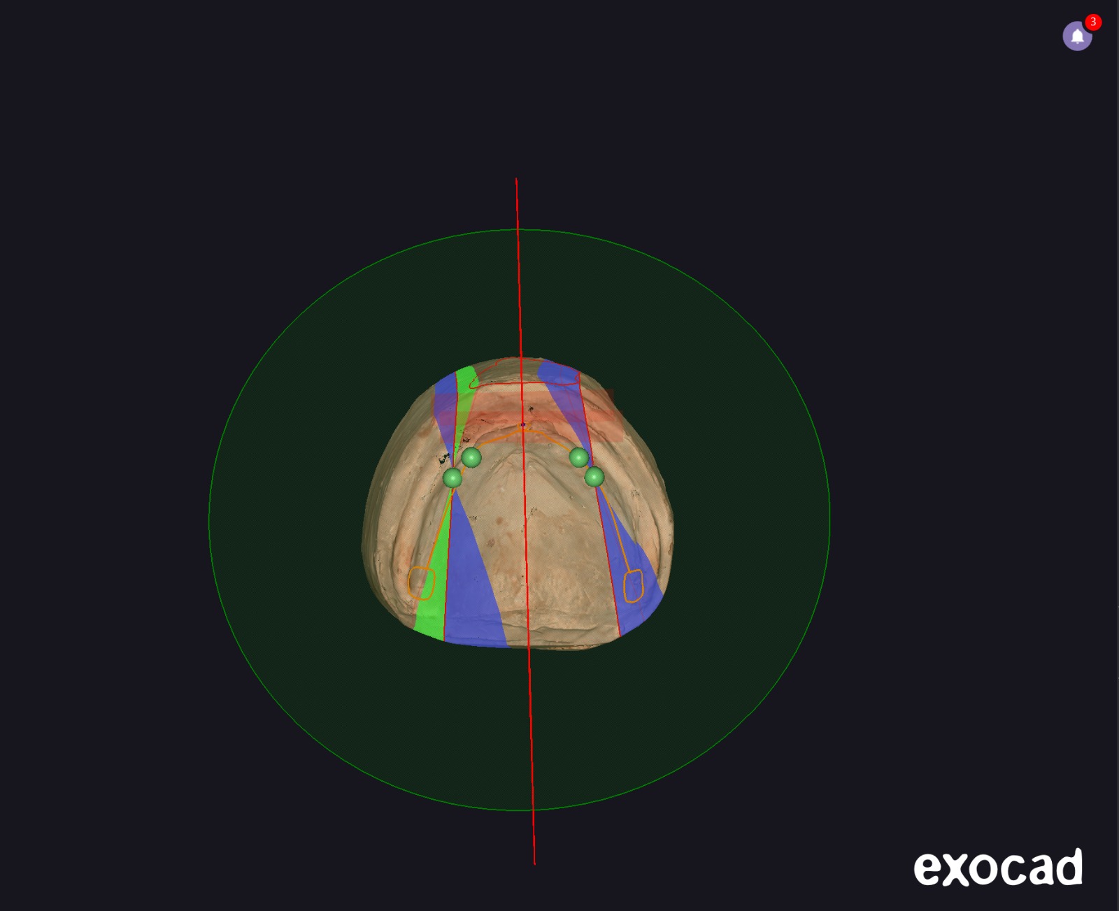
After digitizing the models along with the orientation plan, an analysis was performed for the tooth mounting, defining areas such as: incisive papilla position, canine, premolar and molar positions, palatine raphe, maxillary tuberosities, retromolar trigone, upper/lower occlusal plane, upper and lower labial area limits, and crest lines of the upper and lower models (Figures 8 and 9). With the definition of these areas, it was possible to scan the wax rims and mark the high smile line positions, midline, and canine positions for tooth mounting (Figures 10 and 11) and paciente proof the prostheses with teeth to approval.
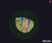
- Digital planning based on the analysis of digitized models for tooth position: determination of the position of the canines, premolars, and molars, identification of the retromolar trigone, definition of the mandibular occlusal planes, boundaries of the mandibular labial areas, and mandibular alveolar ridge lines.
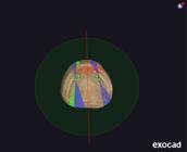
- Digital planning based on the analysis of digitized models for tooth position: determination of the position of the incisive papilla, positioning of the canines, identification of the palatal raphe, maxillary tuberosities, definition of the maxillary occlusal planes; boundaries of the maxillary labial areas, and maxillary alveolar ridge lines.
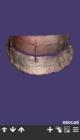
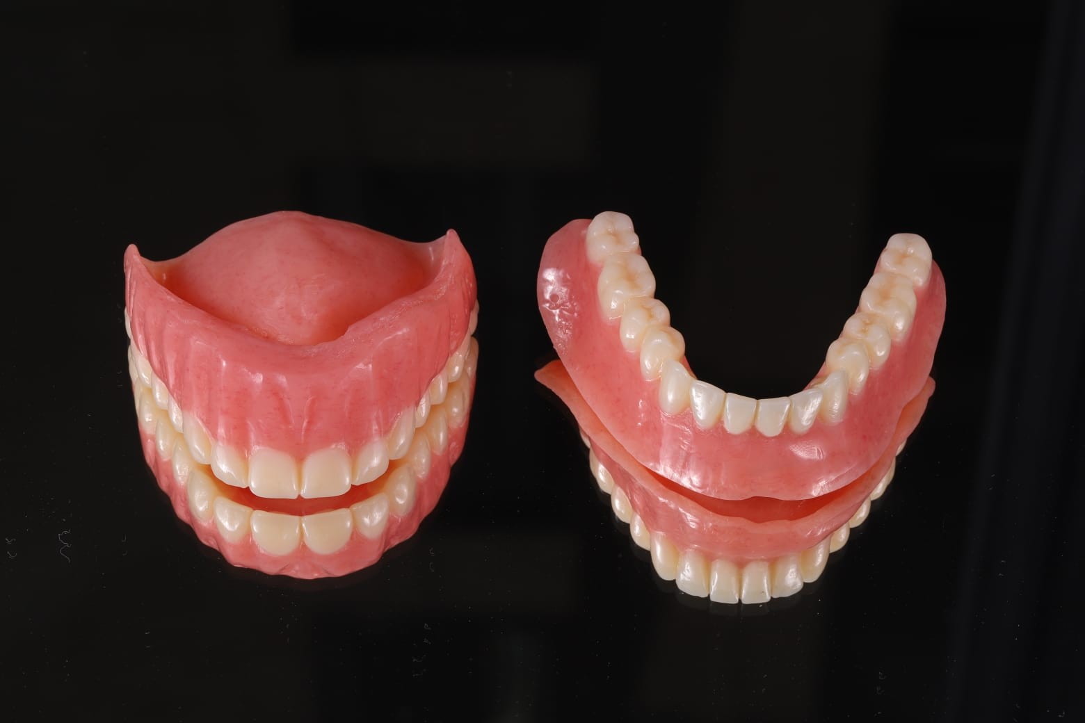
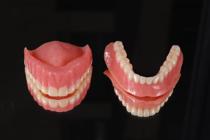
After approval of the patient, milling, texturing, and polishing of monolithic discs that integrates tooth and gingival materials within a single PMMA block, eliminating the need for bonding between separate components.This material is called Ivotion (Ivoclar, Shaan, Liechtenstein) and the colors used was A2 to teeth and color Preference to tissue were performed (Figure 12).
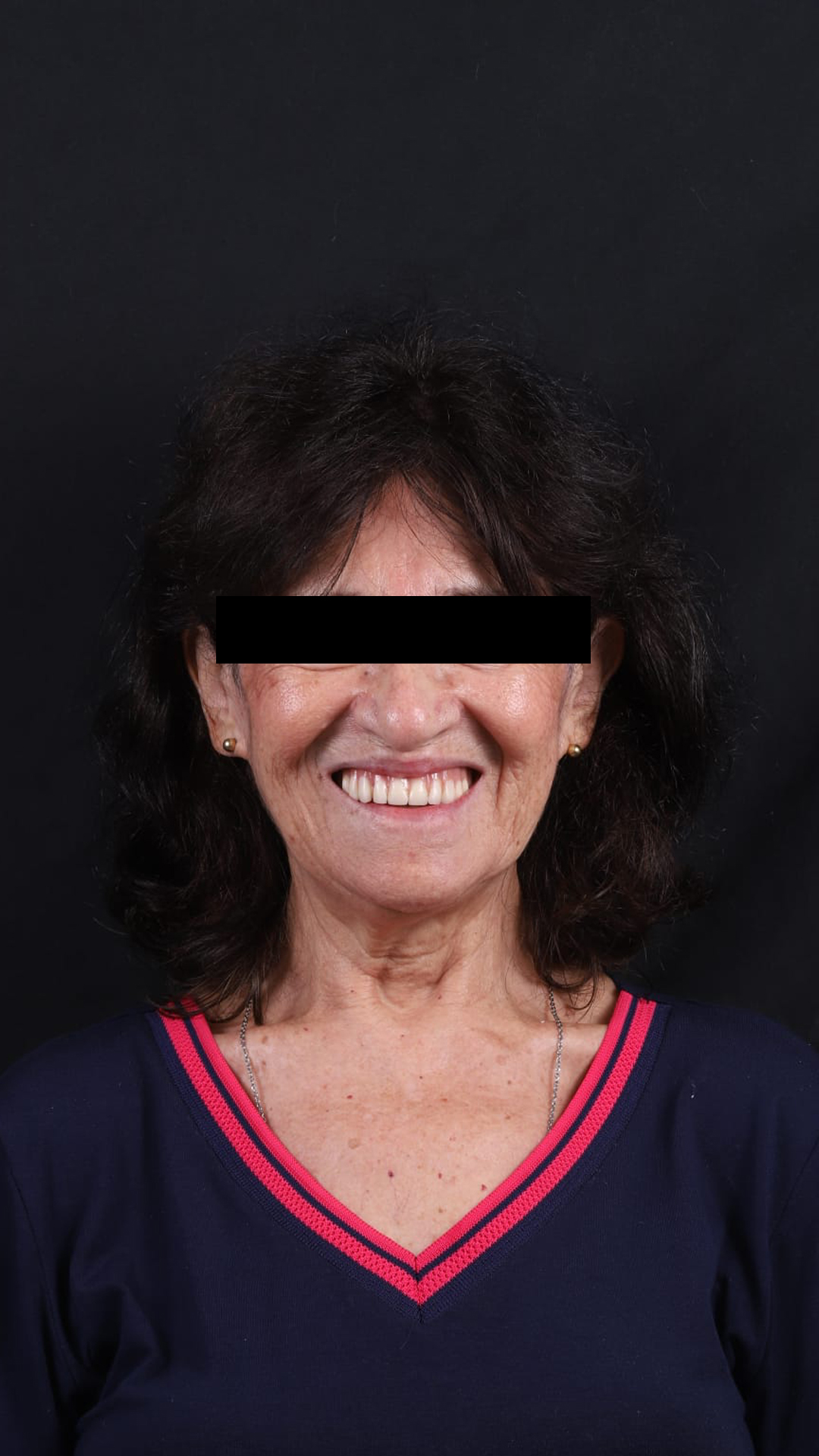
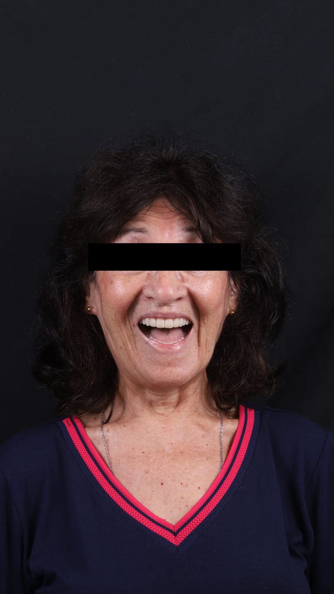
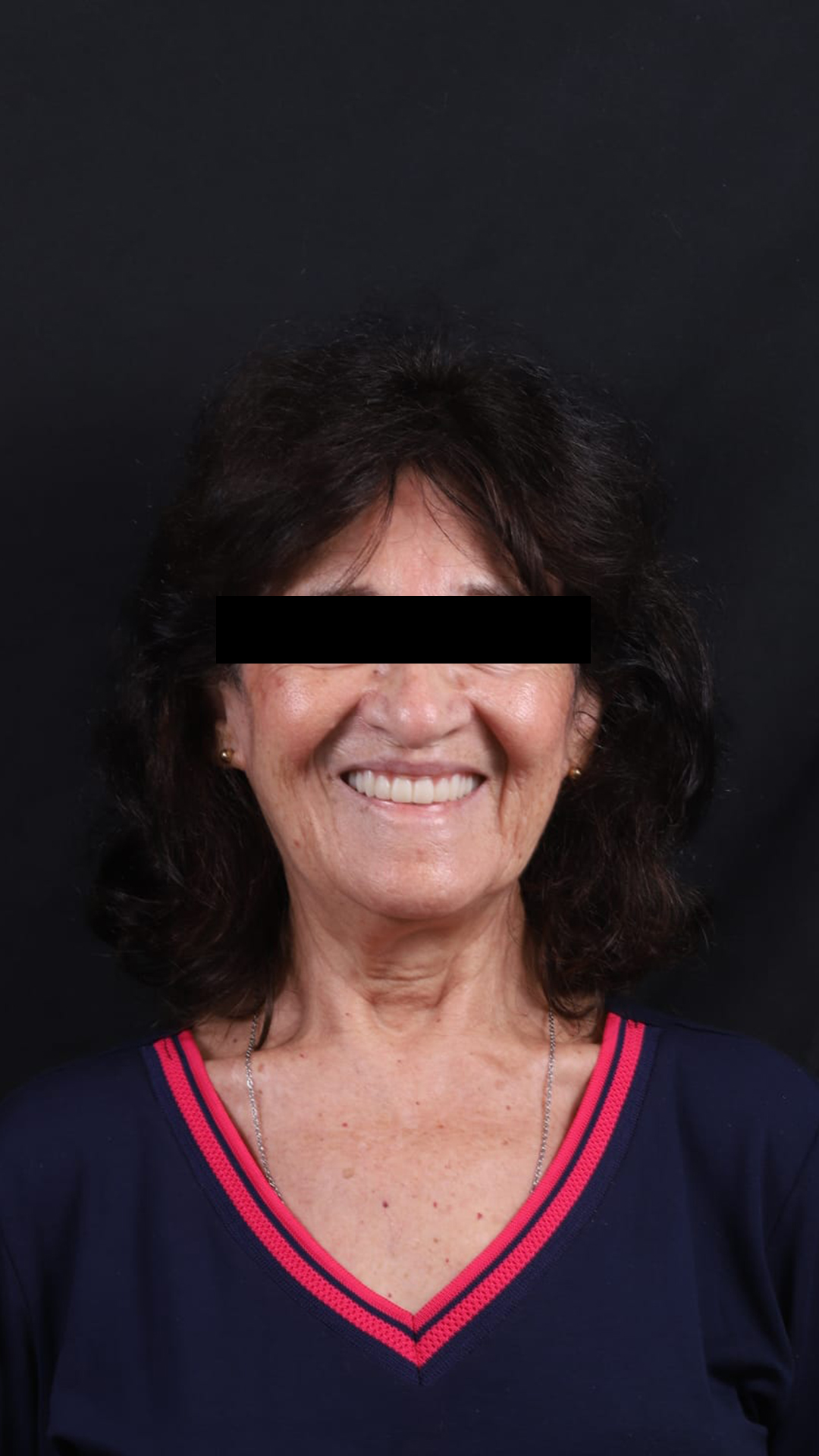
After the prostheses were fabricated, installation was done, which presented satisfactory retention and stability, not requiring adjustments initially. Patient follow-up and adaptation sessions to the prostheses were carried out in the following weeks, where minor adjustments were made. The patient reported esthetic and functional satisfaction with the rehabilitation (Figures 13 and 14).

- Final outcome of the digital complete dentures after insertion, shown with the patient’s full smile.
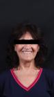
- Final outcome of the digital complete dentures after insertion, shown with the patient’s relaxed smile.
DISCUSSION
Patient satisfaction is generally considered an important indicator of success in prosthetic rehabilitation, particularly in edentulous patients treated with complete dentures. Comfort, retention, stability, aesthetics, and masticatory function are among the main factors influencing patients' perception of treatment results. Digital workflows have been increasingly incorporated into the prosthetic fabrication process to achieve greater stability and predictability of treatment [10-12].
In the digital workflow, one of the systems introduced is the CAD/CAM system as an alternative technique to conventional techniques for the fabrication of complete dentures. In this workflow, the denture base and the arrangement and anatomy of the teeth can be virtually designed and subsequently manufactured using additive or subtractive processes [1,4]. One of the advantages frequently reported in the literature is the potential reduction in the number of clinical and laboratory steps, which can shorten treatment time and improve workflow efficiency [4,8,13,14]. Furthermore, digital systems allow precise control of tooth positioning during the design phase, since each tooth is positioned within a coordinate system defined in the software environment, which can contribute to a balanced occlusion and greater denture stability [3,4].
While digital workflows offer several advantages, some clinical limitations remain, total prosthesis dentures are mucosally supported, a faithful and precise copy of the supporting tissues is necessary to achieve a peripheral seal, allowing for stability and retention. For this retention to be formed, the denture base must extend across the entire fibromucosal support area, avoiding excessive pressure on the underlying tissues [1]. Thus, although intraoral scanners demonstrate high accuracy in recording hard tissues, their ability to capture mobile mucosal tissues and functional border areas remains limited, which may impair the retention of the prosthetic piece [6,7].
One explanation for this limitation is the lack of controlled tissue compression during optical scanning procedures, which is normally achieved during conventional impression techniques. Consequently, some authors have proposed hybrid workflows that combine conventional functional impressions with subsequent digitalization of the models [8]. In the present case, functional impressions were obtained using conventional techniques and the resulting casts were digitized with a benchtop scanner. This approach enabled better control of the peripheral seal while preserving the advantages of digital design and manufacturing. Hybrid workflows may therefore represent a useful clinical strategy when the limitations of intraoral scanning in edentulous arches are taken into account [13,14].
Another relevant aspect in the digital fabrication of dental prostheses is the manufacturing method. Currently, prostheses can be manufactured using additive manufacturing or subtractive manufacturing (milling) [3,13,15]. In the present study, subtractive manufacturing was selected due to the characteristics of the material used and the mechanical properties associated with pre-polymerized polymethylmethacrylate (PMMA) discs.
In the clinical trial by Haboheikal et al., a randomized evaluation of patient satisfaction regarding the retention of printed, milled, and conventional prostheses was performed. There was no statistically significant difference in patient satisfaction scores among the three types of complete dentures for all five domains evaluated. Despite this, some patients showed greater satisfaction with the prostheses. Patients were more satisfied with printed prostheses, followed by conventional prostheses, and less satisfied with milled prostheses. This can be attributed to the inherent limitations of milling technology, which cannot mill severe retentions greater than 25-30 degrees, depending on the milling machine used, which has limited movement to five axes, and the size of the drill bit.This may have affected retention in the group with milled prostheses and, consequently, compromised patient satisfaction [16].
Unlike conventional acrylic resins, which undergo polymerization during laboratory processing, CAD/CAM milled prostheses are produced from industrially polymerized PMMA discs. These materials have higher density and lower porosity, which can contribute to better dimensional stability and less distortion of the prosthesis base. Previous studies have reported that milled prosthesis bases can exhibit better mechanical properties and greater retention compared to conventional acrylic prostheses [5,15]. In addition, milled prostheses may demonstrate improved adaptation due to the absence of polymerization shrinkage during laboratory processing. AlHelal et al. [5] reported increased retention of maxillary dentures fabricated using CAD/CAM milling compared with conventional polymethyl methacrylate dentures.
The prostheses used in this study were milled from a monolithic PMMA disc (Ivotion, Ivoclar, Schaan, Liechtenstein), which integrates tooth-colored and gingiva-colored materials within a single disc. During industrial processing, these materials are polymerized simultaneously, forming a direct chemical bond between them. As a result, the use of additional adhesive systems to bond the denture teeth to the base is eliminated, which may reduce the risk of adhesive failures [9]. This monolithic configuration has been associated with improved mechanical reliability [17].
In clinical practice, the applicability of digital workflows may vary depending on patient-related factors such as alveolar ridge morphology, degree of alveolar resorption, and neuromuscular control may influence treatment planning and clinical outcomes [8,13]. Subtractive manufacturing also presents inherent limitations. Because the prosthesis is milled from a solid disc, the process inevitably generates material waste. Another limitation involves geometric constraints related to the diameter of milling burs and the axis angulation of some milling machines, which may limit the reproduction of extremely fine anatomical details. The need for specialized equipment and laboratory infrastructure may also represent a limiting factor in certain clinical contexts [13,15].
In the present case, the digital workflow allowed the fabrication of complete dentures with satisfactory retention and stability. No adjustments were required at the time of prosthesis delivery, and only minor occlusal refinements were necessary during follow-up appointments. The patient reported improvements in comfort, masticatory function, and esthetic appearance after rehabilitation [15].
Within the limitations of this clinical report, the digital workflow allowed the fabrication of complete dentures with satisfactory retention, stability, and patient comfort. Nevertheless, further clinical studies with larger samples and longer follow-up periods are necessary to better understand the long-term clinical performance of digitally fabricated complete dentures and to establish clearer indications for the use of digital workflows in edentulous rehabilitation [10,12].
CONCLUSION
Incorporating CAD/CAM technology into the design and manufacture of complete dentures streamlines clinical and laboratory processes, offers flexible workflow options and improves communication between the patient, dentist and laboratory technician. In addition, digital devices allow for better planning and predictability of the clinical case, providing the information and resources needed to guarantee patient satisfaction, since the process requires fewer appointments and provides prostheses with precise retention, stability and adaptation, and which generate fewer adjustments than prostheses produced by the traditional method. However, it is recommended that clinical studies should be carried out with long-term follow-up to better assess biomechanical performance and longevity when compared to the traditional manufacturing technique.
Acknowledgements
The authors would like to thank the patient and Ivoclar and exocad for their valuable collaboration during the development of the clinical case.
How to cite:
Funding
Regulatory Statement
Data availability
No datasets were generated or analyzed during the current case report.
REFERENCES
- 1 Maeda Y, Minoura M, Tsutsumi S, Okada M, Nokubi TA. CAD/CAM system for removable denture. Int J Prosthodont. 1994;7(1):17-21. PMid:8179777.
-
2 Goodacre CJ, Garbacea A, Naylor WP, Daher T, Marchack CB, Lowry J. CAD/CAM fabricated complete dentures: concepts and clinical methods of obtaining required morphological data. J Prosthet Dent. 2012;107(1):34-46. https://doi.org/10.1016/S0022-3913(12)60015-8 PMid:22230914.
» https://doi.org/10.1016/S0022-3913(12)60015-8 - 3 Kattadiyil MT, Goodacre CJ, Baba NZ. CAD/CAM complete dentures: a review of two commercial fabrication systems. J Prosthet Dent. 2015;114(4):489-95.
-
4 Srinivasan M, Kalberer N, Naharro M, Marchand L, Lee H, Müller F. CAD-CAM milled dentures: the Geneva protocols for digital dentures. J Prosthet Dent. 2020;123(1):27-37. https://doi.org/10.1016/j.prosdent.2018.12.008 PMid:31079883.
» https://doi.org/10.1016/j.prosdent.2018.12.008 -
5 AlHelal A, AlRumaih HS, Kattadiyil MT, Baba NZ, Goodacre CJ. Comparison of retention between maxillary milled and conventional denture bases: a clinical study. J Prosthet Dent. 2017;117(2):233-8. https://doi.org/10.1016/j.prosdent.2016.08.007 PMid:27765399.
» https://doi.org/10.1016/j.prosdent.2016.08.007 -
6 Rasaie V, Abduo J, Hashemi S. Accuracy of intraoral scanners for recording the denture bearing areas: a systematic review. J Prosthodont. 2021;30(6):520-39. https://doi.org/10.1111/jopr.13345 PMid:33554361.
» https://doi.org/10.1111/jopr.13345 -
7 Srivastava G, Padhiary SK, Mohanty N, Molinero-Mourelle P, Chebib N. Accuracy of intraoral scanners for recording completely edentulous arches: a systematic review. Dent J. 2023;11(10):241. https://doi.org/10.3390/dj11100241 PMid:37886926.
» https://doi.org/10.3390/dj11100241 - 8 Peroz I, Blankenstein F, Lange KP, Naumann M. Restoring the edentulous maxilla with complete dentures: traditional versus digital workflow. J Prosthet Dent. 2022;128(4):640-7.
- 9 Frei R. Monolithic digital dentures in impressive material quality. Liechtenstein: Ivoclar Vivadent; 2020.
-
10 Verkhovskiy AE, Apresyan S, Stepanov A. Overview of the clinical and laboratory evaluation results of digital methods for fabrication of complete removable acrylic dentures. Russ J Dent. 2024;27(6):591-602. https://doi.org/10.17816/dent368253
» https://doi.org/10.17816/dent368253 -
11 Chappuis-Chocano AP, Venante HS, Costa RMB, Pordeus MD, Ferreira-Júnior JF, Porto VC. Evaluation of the clinical performance of dentures manufactured by computer-aided technology and conventional techniques: a systematic review. J Prosthet Dent. 2023;129(4):547-53. https://doi.org/10.1016/j.prosdent.2021.06.029 PMid:34332774.
» https://doi.org/10.1016/j.prosdent.2021.06.029 -
12 Srinivasan M, Cantin Y, Mehl A, Müller F. Digital complete dentures: a systematic review of clinical outcomes. J Dent. 2023;132:104470. https://doi.org/10.1016/j.jdent.2023.104470
» https://doi.org/10.1016/j.jdent.2023.104470 -
13 Thu KM, Molinero-Mourelle P, Yeung AWK, Abou-Ayash S, Lam WYH. Which clinical and laboratory procedures should be used to fabricate digital complete dentures? A systematic review. J Prosthet Dent. 2024;132(5):922-38. https://doi.org/10.1016/j.prosdent.2023.07.027 PMid:37689573.
» https://doi.org/10.1016/j.prosdent.2023.07.027 -
14 Mangano FG, Margiani B, Admakin O. A novel full-digital protocol (SCAN-PLAN-MAKE-DONE®) for the design and fabrication of complete dentures: a prospective clinical study. BMC Oral Health. 2022;22:1-10. http://doi.org/10.1186/s12903-022-02167-7.
» https://doi.org/10.1186/s12903-022-02167-7 -
15 Revilla-León M, Özcan M. Additive and subtractive manufacturing technologies used for processing polymers in prosthetic dentistry: current status and future perspectives. J Prosthodont. 2023;32(5):369-80. https://doi.org/10.1111/jopr.13568
» https://doi.org/10.1111/jopr.13568 -
16 Heikal MMA, Abdel Nabi N, Wagdy Elkerdawy M. A study comparing patient satisfaction and retention of CAD/CAM milled complete dentures and 3D printed CAD/CAM complete dentures versus conventional complete dentures: a randomized clinical trial. Braz Dent Sci. 2022;25(1):e2785. https://doi.org/10.4322/bds.2022.e2785
» https://doi.org/10.4322/bds.2022.e2785 -
17 Silva NRFA, Kukucka ED. Innovative subtractive production of a digital removable complete denture from start to finish: a JPD digital video presentation. J Prosthet Dent. 2022;127(1):1-5. https://doi.org/10.1016/j.prosdent.2021.12.002 PMid:35027149.
» https://doi.org/10.1016/j.prosdent.2021.12.002
Conflict of Interest
Correspondence
Guilherme de Siqueira Ferreira Anzaloni Saavedra (Corresponding address) Universidade Estadual de São Paulo, Departamento de Materiais Odontológicos e Prótese, São Jose dos Campos, SP, Brazil. Email: guilherme.saavedra@unesp.brEditor-in-chief:
Editor: