
 Effect of the Blooming Artifact Reduction filter on the detection of voids in root canal fillings using cone-beam computed tomography images
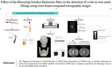
Este estudo avaliou a efetividade do filtro Blooming Artifact Reduction (BAR) (e-Vol DX Software, CDT, Bauru, SP, Brasil) para a detecção de espaços vazios em canais radiculares obturados com diferentes cimentos, utilizando dois aparelhos de TCFC e a micro-CT como referência. Vinte incisivos inferiores com canais radiculares ovais longos foram obturados com AH Plus Jet (AHPJ) ou Bio-C Sealer (BCS). Cada dente foi posicionado no alvéolo mandibular de um crânio seco, recoberto com material Mix-D e escaneado nos aparelhos de TCFC VeraView X800 e OP300 Maxio. As imagens de micro-CT foram utilizadas como referência. Três examinadores calibrados avaliaram as imagens de TCFC, sem e com aplicação do filtro BAR (intensidade nível 1 para o OP300 Maxio e nível 3 para o VeraView X800), para detecção de espaços vazios usando uma escala de cinco pontos. Os valores diagnósticos (área sob a curva ROC, sensibilidade e especificidade) foram comparados por meio de análise de variância multifatorial (p=0,05). A concordância intra e interobservador foi avaliada pelo índice Kappa ponderado. No OP300, o uso do filtro BAR 1 aumentou significativamente os valores da curva ROC para detecção de espaços vazios no terço apical com AHPJ e BCS (p<0,05), além de elevar a especificidade para detecção no terço apical com AHPJ (p<0,05). No VeraView X800, os valores da curva ROC e da sensibilidade não foram afetados (p>0,05), porém o uso do filtro BAR 3 aumentou significativamente a especificidade para detecção de espaços vazios nos terços cervical/médio com BCS (p<0,05). O desempenho diagnóstico do filtro BAR é dependente do aparelho de TCFC. Nos exames do OP300, ele melhora a detecção de espaços vazios no terço apical com AHPJ e BCS. Nos exames do VeraView X800, aumenta a especificidade para detecção nos terços cervical/médio com BCS.
Autoria
person Airton Oliveira Santos-Junior
schoolDepartment of Restorative Dentistry, São Paulo State University (UNESP), School of Dentistry, Araraquara, São Paulo, BrazilSão Paulo State UniversityBrazilAraraquara, São Paulo, BrazilDepartment of Restorative Dentistry, São Paulo State University (UNESP), School of Dentistry, Araraquara, São Paulo, Brazil
person Rocharles Cavalcante Fontenele
schoolOMFS IMPATH Research Group, Department of Imaging and Pathology, Faculty of Medicine, University of Leuven, Leuven, BelgiumUniversity of LeuvenBelgiumLeuven, BelgiumOMFS IMPATH Research Group, Department of Imaging and Pathology, Faculty of Medicine, University of Leuven, Leuven, BelgiumschoolDepartment of Stomatology, Public Health and Forensic Dentistry, Division of Oral Radiology, School of Dentistry of Ribeirão Preto, University of São Paulo (USP), Ribeirão Preto, São Paulo, BrazilUniversity of São PauloBrazilRibeirão Preto, São Paulo, BrazilDepartment of Stomatology, Public Health and Forensic Dentistry, Division of Oral Radiology, School of Dentistry of Ribeirão Preto, University of São Paulo (USP), Ribeirão Preto, São Paulo, Brazil
person Jáder Camilo Pinto
schoolDepartment of Dentistry, Centro Universitário Presidente Antônio Carlos (UNIPAC), Barbacena, Minas Gerais, BrazilCentro Universitário Presidente Antônio CarlosBrazilBarbacena, Minas Gerais, BrazilDepartment of Dentistry, Centro Universitário Presidente Antônio Carlos (UNIPAC), Barbacena, Minas Gerais, BrazilschoolDepartment of Dentistry - Centro Universitário Presidente Tancredo de Almeida Neves (UNIPTAN), São João del Rei, Minas Gerais, BrazilCentro Universitário Presidente Tancredo de Almeida NevesBrazilSão João del Rei, Minas Gerais, BrazilDepartment of Dentistry - Centro Universitário Presidente Tancredo de Almeida Neves (UNIPTAN), São João del Rei, Minas Gerais, Brazil
person André Ferreira Leite
schoolDepartment of Dentistry, University of Brasília (UnB), Campus Darcy Ribeiro, Asa Norte, Brasília, Distrito Federal, BrazilUniversity of BrasíliaBrazilBrasília, Distrito Federal, BrazilDepartment of Dentistry, University of Brasília (UnB), Campus Darcy Ribeiro, Asa Norte, Brasília, Distrito Federal, Brazil
person Fernanda Ferrari Esteves Torres
schoolDepartment of Restorative Dentistry, São Paulo State University (UNESP), School of Dentistry, Araraquara, São Paulo, BrazilSão Paulo State UniversityBrazilAraraquara, São Paulo, BrazilDepartment of Restorative Dentistry, São Paulo State University (UNESP), School of Dentistry, Araraquara, São Paulo, Brazil
person Karina Ines Medina Carita Tavares
schoolDepartment of Restorative Dentistry, São Paulo State University (UNESP), School of Dentistry, Araraquara, São Paulo, BrazilSão Paulo State UniversityBrazilAraraquara, São Paulo, BrazilDepartment of Restorative Dentistry, São Paulo State University (UNESP), School of Dentistry, Araraquara, São Paulo, Brazil
person Marcelo Gonçalves
schoolDepartment of Diagnosis and Surgery, São Paulo State University (UNESP), School of Dentistry, Araraquara, São Paulo, BrazilSão Paulo State UniversityBrazilAraraquara, São Paulo, BrazilDepartment of Diagnosis and Surgery, São Paulo State University (UNESP), School of Dentistry, Araraquara, São Paulo, Brazil
person Juliane Maria Guerreiro-Tanomaru
schoolDepartment of Restorative Dentistry, São Paulo State University (UNESP), School of Dentistry, Araraquara, São Paulo, BrazilSão Paulo State UniversityBrazilAraraquara, São Paulo, BrazilDepartment of Restorative Dentistry, São Paulo State University (UNESP), School of Dentistry, Araraquara, São Paulo, Brazil
person Mário Tanomaru-Filho
schoolDepartment of Restorative Dentistry, São Paulo State University (UNESP), School of Dentistry, Araraquara, São Paulo, BrazilSão Paulo State UniversityBrazilAraraquara, São Paulo, BrazilDepartment of Restorative Dentistry, São Paulo State University (UNESP), School of Dentistry, Araraquara, São Paulo, Brazil
Correspondence: Prof. Dr. Mário Tanomaru-Filho. Araraquara Dental School, São Paulo State University - UNESP. Department of Restorative Dentistry. Rua Humaitá, 1680, CEP 14801-903, Araraquara, SP, Brazil; Phone: +55 16 3301-6390; E-mail:
tanomaru@uol.com.br
tanomaru@uol.com.br
Statement of Conflict of Interest
The authors declare that they have no conflict of interest.
Responsible editor
Manoel Damião de Sousa-Neto
SCIMAGO INSTITUTIONS RANKINGS
Department of Restorative Dentistry, São Paulo State University (UNESP), School of Dentistry, Araraquara, São Paulo, BrazilSão Paulo State UniversityBrazilAraraquara, São Paulo, BrazilDepartment of Restorative Dentistry, São Paulo State University (UNESP), School of Dentistry, Araraquara, São Paulo, Brazil
OMFS IMPATH Research Group, Department of Imaging and Pathology, Faculty of Medicine, University of Leuven, Leuven, BelgiumUniversity of LeuvenBelgiumLeuven, BelgiumOMFS IMPATH Research Group, Department of Imaging and Pathology, Faculty of Medicine, University of Leuven, Leuven, Belgium
Department of Stomatology, Public Health and Forensic Dentistry, Division of Oral Radiology, School of Dentistry of Ribeirão Preto, University of São Paulo (USP), Ribeirão Preto, São Paulo, BrazilUniversity of São PauloBrazilRibeirão Preto, São Paulo, BrazilDepartment of Stomatology, Public Health and Forensic Dentistry, Division of Oral Radiology, School of Dentistry of Ribeirão Preto, University of São Paulo (USP), Ribeirão Preto, São Paulo, Brazil
Department of Dentistry, Centro Universitário Presidente Antônio Carlos (UNIPAC), Barbacena, Minas Gerais, BrazilCentro Universitário Presidente Antônio CarlosBrazilBarbacena, Minas Gerais, BrazilDepartment of Dentistry, Centro Universitário Presidente Antônio Carlos (UNIPAC), Barbacena, Minas Gerais, Brazil
Department of Dentistry - Centro Universitário Presidente Tancredo de Almeida Neves (UNIPTAN), São João del Rei, Minas Gerais, BrazilCentro Universitário Presidente Tancredo de Almeida NevesBrazilSão João del Rei, Minas Gerais, BrazilDepartment of Dentistry - Centro Universitário Presidente Tancredo de Almeida Neves (UNIPTAN), São João del Rei, Minas Gerais, Brazil
Department of Dentistry, University of Brasília (UnB), Campus Darcy Ribeiro, Asa Norte, Brasília, Distrito Federal, BrazilUniversity of BrasíliaBrazilBrasília, Distrito Federal, BrazilDepartment of Dentistry, University of Brasília (UnB), Campus Darcy Ribeiro, Asa Norte, Brasília, Distrito Federal, Brazil
Department of Diagnosis and Surgery, São Paulo State University (UNESP), School of Dentistry, Araraquara, São Paulo, BrazilSão Paulo State UniversityBrazilAraraquara, São Paulo, BrazilDepartment of Diagnosis and Surgery, São Paulo State University (UNESP), School of Dentistry, Araraquara, São Paulo, Brazil
Figuras | Tabelas
 Thumbnail
 Thumbnail
 Thumbnail
 Thumbnail
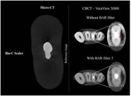 Thumbnail
imageopen_in_new

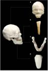
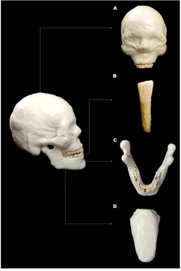
imageFigure 1 Anthropomorphic phantom consisting of a dry human skull with the mandible, both covered in Mix-D, along with a tongue model made with Mix-D. (A) Frontal view of the dry human skull; (B) Frontal view of the mandibular left central incisor; (C) Occlusal view of the mandible; (D) Tongue model made with Mix-D. open_in_new
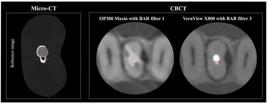
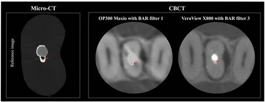
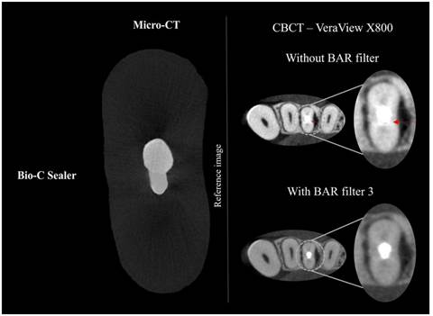
imageFigure 2 Axial reconstructions of the same specimen obtained using micro-CT as a reference and CBCT scans from the OP300 Maxio (BAR filter intensity 1) and VeraView X800 (BAR filter intensity 3), with red arrows indicating voids within the filling material. open_in_new
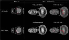
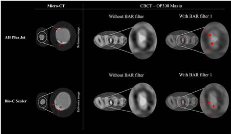
imageFigure 3 Examples of voids located in the apical third of long-oval root canals filled with AH Plus Jet and Bio-C Sealer. Axial reconstructions of CBCT scans without the BAR filter do not reveal these voids. However, when the BAR filter is applied at intensity level 1, the voids become clearly visible, as indicated by the red arrows. Their presence is confirmed in the reference micro-CT images, where the same regions are also highlighted with red arrows. open_in_new
imageFigure 4 An example of an axial reconstruction of the CBCT scan showing a false-positive detection of a void in the cervical/middle thirds of a canal filled with Bio-C Sealer, as indicated by the red arrow. After applying the BAR filter at intensity level 3, the absence of the void was confirmed, as also seen in the reference micro-CT image. open_in_new
table_chartTable 1
Acquisition parameters of the cone-beam computed tomography dataset
| CBCT device | kVp | mA | FOV (cm) | Voxel size (mm3) |
|---|---|---|---|---|
| VeraView X800 | 70 | 5 | 40 x 40 | 0.08 |
| OP300 Maxio | 90 | 8 | 50 x 50 | 0.08 |
-
CBCT: cone-beam computed tomography; kVp: kilovoltage-peak; mA: milliampere; FOV: field of view; cm: centimeters; mm3: cubic millimeters.
table_chartTable 2
Distribution of teeth (percentage) regarding the presence or absence of voids in different root canal thirds based on sealer type in micro-CT assessment.
| Sealer | Cervical/ Middle | Apical | ||
|---|---|---|---|---|
| Absence | Presence | Absence | Presence | |
| AH Plus Jet (10 Teeth) | 3 (30) | 7 (70) | 4 (40) | 6 (60) |
| Bio-C Sealer (10 Teeth) | 3 (30) | 7 (70) | 4 (40) | 6 (60) |
table_chartTable 3
Mean ± standard deviation (SD) of the area under the receiver operating curve (AUC), sensitivity, and specificity values for the detection of voids based on BAR use (BAR filter intensity level 1), sealer, and root third in the OP300 Maxio CBCT device.
| BAR | Sealer | Root third | AUC | Sensitivity | Specificity |
|---|---|---|---|---|---|
| Without BAR | AH Plus Jet | Cervical/Middle | 0.60 (0.01) | 0.39 (0.00) | 0.86 (0.01) |
| Apical | 0.53 (0.02) | 0.40 (0.34) | 0.80 (0.35) | ||
| Bio C-Sealer | Cervical/Middle | 0.57 (0.06) | 0.73 (0.14) | 0.50 (0.00) | |
| Apical | 0.53 (0.02) | 0.45 (0.48) | 0.58 (0.29) | ||
| With BAR filter 1 | AH Plus Jet | Cervical/Middle | 0.61 (0.03) | 0.63 (0.21) | 0.67 (0.17) |
| Apical | 0.72 (0.07) *# | 0.47 (0.11) | 1.00 (0.00) + | ||
| Bio C-Sealer | Cervical/Middle | 0.53 (0.00) | 0.67 (0.04) | 0.50 (0.00) | |
| Apical | 0.65 (0.03) *# | 1.00 (0.00) | 0.42 (0.14) |
-
# indicates a statistically significant difference in the use of BAR within the sealer and root third investigated (p<0.05).
-
* indicates a statistically significant difference between the root thirds within both sealer and BAR conditions (p<0.05).
-
+ indicates a statistically significant difference between the sealers within both root third and BAR conditions (p<0.05).
table_chartTable 4
Mean ± standard deviation (SD) of the area under the receiver operating curve (AUC), sensitivity, and specificity values for the detection of voids based on BAR use (BAR filter intensity level 3), sealer, and root third in the VeraView X800 CBCT device
| BAR | Sealer | Root third | AUC | Sensitivity | Specificity |
|---|---|---|---|---|---|
| Without BAR | AH Plus Jet | Cervical/Middle | 0.67 (0.08) | 0.51 (0.17) | 0.80 (0.00) |
| Apical | 0.62 (0.04) | 0.60 (0.20) | 0.70 (0.10) | ||
| Bio C-Sealer | Cervical/Middle | 0.56 (0.04) | 0.61 (0.29) | 0.58 (0.29) | |
| Apical | 0.54 (0.04) | 0.56 (0.20) | 0.50 (0.00) | ||
| With BAR filter 3 | AH Plus Jet | Cervical/Middle | 0.66 (0.10) | 0.64 (0.27) | 0.67 (0.11) |
| Apical | 0.56 (0.00) | 0.40 (0.00) | 0.80 (0.00) | ||
| Bio C-Sealer | Cervical/Middle | 0.82 (0.17) | 0.66 (0.31) | 1.00 (0.00) # | |
| Apical | 0.72 (0.16) | 0.61 (0.38) | 0.83 (0.14) |
-
# indicates a statistically significant difference in the use of BAR within the sealer and root third investigated (p<0.05).
Como citar
location_on
Fundação Odontológica de Ribeirão Preto
Av. do Café, S/N, 14040-904 Ribeirão Preto SP Brasil, Tel.: (55 16) 3602-3982, Fax: (55 16) 3633-0999 -
Ribeirão Preto -
SP -
Brazil
E-mail: bdj@forp.usp.br
E-mail: bdj@forp.usp.br
rss_feed
Acompañe los números de esta revista en su lector de RSS
Versão para download de PDF
Artículos relacionados
Lista de links para artigos relacionados. Os links abrem em nova aba.
Versões e tradução automática
Escolha a versão original do texto ou utilize um serviço de tradução automática.
Versão original do texto
Como citar
Escolha um formato para exportar ou selecione um estilo de citação. O conteúdo abaixo pode ser atualizado após a seleção.