The Use of Mesenchymal Stromal/Stem Cells (MSC) for Periodontal and Peri-implant Regeneration: Scoping Review
Abstract
The necessity for regenerating peri-implant and periodontal tissues is increasingly apparent. Periodontal diseases can result in a significant loss of clinical attachment level, and tissue regeneration stands as the ultimate goal of periodontal therapy. With the rise of osseointegration, the prosthetic rehabilitation of missing teeth using dental implants has surged, leading to a frequent need for alveolar bone regeneration around implants. This review assessed studies reporting various sources of mesenchymal stromal/stem cells (MSC) and their potential in regenerating periodontal and peri-implant bone tissue. A search was conducted across seven databases spanning the past decade. Three authors independently screened all identified titles and abstracts for eligibility, generating tables to summarize included studies in animals and humans separately. A total of 55 articles were chosen for final evaluation, showcasing five origins of MSC used in humans and animals for regenerating periodontal tissues and peri-implant bone, using different types of scaffolds. Overall, research from the past decades supports the effectiveness of MSC in promoting periodontal and peri-implant regeneration. However, the impact of MSC on regenerative therapies in humans is still in its initial stages. Future research should optimize MSC application protocols by combining techniques, such as the use of nanomedicine and 3D printing for tissue engineering. Clinical studies should also understand the long-term effects and compare MSC therapies with current treatment modalities. By addressing these areas, the scientific community can ensure that MSC therapies are both safe and effective, ultimately enhancing therapeutic strategies and treatment outcomes in Periodontology and Implantology.
Key Words:
Stem cells; stromal cells; peri-implant regeneration; intrabony defect; furcation
Resumo
A necessidade de regenerar tecidos peri-implantares e periodontais é cada vez mais evidente. As doenças periodontais podem resultar em uma perda significativa do nível de fixação clínica, e a regeneração tecidual se apresenta como o objetivo final da terapia periodontal. Com o surgimento da osseointegração, a reabilitação protética de dentes ausentes usando implantes dentários tem aumentado, levando a uma necessidade frequente de regeneração do osso alveolar ao redor dos implantes. Esta revisão busca avaliar diversas fontes de células-tronco mesenquimais e seu potencial na regeneração de tecidos ósseos periodontais e peri-implantares. Foi realizada uma busca em sete bases de dados ao longo da última década. Três autores avaliaram de forma independente todos os títulos e resumos identificados quanto à elegibilidade, gerando tabelas para resumir estudos incluídos em animais e humanos separadamente. Um total de 55 artigos foram escolhidos para avaliação final, demonstrando cinco origens de células-tronco mesenquimais usadas em humanos e animais para regenerar tecidos periodontais e osso peri-implantar, utilizando diferentes tipos de suportes. Pesquisas das últimas décadas demonstram a eficácia das células-tronco mesenquimais na promoção da regeneração periodontal e peri-implantar. No entanto, o impacto das células-tronco mesenquimais nas terapias regenerativas em humanos ainda está em seus estágios iniciais. Pesquisas futuras devem otimizar os protocolos de aplicação das células-tronco mesenquimais combinando técnicas, como o uso de nanomedicina e impressão 3D para engenharia tecidual. Estudos clínicos também devem compreender os efeitos a longo prazo e comparar as terapias com células-tronco mesenquimais com as modalidades de tratamento atuais. Ao abordar essas áreas, a comunidade científica pode garantir que as terapias com células-tronco mesenquimais sejam seguras e eficazes, aprimorando as estratégias terapêuticas em Periodontia e Implantodontia.
Introduction
Following non-surgical or conventional periodontal surgical therapy, is usual to have a persistent residual deep pocket depth in sites where deep intrabony or furcation defects exist. Periodontal ressective surgery can eliminate these deep defects, however it is associated with substantial loss of clinical attachment 1.
Human histological studies 2,3 have shown that after conventional periodontal therapy, healing is characterized by the formation of a long epithelial attachment along the root surface, often with limited periodontal regeneration, mostly in the apical part of the defect.
However, over the years, new biologically guided surgical approaches and biomaterials used in periodontal regeneration therapy, have contributed to expanding its indications 1. In this context, periodontal regeneration implies that CAL (clinical attachment level) gain is achieved by the formation of a functional new cementum on the previously affected root surface, in conjunction with new alveolar bone and periodontal ligament 1,4.
Dental implant therapy has become an integral part of clinical dentistry for rehabilitating patients who lost teeth due to periodontal disease or another factor. However, it is not unusual to face situations where horizontal or vertical bone deficiencies of the alveolar process are present and may jeopardize the results for implant success and survival 5.
When implants are placed and a bone defect results, exposing part of the endosseous surface of the implant, the literature indicates that guided bone regeneration is a successful technique for predictable bone formation around implants 5,6,7. In such clinical scenarios, the combination of a membrane and bone graft or bone substitute is generally recommended for guided bone regeneration procedures to provide adequate support and enhance bone ingrowth into the defect 5,7. To avoid or decrease the morbidity associated with the harvesting of autogenous bone graft, the use of bone substitute materials is therefore recommended for bone regeneration at exposed implant surfaces to treat dehiscence bone defects simultaneously with implant placement 6,7
Current research in regenerative therapy aims to develop effective strategies to enhance the body’s ability to regenerate lost tissues, improve treatment predictability, and, at the same time, reduce surgical morbidity. In this context, efforts are focusing on growth and differentiation factors and their delivery systems, trying to identify bioactive molecules that can regulate wound and tissue regeneration, and stimulate tissue growth in the area to be regenerated around teeth and implants 7.
Tissue engineering relies on three main parameters: the type of cells that can be used, the scaffolds where the cells should be seeded, and the growth factor/molecular signals that should be applied 8. In this context, different sources of mesenchymal stromal cells (MSC) have been used to make alveolar bone tissue engineering and periodontal tissue engineering. Studies have reported significant periodontal regeneration using MSC derived from bone marrow, dental pulp, or periodontal ligament 8,9,10,11. Gingival fibroblasts have potentials that could be used to increase cellularity and regenerative potential since they seem to meet the requirements of MSC 8,12,13.
When treating intra-bony periodontal defects, studies using mesenchymal stem cells have demonstrated significant reduction in pocket depth and CAL gains, suggesting an interesting outcome in these situations 13,14
Therefore, taking into consideration these promising results reported in previous studies, this review aims to summarize the potential of MSC cells for use in the regeneration of bone around implants and periodontal regeneration previously described in the literature.
Materials and methods
This scoping review was conducted following the recommendations of the Preferred Reporting Items for Systematic Reviews and Meta-Analyses - Extension for Scoping Reviews (PRISMA-ScR).
A PICO search strategy was used to define the question of this review: Population: “in vivo” (human and animal), Intervention: Mesenchymal cells for periodontal and peri-implant osteogenesis, Comparison: Type of osteogenic mesenchymal cell for periodontal and peri-implant regeneration, Outcomes: Types of mesenchymal cells present that may lead to better periodontal and peri-implant regeneration.
The search strategy was applied to seven databases PubMed/MEDLINE, Scopus, Cochrane Central Register of Controlled Trials, Embase, LILACS, ClinicalTrials.gov, and Web of Science, with a time interval of the last ten years, without language restriction, and gray literature was not included. Three separate keywords were used for the search: (stem cells) AND (peri-implant regeneration), (stem cells) AND (intrabony defect), (stem cells) AND (furcation) for each database. Inclusion and exclusion criteria for the review were determined before the reading of titles, abstracts, and full texts. Inclusion criteria: clinical trials, randomized controlled trials, control cases, case series, search time of ten years. Exclusion criteria: in vitro studies, review, systematic reviews.
The manuscripts were then uploaded to the Rayyan website (https://rayyan.qcri.org/welcome). All duplicates of the manuscripts were found and deleted using the Rayyan website tool. The application of the inclusion and exclusion criteria for each article was initially carried out by three blind researchers (KC, EK, GV) through screening of titles and abstracts. Conflicts were resolved by discussion between the three reviewers as well as the eligibility of the analysis.
Results
A total of 788 abstracts (167 in Pudmed/MEDLINE, 30 in Scopus, 41 in the Cochrane Central Register of Controls, 4 in LILACS, 2 in Clinical Trials.gov, 202 in Embase, and 342 in Web of Science) between 2004 and 2022 have been retrieved from the systematic search. Excluding duplicate manuscripts, totaling 480 abstracts to be reviewed. The titles and abstracts were independently reviewed by three reviewers to determine which of the 480 articles would proceed to full-text analysis, within the predetermined inclusion and exclusion criteria in the search project. Following this, 417 manuscripts were removed for not meeting the inclusion criteria or being outside the scope of the research. Concluding this phase, 63 abstracts were selected for full-text analysis. Of these 63 manuscripts, 48 were animal studies and 15 were human studies. Following the complete reading, 7 of the 63 manuscripts were excluded, leaving a final set of 55 articles, 42 in animals and 13 in humans, to comprise the final set of manuscripts included in this scoping review. A workflow diagram was created to visualize the work steps from identification, and screening to final inclusion (Figure 1).
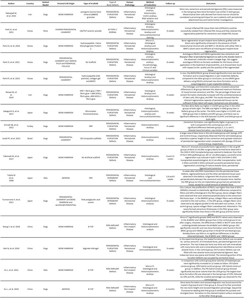
Tables were created separating these two types of studies (animal and human). In the animal studies table, authors, country, cell origin, scaffold type, bone defect type, human pathology, evaluated methodology, follow-up time, and results were listed. In the human studies table, authors, country, cell origin, study type, sample size, defect type, evaluation method, follow-up time, and results were listed (Box 1 and Box 2).
Studies Using Animal Models
Animal Models and Type of Bone Defects
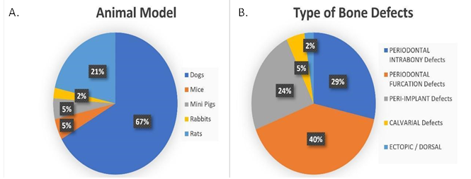
Dogs were the animal model most used to quantify bone defect repair, the experimental subjects were treated with scaffolds associated with some types of stromal cells (28 out of 42 experiments) 18,19,20,21,22,23,24,25,26,27,28,29,30,31,32,33,34,35,36,37,38,39,40,41,42,43,44,45. Rats were used in 9 out of 42 experiments 46,47,48,49,50,51,52,53,54, while Mice and Minipigs represent 10% (5% each) 55,56,57,58 of the total amount of manuscripts and only 2% of the manuscripts used Rabbits as animal models 59. The types of bone defects could not be compared among the experiments, because of the different methods that were applied to build them, but, out of 42 experiments, 17 were simulating periodontal furcation defects 19,24,25,26,27,28,29,30,31,32,33,34,35,36,39,50,58, 12 periodontal 23,37,41,42,43,45,46,48,49,51,54 and 10 peri-implant defects 18,20,21,22,38,40,44,52,57,59. Also aiming for oral bone repair 2 authors described calvarial defects 47,55 and 1 showed an ectopic/dorsal site 56. These findings may reflect that the jaws are the main site to represent a periodontal or peri-implant defect. These results can be observed in Figure 2.


The Human Pathology
Most of the objectives of the studies were focused on Inflammatory Periodontal Disease, 33 out of 42 19,20,23,24,25,26,27,28,29,30,31,32,33,34,35,36,37,39,41,42,43,44,45,46,47,48,49,50,51,52,53,54,55,56. Inflammatory Peri-Implant Disease was the second porpoise most studied found in this review, 8 out of 42 18,21,22,38,40,44,52,54,59. One study was engaged to answer for oral bone with no specific site 47 (Figure 3).
Stromal Cell Origin
The main finding of this review was the type of Stromal Cell Origin that leads the periodontal and peri-implant research. The most used Stromal Cell Origin was the bone marrow stromal cells (31%) 21,24,27,28,29,32,33,34,36,37,38,39,44,45,52,60,61,62 followed by a periodontal ligament (19%) 23,30,31,35,37,41,42,43,53,54,63,64,65,66,67) and adipose origin (17%) 20,26,46,51,52,59 A representative percentage of 10% of human periodontal ligament stromal cells 19,49,55,56 and 5% 46,60,61 human bone marrow, shows the heterologous part of the intention of the authors in using this way of treatment with stromal cells 3% were derived from umbilical cord 22, 2 % of the manuscripts described the dental pulp stromal cells for their experiments 10,25,30,41,68,69,70 and the others divided an equal percentage of 1% of the total number animal experiments 30,58,71, as shown in Figure 4.


Method of evaluation
The evaluations in most of the manuscripts were conducted with micro-CT and histological analysis methods, such as histomorphometry and Immunohistochemistry had an important place in the methods of evaluation for many articles. All methods of evaluation were employed to compare and establish significant results for in vivo periodontal/peri-implant defect repair using stromal cells associated with some type of scaffold (Box 1).


Clinical Studies
Study Design
All manuscripts included as a Clinical Study were counted in a total of 13. This scope review described the design of studies as: Randomized Clinical Trials, representing 5 of the total manuscripts 60,63,64,67,69; Case Reports, representing 3 manuscripts 62,66,71; Clinical trials, representing 2 manuscripts 61,65; Case Series, representing 2 Manuscripts 10,68 and 1 Pilot Clinical Study 70 (Figure 5).

Type of Bone Defect
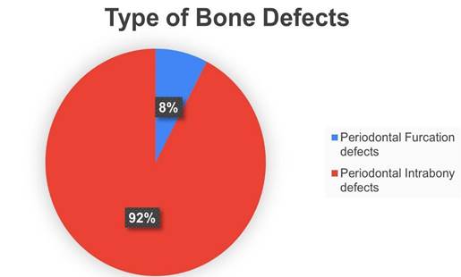
The bone defects created for the experiments were periodontal in total, but almost all of them were treating periodontal intrabony defects 60,61,62,63,64,65,66,67,68,69,70,10 during the clinical manuscripts. Only one out of 13 was described as treating a periodontal furcation defect 71 (Figure 6).
Stromal Cell Origin
Regarding the origin of the stromal cell used in the techniques described for clinical treatments the most used stromal cells were the periodontal ligament 5) (63,64,65,66,67, followed by dental pulp 4) (10,68,69,70, bone marrow 3) (60,61,62 and finally by embryonic stromal cells 70 (Figure 7).
Outcomes
All manuscripts included in this scoping review showed the results during bone periodontal/peri-implant repair using any kind of stromal cell were listed with other information and their outcomes in Box 1(Animals) and observations Box 2(Human).

Discussion
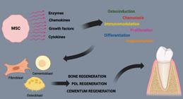
This scoping review compiled the use of animal models in the study of mesenchymal stem cells (MSCs) for tissue engineering in the treatment of peri-implant alterations, intraosseous defects, and periodontal diseases, in addition to reporting the use of stem cells in humans in periodontics. The success of periodontal regeneration via tissue engineering depends on factors such as a supply of progenitor cells capable of differentiating into specific phenotypes, appropriate signals for cellular differentiation, and a supporting structure. MSCs are prime candidates due to their ability to differentiate into various cell types (Graphic Abstract). MSCs must be capable of differentiating into osteogenic, chondrogenic, and adipogenic cells, in addition to their immunosuppressive properties 72. In periodontitis, MSCs can modulate the immune response, reducing inflammation and promoting healing 73. However, the constant exposure of periodontal tissues to a complex microbiota creates a continuous inflammatory environment that can affect the function of periodontal ligament stem cells but can also activate mechanisms of resolution and return to homeostasis 74.
The transition from the inflammatory phase to the proliferative phase is crucial for tissue healing and regeneration, involving the recruitment of fibroblasts from the wound edges, and the epithelial-mesenchymal transition involves the formation of epithelium from keratinocytes. Periodontal ligament stem cells (PDL) are multipotent progenitor cells and, therefore, capable of proliferating and differentiating into the various cell types necessary for the periodontium, indicating that periodontal tissues can regenerate if these cells can be mobilized 75.
In peri-implantitis, the ability of MSCs to modulate the immune response can reduce inflammation around the implant and promote tissue regeneration, in addition to forming new bone and blood vessels, ensuring their survival and proper integration with existing periodontal structures 76,77,78. MSCs can be combined with biomaterials to enhance their regenerative potential, as these can serve as structural support and improve the delivery and efficacy of MSCs at the treatment site, as demonstrated by the different types of scaffolds used for the treatment of periodontal and peri-implant diseases in the research 79. Different animal models, such as rats, dogs, pigs, and mice, are used to study tissue engineering with MSCs, each offering distinct advantages. This review observed 28 studies with dog models 18,19,20,21,22,23,24,25,26,27,28,29,30,31,32,33,34,35,36,37,38,39,40,41,42,43,44,45, 9 with rats 46,47,48,49,50,51,52,53,54, 2 with mice 55,56, 1 with rabbits 59, and 2 with minipigs 59.
Studies using canine models are predominant due to their anatomical and physiological similarities of periodontal tissues with humans, including factors such as bone density, cell renewal rate, and healing processes 18,57. The spacious oral cavity of dogs facilitates manipulation and creation of significant periodontal defects, allowing the use of scaffolds of different sizes and shapes 25,26. Their immune system, like humans, permits comparative observation of the effects of mesenchymal stem cells 19,31. The long lifespan of dogs allows for long-term effect studies on periodontal regeneration 20,21. The canine oral microbiota, although not identical, is more similar to that of humans, influencing regenerative treatments 29,41. The progression of periodontal disease in dogs is comparable to that in humans, making them ideal models for studies and therapeutic interventions 36,59. Despite requiring more resources, canine models provide an approach that is closer to human conditions, allowing for more predictive extrapolation of research results to potential clinical applications.
The MSCs used in tissue engineering can be obtained from various tissues, including bone marrow, adipose tissue, dental pulp, periodontal ligament, and umbilical cord among others. The choice of MSC source is critical, as it can influence their differentiation capabilities, immunomodulatory properties, and overall therapeutic potential. This review has recorded the use of MSCs from periodontal ligaments in 17 studies 19,23,30,31,35,37,41,42,43,44,48,49,50,55,56,57, bone marrow in 17 studies 21,24,27,28,29,32,33,34,36,37,38,39,44,45,52,60,61,62, adipose tissue in 7 studies 18,20,26,46,51,52,59, umbilical cords in one study 22, dental pulp in tree study 10,25,30,41,68,69,70, and one study on embryonic stem cells 58. These selections often reflect a balance between the high regenerative potential of the cells and practical considerations such as cell availability and the invasiveness of the harvesting procedure.
The study by Pinheiro et al . 2019 highlighted that mesenchymal stem cells (MSCs) can be obtained less invasively from sites such as the umbilical cord, the orbicularis oris muscle, and deciduous dental pulp. These cells exhibit excellent osteogenic potential due to their neural crest characteristics, which predispose them to greater osteogenic differentiation. The use of dental pulp stem cells has shown satisfactory clinical results in the bone consolidation of cleft lip and palate, with benefits such as shorter operative time, reduced postoperative pain, costs, and hospitalization time. The results indicate that regeneration with this type of cell can be promising 81.
This scoping review highlights the efficacy of MSCs obtained from the periodontal ligament, bone marrow, and adipose tissue, which have been more largely used and validated in the literature reviewed for treating periodontal disease 19,23,30,31,35,37,41,42,43,44,48,49,50,55,56,57,21,24,27,28,29,32,33,34,36,37,38,39,44,45,52,60,61,62,20,26,46,51,52,59.
The PDLSCs are especially suited for this role, given their involvement in the maintenance and repair of periodontal tissues and they can be harvested from extracted teeth, offering a source of cells without the need for additional invasive procedures. These PDLSCs have shown the potential to differentiate into various cell types that constitute the periodontium, which is vital for periodontal regeneration 19,23,30,31,35,37,41,42,43,44,48,49,50,55,56,57,63,64,65,66,67. Bone marrow-derived stem cells (BMSCs) are recognized for their high differentiation potential, supporting the formation of bone, cartilage, and a functional periodontal ligament, in addition to their ability to modulate the immune response, an advantage in controlling associated inflammation. to periodontal diseases 21,24,27,28,29,32,33,34,36,37,38,39,44,45,52,60,61,62. The third most used cell source of MSC was the adipose-derived stem cells (ADSCs). They can be isolated in large quantities from adipose tissue and have been demonstrated to differentiate into cell types crucial for periodontal regeneration, such as osteogenic lineage 18,20,26,46,51,52,59.
The review also showed MSC sources referenced (PLSC, BMSC, ADSC) are capable of secreting bioactive factors that modulate the inflammatory environment and recruit other progenitor cells 19,23,30,31,35,37,4,42,43,44,48,49,50,55,46,57,63,64,65,66,67,21,24,27,28,29,32,33,34,36,67,68,39,44,45,52,60,61,62,18,20,26,46,51,52,59, but these characteristics are inherent to all MSCs and have been detailed by authors who also utilized other sources like umbilical cord, dental pulp, and embryonic stem cells to make the treatment of periodontal disease in animal model studies and got success in their results 22,10,25,30,41,68,69,70,58.
A single article from this review focused on the regeneration of intraosseous periodontal defects in rats using exosomes embedded in a collagen matrix, vesicles that replicate the therapeutic capacity of mesenchymal cells in the studied animal model 48. Significant bone and periodontal tissue regeneration can be observed, indicating improved migration, proliferation, matrix synthesis, and differentiation into new bone as well as restoration of the periodontal ligament. This demonstrates a promising path in the use of exosomes from human mesenchymal cells in periodontal regeneration.
Signaling vesicles, highlighted for their regenerative capacity, have received special attention recently. Pinheiro et al, 2023 82, emphasized in a brief review how these extracellular vesicles can positively influence bone tissue regeneration, following the stages of inflammation, repair, and remodeling, and involving multiple signaling pathways in bone. A subsequent review highlighted those therapeutic exosomes, generally obtained from dental pulp, periodontal ligament cells, gingival fibroblasts, stem cells from exfoliated deciduous teeth, and the apical papilla, facilitated the regenerative potential of tissues such as bone and periodontal tissue, as well as assisting in orthodontic treatments and alleviating temporomandibular joint disorders and others. The 113 articles analyzed confirmed the efficacy of exosomes in 100% of the cases, indicating that, in bone regeneration, they are as effective or more effective than rhBMP2, with the additional advantage of reducing inflammation. This finding underscores the still unexplored potential of exosomes in oral regeneration 83.
The importance of scaffolds in tissue engineering, especially in tissue regeneration mediated by mesenchymal stem cells, should be emphasized. Scaffolds are critical for tissue engineering and can be made from materials such as natural and synthetic polymers, hydrogels, and ceramics with distinct properties. They are essential for providing support for the adhesion, proliferation, and differentiation of MSCs. The choice of scaffold material is crucial, as its specific properties directly influence the success of tissue regeneration. This review evaluates successful scaffolds, guiding future selections for periodontal regeneration (Box 1). Overall, the reviewed studies demonstrate that the combination of MSCs with scaffolds leads to better tissue regeneration compared to the use of scaffolds alone for the treatment of periodontal disease 46, and the choice of scaffold type can influence the mechanical properties of the regenerated tissue 47.
This review's findings are instrumental for selecting the appropriate animal model, MSC source, and scaffold material for further periodontal tissue engineering research. The comparison of various evaluation methods across studies, such as micro-CT scans and histomorphometric analysis, contributes to standardizing outcome measures in this field and all this data can be observed in Box 1.
When human studies were evaluated for this scoping review, a diverse range of study designs, cell origins, and methodological assessments were observed across different countries, highlighting the global effort in utilizing MSCs for periodontal repair 10,60,62,63,64,65,66,67,68,69,70,71.
The clinical application of MSCs in periodontal therapy described in this manuscript explored promising outcomes across various study designs and evaluation methods. A significant contribution to this field was made by four authors who conducted a randomized clinical trial 60,63,64,67,69. Other studies such as case series, cohort studies, and clinical case reports have also been described 62,66,71,61,65,10,68,70 opening new avenues for the use of tissue engineering strategies using MSCs and scaffolds to regenerate tissues affected by periodontal disease. While there is a generally positive trend towards using MSCs, varying degrees of clinical success were observed in this scoping review it is a necessity for further standardized long-term studies development to confirm their therapeutic efficacy fully.
Promising results have been described in this manuscript for the clinical application of MSCs with various study designs and evaluation methods such as randomized clinical trials 60,63,64,67,69, case series, cohort studies, and clinical case reports 62,66,71,61,65,10,68,70, opening new avenues for the use of MSCs and scaffolds to regenerate periodontal tissues. However, the development of more standardized long-term studies is necessary to confirm the therapeutic efficacy.
When comparing studies on periodontal regeneration using animal models and those conducted in humans, several key differences emerge. Animal models, often using rodents, dogs, or non-human primates, provide a controlled environment where variables can be tightly regulated, allowing for a detailed examination of cellular and molecular mechanisms involved in periodontal regeneration. These models enable invasive procedures and frequent sampling that are not feasible in human studies, facilitating a deeper understanding of tissue responses and healing processes. However, the anatomical, physiological, and host-microbe interaction differences between animal models and humans can limit the direct applicability of findings to clinical practice. Human studies, on the other hand, offer the advantage of directly assessing the efficacy and safety of regenerative therapies in the target population. These studies are critical for evaluating clinical outcomes, patient-related factors, and long-term benefits of treatment, yet they often face ethical constraints, variability in patient conditions, and limited opportunities for invasive monitoring. However, clinical trials are costly and time-consuming. Moreover, human studies involving MSCs encounter numerous regulatory challenges to be implemented. Thus, while animal models are invaluable for foundational research and mechanistic insights, human studies are essential for translating these findings into effective clinical treatments.
Research from the past decades supports the effectiveness of MSC in promoting periodontal and peri-implant regeneration. However, the impact of MSC on regenerative therapies in humans is still in its initial steps. Many existing studies are preclinical, some are in early trial stages, and a few are well-designed longitudinal clinical trials. Future research should optimize MSC application protocols by combining techniques, such as the use of nanomedicine and 3D printing for tissue engineering. Clinical studies should also understand the long-term effects and compare MSC therapies with current treatment modalities. By addressing these areas, the scientific community can ensure that MSC therapies are both safe and effective, ultimately enhancing therapeutic strategies and treatment outcomes in Periodontology and Implantology.
References
-
1 -Stavropoulos A, Bertl K, Spineli LM, Sculean A, Cortellini P, Tonetti M. Medium- and long-term clinical benefits of periodontal regenerative/reconstructive procedures in intrabony defects: Systematic review and network meta-analysis of randomized controlled clinical studies. J Clin Periodontol. 2021;48(3):410-430. doi:10.1111/jcpe.13409.
» https://doi.org/10.1111/jcpe.13409 -
2 - Caton J, Nyman S, Zander H. Histometric evaluation of periodontal surgery. II. Connective tissue attachment levels after four regenerative procedures. J Clin Periodontol. 1980;7(3):224-231. doi: 10.1111/j.1600-051x.1980.tb01965. x.
» https://doi.org/10.1111/j.1600-051x.1980.tb01965. x -
3 -Wilson TG Jr, Carnio J, Schenk R, Myers G. Absence of histologic signs of chronic inflammation following closed subgingival scaling and root planing using the dental endoscope: human biopsies - a pilot study. J Periodontol. 2008 Nov;79(11):2036-41. doi: 10.1902/jop.2008.080190. PMID: 18980510.
» https://doi.org/10.1902/jop.2008.080190 -
4 -Cortellini P, Stalpers G, Mollo A, Tonetti MS. Periodontal regeneration versus extraction and dental implant or prosthetic replacement of teeth severely compromised by attachment loss to the apex: A randomized controlled clinical trial reporting 10-year outcomes, survival analysis and mean cumulative cost of recurrence. J Clin Periodontol. 2020;47(6):768-776. doi:10.1111/jcpe.13289.
» https://doi.org/10.1111/jcpe.13289 -
5 -Kuchler U, von Arx T. Horizontal ridge augmentation in conjunction with or prior to implant placement in the anterior maxilla: a systematic review. Int J Oral Maxillofac Implants. 2014;29Suppl:14-24. doi: 10.11607/jomi.2014suppl.g1.1.
» https://doi.org/10.11607/jomi.2014suppl.g1.1 - 6 -Jensen SS, Terheyden H. Bone augmentation procedures in localized defects in the alveolar ridge: clinical results with different bone grafts and bone-substitute materials. Int J Oral Maxillofac Implants. 2009;24Suppl:218-236.
-
7 -Benic GI, Hämmerle CH. Horizontal bone augmentation by means of guided bone regeneration. Periodontol2000. 2014;66(1):13-40. doi:10.1111/prd.12039.
» https://doi.org/10.1111/prd.12039 - 8 - Lymperi S, Ligoudistianou C, Taraslia V, Kontakiotis E, Anastasiadou E. Dental stem cells and their applications in dental tissue engineer- ing. Open Dent J. 2013;7:76-81.
-
9 - Sadek KM, El Moshy S, Radwan IA, Rady D, Abbass MMS, El-Rashidy AA, Dörfer CE, Fawzy El-Sayed KM. Molecular Basis beyond Interrelated Bone Resorption/Regeneration in Periodontal Diseases: A Concise Review. Int J Mol Sci. 2023Feb 27;24(5):4599. doi: 10.3390/ijms24054599.
» https://doi.org/10.3390/ijms24054599 -
10 - Nuñez J, Sanz-Blasco S, Vignoletti F, Muñoz F, Arzate H, Villalobos C, Nuñez L, Caffesse RG, Sanz M. Periodontal regeneration following implantation of cementum and periodontal ligament-derived cells. J Periodontal Res. 2012Feb;47(1):33-44. doi: 10.1111/j.1600-0765.2011.01402.x.
» https://doi.org/10.1111/j.1600-0765.2011.01402.x -
11 - Aimetti M, Ferrarotti F, Gamba MN, Giraudi M, Romano F. Regenerative Treatment of Periodontal Intrabony Defects Using Autologous Dental Pulp Stem Cells: A 1-Year Follow-Up Case Series. Int J Periodontics Restorative Dent. 2018Jan/Feb;38(1):51-58. doi: 10.11607/prd.3425.
» https://doi.org/10.11607/prd.3425 - 12 -Tassi SA, Sergio NZ, Misawa MY, Villar CC. Efficacy of stem cells on periodontal regeneration: systematic review of pre-clinical studies. J Periodontal Res. 2017;52:793-812.
-
13 - Murphy KG, Gunsolley JC. Guided tissue regeneration for the treatment of periodontal intrabony and furcation defects. A systematic review. Ann Periodontol. 2003Dec;8(1):266-302. doi: 10.1902/annals.2003.8.1.266.
» https://doi.org/10.1902/annals.2003.8.1.266 -
14 - Roi A, Roi C, Negruțiu ML, Rusu LC, Riviș M. Mesenchymal Stem Cells Derived from Human Periapical Cysts and Their Implications in Regenerative Medicine. Biomedicines. 2023Aug 31;11(9):2436. doi: 10.3390/biomedicines11092436.
» https://doi.org/10.3390/biomedicines11092436 - 15 -Arksey H, O’Malley L. Scoping studies: towards a me-thodological framework. Int J Soc Res Methodol. 2005; 8(1):19 -32.
-
16 - Grant MJ, Booth A. A typology of reviews: an analysis of 14 review types and associated methodologies. Health Info Libr J. 2009Jun;26(2):91-108. doi: 10.1111/j.1471-1842.2009.00848.x.
» https://doi.org/10.1111/j.1471-1842.2009.00848.x -
17 - Pham MT, Rajić A, Greig JD, Sargeant JM, Papadopoulos A, McEwen SA. A scoping review of scoping reviews: advancing the approach and enhancing the consistency. Res Synth Methods. 2014Dec;5(4):371-85. doi: 10.1002/jrsm.1123.
» https://doi.org/10.1002/jrsm.1123 - 18 - Bressan E, Botticelli D, Sivolella S, Bengazi F, Guazzo R, Sbricoli L, Ricci S, Ferroni L, Gardin C, Velez JU, Zavan B. Adipose-Derived Stem Cells as a Tool for Dental Implant Osseointegration: an Experimental Study in the Dog. Int J Mol Cell Med. 2015Fall;4(4):197-208. PMID: 27014644; PMCID: PMC4769597.
-
19 Chantarawaratit P, Sangvanich P, Banlunara W, Soontornvipart K, Thunyakitpisal P. Acemannan sponges stimulate alveolar bone, cementum and periodontal ligament regeneration in a canine class II furcation defect model. J Periodontal Res. 2014Apr;49(2):164-78. doi: 10.1111/jre.12090.
» https://doi.org/10.1111/jre.12090 -
20 -Ding L, Tang S, Liang P, Wang C, Zhou PF, Zheng L. Bone Regeneration of Canine Peri-implant Defects Using Cell Sheets of Adipose-Derived Mesenchymal Stem Cells and Platelet-Rich Fibrin Membranes. J Oral Maxillofac Surg. 2019Mar;77(3):499-514. doi: 10.1016/j.joms.2018.10.018.
» https://doi.org/10.1016/j.joms.2018.10.018 -
21 - Han X, Liu H, Wang D, Su F, Zhang Y, Zhou W, Li S, Yang R. Alveolar bone regeneration around immediate implants using an injectable nHAC/CSH loaded with autogenic blood-acquired mesenchymal progenitor cells: an experimental study in the dog mandible. Clin Implant Dent Relat Res. 2013Jun;15(3):390-401. doi: 10.1111/j.1708-8208.
» https://doi.org/10.1111/j.1708-8208 - 22 - Hao PJ, Wang ZG, Xu QC, Xu S, Li ZR, Yang PS, Liu ZH. Effect of umbilical cord mesenchymal stem cell in peri-implant bone defect after immediate implant: an experiment study in beagle dogs. Int J Clin Exp Med. 2014Nov 15;7(11):4131-8.
-
23 - Inukai T, Katagiri W, Yoshimi R, Osugi M, Kawai T, Hibi H, Ueda M. Novel application of stem cell-derived factors for periodontal regeneration. Biochem Biophys Res Commun. 2013Jan 11;430(2):763-8. doi: 10.1016/j.bbrc.2012.11.074.
» https://doi.org/10.1016/j.bbrc.2012.11.074 -
24 -Kawaguchi H, Hirachi A, Hasegawa N, Iwata T, Hamaguchi H, Shiba H, Takata T, Kato Y, Kurihara H. Enhancement of periodontal tissue regeneration by transplantation of bone marrow mesenchymal stem cells. J Periodontol. 2004Sep;75(9):1281-7. doi: 10.1902/jop.2004.75.9.1281.
» https://doi.org/10.1902/jop.2004.75.9.1281 -
25 - Khorsand A, Eslaminejad MB, Arabsolghar M, Paknejad M, Ghaedi B, Rokn AR, Moslemi N, Nazarian H, Jahangir S. Autologous dental pulp stem cells in regeneration of defect created in canine periodontal tissue. J Oral Implantol. 2013Aug;39(4):433-43. doi: 10.1563/AAID-JOI-D-12-00027.
» https://doi.org/10.1563/AAID-JOI-D-12-00027 -
26 - Mansour AM, Yahia S, Elsayed HRH, El-Attar SAE, Grawish ME, El-Hawary YM, El-Sherbiny IM. Efficacy of biocompatible trilayers nanofibrous scaffold with/without allogeneic adipose-derived stem cells on class II furcation defects of dogs' model. Clin Oral Investig. 2022Mar;26(3):2537-2553. doi: 10.1007/s00784-021-04222-x.
» https://doi.org/10.1007/s00784-021-04222-x -
27 - Nagahara T, Yoshimatsu S, Shiba H, Kawaguchi H, Takeda K, Iwata T, Mizuno N, Fujita T, Kurihara H. Introduction of a mixture of β-tricalcium phosphate into a complex of bone marrow mesenchymal stem cells and type I collagen can augment the volume of alveolar bone without impairing cementum regeneration. J Periodontol. 2015Mar;86(3):456-64. doi: 10.1902/jop.2014.140384.
» https://doi.org/10.1902/jop.2014.140384 -
28 - Paknejad M, Eslaminejad MB, Ghaedi B, Rokn AR, Khorsand A, Etemad-Moghadam S, Alaeddini M, Dehghan MM, Moslemi N, Nowzari H. Isolation and Assessment of Mesenchymal Stem Cells Derived From Bone Marrow: Histologic and Histomorphometric Study in a Canine Periodontal Defect. J Oral Implantol. 2015Jun;41(3):284-91. doi: 10.1563/AAID-JOI-D-13-00220.
» https://doi.org/10.1563/AAID-JOI-D-13-00220 -
29 -Park JH, Jung YE, Kim MJ, Hwang SJ. Periimplant bone regeneration in hydroxyapatite block grafts with mesenchymal stem cells and bone morphogenetic protein-2. Tissue Eng Regen Med. 2016Jan 5;13(4):437-445. doi: 10.1007/s13770-015-0049-8..
» https://doi.org/10.1007/s13770-015-0049-8. -
30 -Park JY, Jeon SH, Choung PH. Efficacy of periodontal stem cell transplantation in the treatment of advanced periodontitis. Cell Transplant. 2011;20(2):271-85. doi: 10.3727/096368910X519292..
» https://doi.org/10.3727/096368910X519292. -
31 -Park SY, Kim KH, Gwak EH, Rhee SH, Lee JC, Shin SY, Koo KT, Lee YM, Seol YJ. Ex vivo bone morphogenetic protein 2 gene delivery using periodontal ligament stem cells for enhanced re-osseointegration in the regenerative treatment of peri-implantitis. J Biomed Mater Res A. 2015Jan;103(1):38-47. doi: 10.1002/jbm.a.35145.
» https://doi.org/10.1002/jbm.a.35145 -
32 - Rezaei M, Jamshidi S, Saffarpour A, Ashouri M, Rahbarghazi R, Rokn AR, Motahari P. Transplantation of Bone Marrow-Derived Mesenchymal Stem Cells, Platelet-Rich Plasma, and Fibrin Glue for Periodontal Regeneration. Int J Periodontics Restorative Dent. 2019Jan/Feb;39(1):e32-e45. doi: 10.11607/prd.3691..
» https://doi.org/10.11607/prd.3691. -
33 -Sakaguchi K, Katagiri W, Osugi M, Kawai T, Sugimura-Wakayama Y, Hibi H. Periodontal tissue regeneration using the cytokine cocktail mimicking secretomes in the conditioned media from human mesenchymal stem cells. Biochem Biophys Res Commun. 2017Feb 26;484(1):100-106. doi: 10.1016/j.bbrc.2017.01.065..
» https://doi.org/10.1016/j.bbrc.2017.01.065. -
34 - Simsek SB, Keles GC, Baris S, Cetinkaya BO. Comparison of mesenchymal stem cells and autogenous cortical bone graft in the treatment of class II furcation defects in dogs. Clin Oral Investig. 2012Feb;16(1):251-8. doi: 10.1007/s00784-010-0486-7.
» https://doi.org/10.1007/s00784-010-0486-7 -
35 -Suaid FF, Ribeiro FV, Gomes TR, Silvério KG, Carvalho MD, Nociti FH Jr, Casati MZ, Sallum EA. Autologous periodontal ligament cells in the treatment of Class III furcation defects: a study in dogs. J Clin Periodontol. 2012Apr;39(4):377-84. doi: 10.1111/j.1600-051X.2012.01858.x.
» https://doi.org/10.1111/j.1600-051X.2012.01858.x -
36 -Takewaki M, Kajiya M, Takeda K, Sasaki S, Motoike S, Komatsu N, Matsuda S, Ouhara K, Mizuno N, Fujita T, Kurihara H. MSC/ECM Cellular Complexes Induce Periodontal Tissue Regeneration. J Dent Res. 2017Aug;96(9):984-991. doi: 10.1177/0022034517708770.
» https://doi.org/10.1177/0022034517708770 -
37 -Tsumanuma Y, Iwata T, Washio K, Yoshida T, Yamada A, Takagi R, Ohno T, Lin K, Yamato M, Ishikawa I, Okano T, Izumi Y. Comparison of different tissue-derived stem cell sheets for periodontal regeneration in a canine 1-wall defect model. Biomaterials. 2011Sep;32(25):5819-25. doi: 10.1016/j.biomaterials.2011.04.071.
» https://doi.org/10.1016/j.biomaterials.2011.04.071 -
38 -Wang F, Zhou Y, Zhou J, Xu M, Zheng W, Huang W, Zhou W, Shen Y, Zhao K, Wu Y, Zou D. Comparison of Intraoral Bone Regeneration with Iliac and Alveolar BMSCs. J Dent Res. 2018Oct;97(11):1229-1235. doi: 10.1177/0022034518772283.
» https://doi.org/10.1177/0022034518772283 -
39 -Wei N, Gong P, Liao D, Yang X, Li X, Liu Y, Yuan Q, Tan Z. Auto-transplanted mesenchymal stromal cell fate in periodontal tissue of beagle dogs. Cytotherapy. 2010Jul;12(4):514-21. doi: 10.3109/14653241003709702.
» https://doi.org/10.3109/14653241003709702 -
40 -Xu L, Qin X, Mozaffari MS, Yan D, Sun X, Cao Y. Hybrid system with stable structure of hard/soft tissue substitutes induces re-osseointegration in a rat model of biofilm-mediated peri-implantitis. J Biomed Mater Res B Appl Biomater. 2022Nov;110(11):2452-2463. doi: 10.1002/jbm.b.35102.
» https://doi.org/10.1002/jbm.b.35102 -
41 -Yang C, Lee JS, Jung UW, Seo YK, Park JK, Choi SH. Periodontal regeneration with nano-hyroxyapatite-coated silk scaffolds in dogs. J Periodontal Implant Sci. 2013Dec;43(6):315-22. doi: 10.5051/jpis.2013.43.6.315.
» https://doi.org/10.5051/jpis.2013.43.6.315 -
42 -Yang H, Li J, Hu Y, Sun J, Guo W, Li H, Chen J, Huo F, Tian W, Li S. Treated dentin matrix particles combined with dental follicle cell sheet stimulate periodontal regeneration. Dent Mater. 2019Sep;35(9):1238-1253. doi: 10.1016/j.dental.2019.05.016.
» https://doi.org/10.1016/j.dental.2019.05.016 -
43 -Yoo SY, Lee JS, Cha JK, Kim SK, Kim CS. Periodontal healing using a collagen matrix with periodontal ligament progenitor cells in a dehiscence defect model in beagle dogs. J Periodontal Implant Sci. 2019Jul 16;49(4):215-227. doi: 10.5051/jpis.2019.49.4.215.
» https://doi.org/10.5051/jpis.2019.49.4.215 -
44 -Yun JH, Han SH, Choi SH, Lee MH, Lee SJ, Song SU, Oh N. Effects of bone marrow-derived mesenchymal stem cells and platelet-rich plasma on bone regeneration for osseointegration of dental implants: preliminary study in canine three-wall intrabony defects. J Biomed Mater Res B Appl Biomater. 2014Jul;102(5):1021-30. doi: 10.1002/jbm.b.33084..
» https://doi.org/10.1002/jbm.b.33084. -
45 -Zang S, Jin L, Kang S, Hu X, Wang M, Wang J, Chen B, Peng B, Wang Q. Periodontal Wound Healing by Transplantation of Jaw Bone Marrow-Derived Mesenchymal Stem Cells in Chitosan/Anorganic Bovine Bone Carrier Into One-Wall Infrabony Defects in Beagles. J Periodontol. 2016Aug;87(8):971-81. doi: 10.1902/jop.2016.150504.
» https://doi.org/10.1902/jop.2016.150504 - 46 - Akita D, Morokuma M, Saito Y, Yamanaka K, Akiyama Y, Sato M, et al. Periodontal tissue regeneration by transplantation of rat adipose-derived stromal cells in combination with PLGA-based solid scaffolds. BIOMEDICAL RESEARCH-TOKYO. 2014;35(2):91-103.
-
47 Al-Hezaimi K, Ramalingam S, Al-Askar M, ArRejaie AS, Nooh N, Jawad F, Aldahmash A, Atteya M, Wang CY. Real-time-guided bone regeneration around standardized critical size calvarial defects using bone marrow-derived mesenchymal stem cells and collagen membrane with and without using tricalcium phosphate: an in vivo micro-computed tomographic and histologic experiment in rats. Int J Oral Sci. 2016Mar 30; 8(1):7-15. doi: 10.1038/ijos.2015.34.
» https://doi.org/10.1038/ijos.2015.34 -
48 -Chew JRJ, Chuah SJ, Teo KYW, Zhang S, Lai RC, Fu JH, Lim LP, Lim SK, Toh WS. Mesenchymal stem cell exosomes enhance periodontal ligament cell functions and promote periodontal regeneration. Acta Biomater. 2019Apr 15;89:252-264. doi: 10.1016/j.actbio.2019.03.021.
» https://doi.org/10.1016/j.actbio.2019.03.021 -
49 -Iwasaki K, Akazawa K, Nagata M, Komaki M, Honda I, Morioka C, Yokoyama N, Ayame H, Yamaki K, Tanaka Y, Kimura T, Kishida A, Watabe T, Morita I. The Fate of Transplanted Periodontal Ligament Stem Cells in Surgically Created Periodontal Defects in Rats. Int J Mol Sci. 2019Jan 7;20(1):192. doi: 10.3390/ijms20010192.
» https://doi.org/10.3390/ijms20010192 -
50 -Nagata M, Iwasaki K, Akazawa K, Komaki M, Yokoyama N, Izumi Y, Morita I. Conditioned Medium from Periodontal Ligament Stem Cells Enhances Periodontal Regeneration. Tissue Eng Part A. 2017May;23(9-10):367-377. doi: 10.1089/ten.TEA.2016.0274.
» https://doi.org/10.1089/ten.TEA.2016.0274 -
51 -Tobita M, Masubuchi Y, Ogata Y, Mitani A, Kikuchi T, Toriumi T, Montenegro Raudales JL, Mizuno H, Suzuki Y, Wakana K, Yoneda H, Kamijo R, Kino-Oka M, Morio T, Okada K, Murakami S, Honda M. Study protocol for periodontal tissue regeneration with a mixture of autologous adipose-derived stem cells and platelet rich plasma: A multicenter, randomized, open-label clinical trial. Regen Ther. 2022 Oct 12;21:436-441. doi: 10.1016/j.reth.2022.09.008.
» https://doi.org/10.1016/j.reth.2022.09.008 -
52 -Xu L, Sun X, Bai J, Jiang L, Wang S, Zhao J, Xia L, Zhang X, Wen J, Li G, Jiang X. Reosseointegration Following Regenerative Therapy of Tissue-Engineered Bone in a Canine Model of Experimental Peri-Implantitis. Clin Implant Dent Relat Res. 2016Apr;18(2):379-91. doi: 10.1111/cid.12308.
» https://doi.org/10.1111/cid.12308 -
53 -Yan XZ, van den Beucken JJ, Cai X, Yu N, Jansen JA, Yang F. Periodontal tissue regeneration using enzymatically solidified chitosan hydrogels with or without cell loading. Tissue Eng Part A. 2015Mar;21(5-6):1066-76. doi: 10.1089/ten.TEA.2014.0319.
» https://doi.org/10.1089/ten.TEA.2014.0319 -
54 -Yu N, Oortgiesen DA, Bronckers AL, Yang F, Walboomers XF, Jansen JA. Enhanced periodontal tissue regeneration by periodontal cell implantation. J Clin Periodontol. 2013Jul;40(7):698-706. doi: 10.1111/jcpe.12113..
» https://doi.org/10.1111/jcpe.12113. -
55 - Hung TY, Lin HC, Chan YJ, Yuan K. Isolating stromal stem cells from periodontal granulation tissues. Clin Oral Investig. 2012Aug;16(4):1171-80. doi: 10.1007/s00784-011-0600-5.
» https://doi.org/10.1007/s00784-011-0600-5 -
56 - Park JC, Kim JM, Jung IH, Kim JC, Choi SH, Cho KS, Kim CS. Isolation and characterization of human periodontal ligament (PDL) stem cells (PDLSCs) from the inflamed PDL tissue: in vitro and in vivo evaluations. J Clin Periodontol. 2011Aug;38(8):721-31. doi: 10.1111/j.1600-051X.2011.01716.x.
» https://doi.org/10.1111/j.1600-051X.2011.01716.x -
57 - Kämmerer PW, Scholz M, Baudisch M, Liese J, Wegner K, Frerich B, Lang H. Guided Bone Regeneration Using Collagen Scaffolds, Growth Factors, and Periodontal Ligament Stem Cells for Treatment of Peri-Implant Bone Defects In Vivo. Stem Cells Int. 2017;2017:3548435. doi: 10.1155/2017/3548435.
» https://doi.org/10.1155/2017/3548435 -
58 - Yang JR, Hsu CW, Liao SC, Lin YT, Chen LR, Yuan K. Transplantation of embryonic stem cells improves the regeneration of periodontal furcation defects in a porcine model. J Clin Periodontol. 2013Apr;40(4):364-71. doi: 10.1111/jcpe.12069.
» https://doi.org/10.1111/jcpe.12069 -
59 -Erdogan Ö, Supachawaroj N, Soontornvipart K, Kheolamai P. Treatment of Peri-Implant Defects in the Rabbit's Tibia with Adipose or Bone Marrow-Derived Mesenchymal Stems Cells. Clin Implant Dent Relat Res. 2016Oct;18(5):1003-1014. doi: 10.1111/cid.12378.
» https://doi.org/10.1111/cid.12378 -
60 -Apatzidou DA, Bakopoulou AA, Kouzi-Koliakou K, Karagiannis V, Konstantinidis A. A tissue-engineered biocomplex for periodontal reconstruction. A proof-of-principle randomized clinical study. J Clin Periodontol. 2021Aug;48(8):1111-1125. doi: 10.1111/jcpe.13474.
» https://doi.org/10.1111/jcpe.13474 -
61 -Baba S, Yamada Y, Komuro A, Yotsui Y, Umeda M, Shimuzutani K, Nakamura S. Phase I/II Trial of Autologous Bone Marrow Stem Cell Transplantation with a Three-Dimensional Woven-Fabric Scaffold for Periodontitis. Stem Cells Int. 2016;2016:6205910. doi: 10.1155/2016/6205910.
» https://doi.org/10.1155/2016/6205910 -
62 -Koo S, Alshihri A, Karimbux NY, Maksoud M. Cellular Allograft in the Treatment of a Severe Periodontal Intrabony Defect: A Case Report. Clin Adv Periodontics. 2012Feb;2(1):35-39. doi: 10.1902/cap.2011.110017.
» https://doi.org/10.1902/cap.2011.110017 -
63 -Abdal-Wahab M, Abdel Ghaffar KA, Ezzatt OM, Hassan AAA, El Ansary MMS, Gamal AY. Regenerative potential of cultured gingival fibroblasts in treatment of periodontal intrabony defects (randomized clinical and biochemical trial). J Periodontal Res. 2020Jun;55(3):441-452. doi: 10.1111/jre.12728.
» https://doi.org/10.1111/jre.12728 -
64 -Chen FM, Gao LN, Tian BM, Zhang XY, Zhang YJ, Dong GY, Lu H, Chu Q, Xu J, Yu Y, Wu RX, Yin Y, Shi S, Jin Y. Treatment of periodontal intrabony defects using autologous periodontal ligament stem cells: a randomized clinical trial. Stem Cell Res Ther. 2016 Feb 19;7:33. doi: 10.1186/s13287-016-0288-1.
» https://doi.org/10.1186/s13287-016-0288-1 -
65 -Iwata T, Yamato M, Washio K, Yoshida T, Tsumanuma Y, Yamada A, Onizuka S, Izumi Y, Ando T, Okano T, Ishikawa I. Periodontal regeneration with autologous periodontal ligament-derived cell sheets - A safety and efficacy study in ten patients. Regen Ther. 2018 Aug 24;9:38-44. doi: 10.1016/j.reth.2018.07.002.
» https://doi.org/10.1016/j.reth.2018.07.002 -
66 Kl V, Ryana H, Dalvi PJ. Autologous periodontal stem cell assistance in periodontal regeneration technique (SAI-PRT) in the treatment of periodontal intrabony defects: A case report with one-year follow-up. J Dent Res Dent Clin Dent Prospects. 2017Spring;11(2):123-126. doi: 10.15171/joddd.2017.022.
» https://doi.org/10.15171/joddd.2017.022 -
67 -Sánchez N, Fierravanti L, Núñez J, Vignoletti F, González-Zamora M, Santamaría S, Suárez-Sancho S, Fernández-Santos ME, Figuero E, Herrera D, García-Sanz JA, Sanz M. Periodontal regeneration using a xenogeneic bone substitute seeded with autologous periodontal ligament-derived mesenchymal stem cells: A 12-month quasi-randomized controlled pilot clinical trial. J Clin Periodontol. 2020Nov;47(11):1391-1402. doi: 10.1111/jcpe.13368.
» https://doi.org/10.1111/jcpe.13368 -
68 - Aimetti M, Ferrarotti F, Mariani GM, Cricenti L, Romano F. Use of Dental Pulp Stem Cells/Collagen Sponge Biocomplex in the Treatment of Non-Contained Intrabony Defects: A Case Series. Clin Adv Periodontics. 2015May;5(2):104-109. doi: 10.1902/cap.2013.130047.
» https://doi.org/10.1902/cap.2013.130047 -
69 - Ferrarotti F, Romano F, Gamba MN, Quirico A, Giraudi M, Audagna M, Aimetti M. Human intrabony defect regeneration with micrografts containing dental pulp stem cells: A randomized controlled clinical trial. J Clin Periodontol. 2018Jul;45(7):841-850. doi: 10.1111/jcpe.12931.
» https://doi.org/10.1111/jcpe.12931 -
70 - Li Y, Zhao S, Nan X, Wei H, Shi J, Li A, Gou J. Repair of human periodontal bone defects by autologous grafting stem cells derived from inflammatory dental pulp tissues. Stem Cell Res Ther. 2016Sep 22;7(1):141. doi: 10.1186/s13287-016-0404-2.
» https://doi.org/10.1186/s13287-016-0404-2 -
71 - Rosen PS, Froum SJ, Cohen DW. Consecutive Case Series Using a Composite Allograft Containing Mesenchymal Cells with an Amnion-Chorion Barrier to Treat Mandibular Class III/IV Furcations. Int J Periodontics Restorative Dent. 2015 Jul-Aug;35(4):453-60. doi: 10.11607/prd.2314..
» https://doi.org/10.11607/prd.2314. -
72 - Han J, Menicanin D, Gronthos S, Bartold PM. Stem cells, tissue engineering and periodontal regeneration. Aust Dent J. 2014 Jun;59 Suppl 1:117-30. doi: 10.1111/adj.12100. Epub 2013 Sep 23. PMID: 24111843.
» https://doi.org/10.1111/adj.12100 -
73 - Jiang, Y., Jiang, L. L., Huang, T. S., Zheng, L., Zhao, J., Zhang, W., ... & Wang, J. H. Mesenchymal stem cell therapy for periodontitis: A review of the literature. Stem Cell Research & Therapy, 202112(1), 16. doi: 10.1186/s13287-020-02017-4.).
» https://doi.org/10.1186/s13287-020-02017-4.) -
74 - Yang Y, Alves T, Miao MZ, Wu YC, Li G, Lou J, Hasturk H, Van Dyke TE, Kantarci A, Wu D. Single-Cell Transcriptomic Analysis of Dental Pulp and Periodontal Ligament Stem Cells. J Dent Res. 2024Jan;103(1):71-80. doi: 10.1177/00220345231205283. Epub 2023 Nov 20. PMID: 37982164; PMCID: PMC10850875.
» https://doi.org/10.1177/00220345231205283 -
75 - Kantarci A. Biological Basis of Periodontal Regeneration. DentClin North Am. 2022 Jan;66(1):1-9. doi: 10.1016/j.cden.2021.08.001. Epub 2021 Sep 8. PMID: 34794547.
» https://doi.org/10.1016/j.cden.2021.08.001 -
76 - Kaigler, D., Silva, E. A., Mooney, D. J. Guided bone regeneration using injectable vascular endothelial growth factor delivery gel. Journal of Periodontology, 201384(2), 230-238. doi: 10.1902/jop.2012.110720.
» https://doi.org/10.1902/jop.2012.110720 -
77 - Najeeb, S., Khurshid, Z., Agwan, M. A. S., Zafar, M. S., & Almas, K. Matrices for periodontal and peri-implant regeneration. Materials, 20169(6), 407. doi: 10.3390/ma9060407).
» https://doi.org/10.3390/ma9060407) -
78 - Gao, F., Chiu, S. M., Motan, D. A. L., Zhang, Z., Chen, L., Ji, H. L., ... & Lian, Q. Mesenchymal stem cells and immunomodulation: current status and future prospects. Cell Death & Disease, 20167(1), e2062. doi: 10.1038/cddis.2015.327. - Dreyer, C. H., & Gruber, R. Bioreactor systems for bone tissue engineering. Tissue Engineering Part B: Reviews, 2019 25(6), 463-475. doi: 10.1089/ten.teb.2018.0342.).
» https://doi.org/10.1038/cddis.2015.327 -
79 - Gao, F., Xu, Z., Liang, Q., Peng, L., Feng, C., & Shuai, C. Bone biomaterials and interactions with stem cells. Bone Research, 20219(1), 1-33. doi: 10.1038/s41413-021-00144-2.
» https://doi.org/10.1038/s41413-021-00144-2 -
80 - Pinheiro CCG, Leyendecker Junior A, Tanikawa DYS, Ferreira JRM, Jarrahy R, Bueno DF. Is There a Noninvasive Source of MSCs Isolated with GMP Methods with Better Osteogenic Potential? Stem Cells Int. 2019 Nov 6;2019:7951696. doi: 10.1155/2019/7951696. PMID: 31781247; PMCID: PMC687536
» https://doi.org/10.1155/2019/7951696 -
81 - Tanikawa DYS, Pinheiro CCG, Almeida MCA, Oliveira CRGCM, Coudry RA, Rocha DL, Bueno DF. Deciduous Dental Pulp Stem Cells for Maxillary Alveolar Reconstruction in Cleft Lip and Palate Patients. Stem Cells Int. 2020 Mar 12;2020:6234167. doi: 10.1155/2020/6234167. PMID: 32256610; PMCID: PMC7091546.
» https://doi.org/10.1155/2020/6234167 -
82 - Pinheiro CCG, Campos HCG, Mattos YGF, Bueno DF. Bone Regeneration Capacity through Microvesicles and Exosomes. J Stem Cell Res Dev Ther 2023, 9: 109doi: 10.24966/SRDT-2060/100109.
» https://doi.org/10.24966/SRDT-2060/100109 -
83 - Miron RJ, Estrin NE, Sculean A, Zhang Y. Understanding exososmes: Part 3-therapeutic + diagnostic potential in dentistry. Periodontol 2000. 2024Mar 28. doi: 10.1111/prd.12557.. Epub ahead of print. PMID: 38546137.
» https://doi.org/10.1111/prd.12557.
-
Acknowledgements - We would like to thank the Foundation for Research Support of the State of São Paulo (FAPESP - Fundação de Amparo à Pesquisa do Estado de São Paulo) for financially supporting this research (grant no 2020/12954-2). This study was supported by the Coordination for the Improvement of Higher Education Personnel (CAPES), Brazil-Finance Code 001.





 CAL: Clinical Attachment Level)PD: Probe Depth