
 Challenges in long-oval canal cleaning: effects of instrumentation kinematics and ultrasonic irrigation
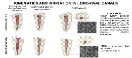
O presente estudo teve como objetivo avaliar, através de imagens de micro-CT, o preparo de canais radiculares ovais-longos utilizando os instrumentos Twisted File Adaptive (TFA) e Reciproc (RC) e a eficácia da limpeza da irrigação ultrassônica (IU) através de uma abordagem de microscopia eletrônica de varredura (MEV) correlacional. Canais distais ovais-longos de trinta e oito molares inferiores foram divididos em dois grupos (n = 19): grupos TFA e RC. Cada espécime foi submetido a três varreduras: pré e pós-instrumentação e pós-IU, e mudanças no volume, superfícies não preparadas e detritos de tecido duro acumulados (DTDA) foram medidos. Os canais radiculares foram divididos ao meio no terço apical e analisados em MEV para avaliação da camada de esfregaço (CE). Os testes de Mann-Whitney e Kruskal-Wallis foram utilizados para análise estatística (p < 0,05), e a correlação entre a DTDA e o CE foi avaliada com o coeficiente de correlação de Pearson. O sistema TFA exibiu maior eficiência na remoção de dentina em comparação ao sistema RC ao considerar todo o canal radicular (p < 0,05), com aproximadamente 26 a 29% da área de superfície não preparada para todo o canal (p> 0,05). Ambos os sistemas produziram o mesmo volume de DTDA (p> 0,05). A IU final levou a uma redução notável nos DTDA (60-70%), exceto no terço apical. Qualitativamente, a análise por MEV revelou remanescentes de dentina não uniformes cobrindo as paredes do canal, particularmente proeminentes no grupo RC. A análise dos dados determinou uma relação positiva moderada entre DTDA e avaliação por MEV. Concluindo, nenhum dos sistemas atingiu o preparo completo das paredes ovais longas do canal radicular, e a IU final reduziu os DTDA. Os DTDA observados por micro-CT foram relacionados à presença da CE avaliada por MEV.
Autoria
person Sandra Soares Kühne Busquim
1
person Laila Gonzales Freire
person Iandara de Lima Scardini
person Marcelo dos Santos
Correspondence: Laila Gonzales Freire, DDS, MSc PhD. Department of Restorative Dentistry, School of Dentistry, University of São Paulo, São Paulo, Brazil. Av. Prof. Lineu Prestes, 2227, Cidade Universitária, São Paulo, Brazil. CEP: 05508-000. E-mail:
lgfreire@usp.brPhone: 55 (11) 3091 7843
lgfreire@usp.brPhone: 55 (11) 3091 7843
Responsible editor
Manoel Damião Sousa-Neto
Figuras | Tabelas
 Thumbnail
 Thumbnail
 Thumbnail
 Thumbnail
imageopen_in_new
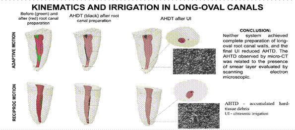
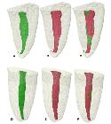
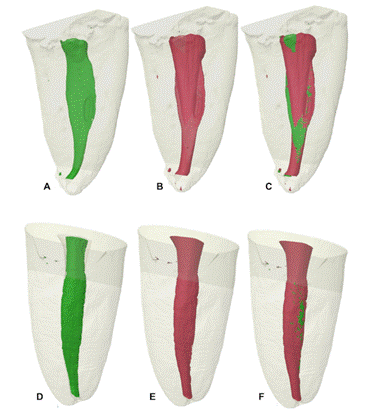
imageFigure 1 Representative 3D reconstructions of the mandibular distal canal of teeth prepared using TFA (A-C) and Reciproc (D-F) systems. In A and D: before root canal preparation; In B and E: after root canal preparation; In C and F: overlay of A or D (green), and B or E (red) images. open_in_new
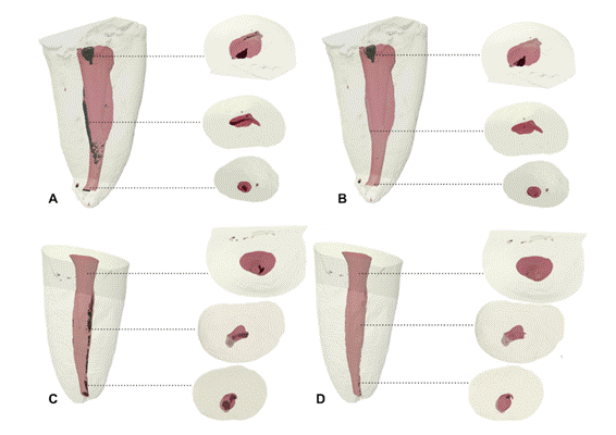
imageFigure 2 Representative 3D reconstructions of the mandibular distal canal of teeth prepared using TFA (A and B) and Reciproc (C and D) systems after UI. In A and C: AHTD (black) after preparation; In B and D: AHTD post-UI. Dotted lines correspond to cross-sectional views of each third of the root canal. open_in_new
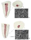
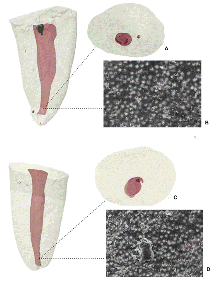
imageFigure 3 3D models of the sample of each group (A and B - TFA; C and D - RC) with the highest AHTD reduction after ultrasonic irrigation in the apical third seen throughout micro-CT (A and C) and the representative microphotograph from SEM B and D. SEM images were obtained using the secondary electron (SE) detector at a magnification of x1000, and a scale bar of 10 micrometers. open_in_new
table_chartTable 1
Mean and standard deviation of morphometric alterations in volume (mm3), surface area (mm2), and percentage of unprepared surfaces. Mean and standard deviation of AHTD volume (mm3) after root canal preparation, after-UI, and the percentage of reduction after-UI for TFA and RC groups.
| Region | TFA | RC | |
|---|---|---|---|
| initial canal volume (mm3) | Entire | 7.296 ± 1.040 | 6.859 ± 0.958 |
| Cervical | 3.933 ± 0.866 | 4.143 ± 0.716 | |
| Medium | 2.069 ± 0.614 | 1.963 ± 0.420 | |
| Apical | 1.255 ± 0.665 | 0.954 ± 0.510 | |
| final canal volume (mm3) | Entire | 13.136 ± 1.796A | 11.086 ± 1.463B |
| Cervical | 7.393 ± 1.547 | 6.605 ± 1.254 | |
| Medium | 3.459 ± 1.054 | 3.071 ± 0.684 | |
| Apical | 2.053 ± 1.441 | 1.644 ± 0.871 | |
| initial surface area (mm2) | Entire | 55.131 ± 13.348 | 46.688 ± 10.907 |
| Cervical | 27.110 ± 8.057 | 23.872 ± 6.333 | |
| Medium | 20.956 ± 4.659 | 18.414 ± 10.256 | |
| Apical | 7.098 ± 3.296 | 7.717 ± 4.971 | |
| final surface area (mm2) | Entire | 68.080 ± 17.338 | 63.824 ± 15.808 |
| Cervical | 33.959 ± 10.622 | 30.947 ± 9.114 | |
| Medium | 23.276 ± 6.543 | 24.451 ± 13.291 | |
| Apical | 10.394 ± 3.700 | 8.362 ± 3.377 | |
| unprepared Surface (%) | Entire | 26,767 ± 13,497 | 29,214 ± 10,096 |
| Cervical | 19,058 ± 12,068 | 21,763 ± 11,878 | |
| Medium | 33,738 ± 17,799 | 33,922 ± 15,619 | |
| Apical | 37,083 ± 21,513 | 44,852 ± 17,241 | |
| AHTD after preparation | Entire | 0.274 ± 0.302a | 0.349 ± 0.343a |
| Cervical | 0.078 ± 0,113c | 0.139 ± 0.263c | |
| Medium | 0.088 ± 0.085e | 0.119 ± 0.132e | |
| Apical | 0.085 ± 0.106g | 0.084 ± 0.065g | |
| AHTD after-UI | Entire | 0.127 ± 0.152b | 0.134 ± 0.130b |
| Cervical | 0.022 ± 0.041d | 0.042 ± 0.049d | |
| Medium | 0.043 ± 0.058f | 0.050 ± 0.063f | |
| Apical | 0.037 ± 0.051g | 0.034 ± 0.035g | |
| AHTD Reduction after-UI (%) | Entire | 71.778 ± 17.146 | 63.543 ± 25.384 |
| Cervical | 71.117 ± 18.936 | 65.582 ± 27.842 | |
| Medium | 69.376 ± 25.495 | 63.759 ± 27.256 | |
| Apical | 67.487 ± 16.570 | 63.440 ± 24.209 |
-
No significant differences between experimental groups (p>0,05)
-
Different uppercase letters show significant differences between groups (p<0,05)
-
Different lowercase letters show intra-group significant differences (p<0,05)
table_chartTable 2
Mean and standard deviation of the accumulated hard tissue debris (AHTD) in the apical third for TFA and RC groups, and the reduction (%) after UI obtained by micro-CT, and smear layer median scores obtained by SEM.
| TFA | RC | |
|---|---|---|
| AHTD after preparation | 0.085 ± 0.106 | 0.084 ± 0.065 |
| AHTD after-UI | 0.037 ± 0.051 | 0.034 ± 0.035 |
| AHTD Reduction after-UI (%) | 67.487 ± 16.570 | 63.440 ± 24.209 |
| Predominant SEM scores | 2.5 (2-3) | 4 (3-5) |
-
r=- 0.91. p<0.01
Como citar
location_on
Fundação Odontológica de Ribeirão Preto
Av. do Café, S/N, 14040-904 Ribeirão Preto SP Brasil, Tel.: (55 16) 3602-3982, Fax: (55 16) 3633-0999 -
Ribeirão Preto -
SP -
Brazil
E-mail: bdj@forp.usp.br
E-mail: bdj@forp.usp.br
rss_feed
Acompañe los números de esta revista en su lector de RSS
Versão para download de PDF
Artículos relacionados
Lista de links para artigos relacionados. Os links abrem em nova aba.
Versões e tradução automática
Escolha a versão original do texto ou utilize um serviço de tradução automática.
Versão original do texto
Como citar
Escolha um formato para exportar ou selecione um estilo de citação. O conteúdo abaixo pode ser atualizado após a seleção.