 Hydroalcoholic Extract from Lantana camara Leaves: Phytochemical Composition and In Vitro Antioxidant and Anti-Inflammatory Activities
Abstract
Lantana camara L. (Verbenaceae) is widely used in traditional medicine to treat wounds, inflammatory conditions, and microbial infections, owing to its phytochemical composition, which includes flavonoids, phenolic acids, and triterpenes, known for their associated antioxidant and anti-inflammatory properties. However, scientific studies exploring its pharmacological potential, particularly for inflammatory skin disorders, remain limited. This study aimed to evaluate the antioxidant and anti-inflammatory activities hydroalcoholic extract from L. camara leaves (LCHA) and characterize its phytochemical composition to support its potential therapeutic applications. Quantitative assays were performed to determine total phenolic, terpene, and saponin contents, while the phytochemical profile of LCHA was elucidated using UFLC-QTOF-MS, revealing the presence of lamiide, geniposide, pectolinarin, luteolin-7-O-glucoside, and rhamnocitrin-O-glucoside. In vitro assays were conducted using peritoneal macrophages and L929 fibroblasts to assess cell viability and to evaluate anti-inflammatory activity through nitric oxide (NO) production and lipid body accumulation. Antioxidant activity was determined via DPPH radical scavenging, phosphomolybdenum reduction, β-carotene/linoleic acid system, and reactive oxygen species (ROS) quantification. LCHA significantly inhibited NO production (100% at the highest concentration) and reduced lipid body formation by 68.61% in macrophages. Moreover, LCHA demonstrated notable antioxidant capacity, including 41.94% inhibition of lipid peroxidation and a 65.05% reduction in ROS levels in activated macrophages. These findings support its traditional use and highlight its potential as a plant-based therapeutic alternative for inflammatory skin disorders, encouraging its further investigation.
Keywords:
Phytochemistry; Lantana camara; Nitric oxide; Skin disorders; Inflammation.
HIGHLIGHTS
Lantana camara hydroalcoholic extract (LCHA) shows antioxidant and anti-inflammatory potential.
LCHA contains lamiide, geniposide, and flavonoid glycosides.
LCHA significantly inhibits nitric oxide and lipid body formation in macrophages.
The extract notable reduces lipid peroxidation and ROS levels in vitro.

INTRODUCTION
Inflammatory skin disorders represent a growing global health concern, affecting millions of people and placing a significant burden on healthcare systems [1]. These conditions, including dermatitis, psoriasis, and premature aging, are often exacerbated by oxidative stress and an excessive inflammatory response, leading to impaired healing and tissue damage [1,2]. Oxidative stress results from an imbalance between the production of reactive oxygen species (ROS) and the body's ability to neutralize them, contributing to chronic inflammation and degenerative skin conditions [3,4]. Additionally, prolonged inflammation can alter the skin barrier, increasing susceptibility to infections and further aggravating tissue damage [5,6].
Conventional treatments, such as corticosteroids and non-steroidal anti-inflammatory drugs (NSAIDs), can provide symptomatic relief but are associated with side effects, including skin atrophy, hypersensitivity reactions, and even cardiovascular effects. Furthermore, the long-term use of these treatments may impair natural skin repair mechanisms and compromise the skin's protective functions [7]. These limitations underscore the need for alternative therapeutic strategies that are both effective and safe for prolonged use, particularly those derived from natural products with known pharmacological potential.
Lantana camara L. (Verbenaceae) is a widely distributed plant in tropical and subtropical regions, traditionally used for treating various ailments, such as wounds, fever, and inflammatory diseases [8,9]. Despite its extensive ethnobotanical use, scientific studies investigating its biological properties and potential therapeutic applications remain scarce. Phytochemical analyses have revealed that L. camara contains a diverse range of bioactive compounds, including flavonoids, triterpenes, saponins and phenolic acids, which are known for their antioxidant and anti-inflammatory properties [10-12]. These compounds have been associated with the inhibition of ROS production and the modulation of inflammatory mediators, suggesting their potential in managing oxidative stress-related skin disorders [5,6,13]
Given the increasing demand for natural products in dermatology and the limited research on L. camara, further studies are necessary to validate its pharmacological potential and explore its applications in dermatological treatments. While many studies have focused on methanolic or ethanolic extracts of different plants parts, the specific biological activities of a hydroalcoholic leaf extract, which is highly relevant for topical preparations, are not as thoroughly understood. In particular, a comprehensive study that connects the detailed phytochemical profile of a hydroalcoholic extract to its specific effects on key cellular mechanisms of inflammation and oxidative stress has not been fully addressed. This study, therefore, focuses on the hydroalcoholic extract to fill this knowledge gap, providing critical insights into its mechanisms of action and highlighting its potential for the development of dermatological formulations. By bridging the gap between traditional use and scientific validation, this study aims to provide critical insights into the potential benefits of L. camara in managing skin disorders linked to oxidative stress and inflammation. Moreover, exploring its therapeutic applications could pave the way for the development of innovative formulations that harness the medicinal potential of L. camara in a scientifically substantiated manner.
MATERIAL AND METHODS
Plant material
Leaves from L. camara were collected in Juiz de Fora, Minas Gerais, Brazil (coordinates 21°77’62’’ south, 43°37’05’’ west), in January 2023. A specimen was deposited in the Leopoldo Krieger Herbarium of the Federal University of Juiz de Fora (CESJ 30671), according to the license number A18AB08 SISGEN/BRASIL.
Extract preparation
The dried and powdered leaves from L. camara (135.98 g) were extracted through exhaustive maceration. The solvent used was a 1:1 mixture of distilled water and ethyl alcohol. After extraction, the resulting liquid was concentrated using the BUCHI® Labortechinik AG (V-700) rotary evaporator at reduced pressure and a water bath (45-55°C), and then lyophilized (Martin Christ® Alpha 1-2 LDplus) to remove the remaining solvent. The extract was named LCHA and resulted in a yield of 10.28%.
Total phenolic content
The total phenolic content was determined using the Folin and Ciocalteu [14] method with some modifications. The calibration curve was prepared using tannic acid as a standard. A stock solution of LCHA at 1 mg/mL was prepared in methanol. The absorbance was read at 770 nm in a UV-Vis spectrophotometer (Thermo Scientific Multiskan GO, software 3.2). The content of phenolic compounds was expressed in μg/mg of plant extract in tannic acid equivalent. Two independent experiments were conducted, both in triplicate.
Terpenes content
The total terpene content was determined using the colorimetric method proposed by Pedrosa and coauthors [15], with adaptations. The calibration curve was prepared using the triterpene lupeol as a standard. A stock solution of LCHA at 1 mg/mL was prepared in ethanol. The absorbance was read at 548 nm in a UV-Vis spectrophotometer (Thermo Scientific Multiskan GO, software 3.2). The total terpene content was expressed as μg/mg of plant extract equivalent to lupeol. Two independent experiments were conducted, both in triplicate.
Saponin content
The saponin content was indirectly determined using the terpene quantification method proposed by Pedrosa and coauthors [15], with adaptations. The terpene quantification was performed after acid hydrolysis executed to release the terpenoid core of the saponins for subsequent quantification. The acid hydrolysis was carried out by the addition of hydrochloric acid to a stock solution of LCHA at 5 mg/mL (water:ethanol), followed by heating and extraction with dichloromethane. The organic phase was completely dried to remove the solvent and the residue was dissolved in ethanol to prepare a 1 mg/mL solution for terpene content determination. The absorbance was measured at 548 nm using a UV-Vis spectrophotometer (Thermo Scientific Multiskan GO, software 3.2). The terpene content after acid hydrolysis was expressed as μg/mg of plant extract equivalent to lupeol. To estimate the theoretical saponin content, the terpene content post-hydrolysis was subtracted from the pre-hydrolysis terpene content. The theoretical content of saponins with terpenoid core was expressed as μg/mg of extract. Two independent experiments were conducted, both in triplicate.
UFLC-QTOF-MS analysis
LCHA was analyzed by ultra-fast liquid chromatography coupled with mass spectrometry (UFLC-QTOF-MS) in positive ion mode, scanning ions in the m/z range of 100-1200. 5 mg was dissolved in a methanol:water mixture (8:2) and sonicated for 15 minutes. The solution was filtered through a 0.22 μm PTFE membrane and directly transferred to a vial. The mobile phase consisted of water (A) and acetonitrile (B), both acidified with 0.1% formic acid. The gradient was as follows: 0-4 min, 5% B; 4-34 min, ramp to 100% B; 34-38 min, held at 100% B; and returned to initial conditions (5% B) over 8 minutes, with an additional 4 minutes for re-equilibration. The injection volume was 3.0 μL, flow rate 0.5 mL/min, and run time of 50 minutes. The analysis was performed using a C18 reverse-phase column (4.6 x 150 mm, 2.6 μm) maintained at 40°C. Ionization was performed by electrospray ionization (ESI) with a capillary voltage of 3.5 kV and a temperature of 325°C. Fragmentation profiles were compared with literature data to identify sample constituents. The analysis was conducted in collaboration with Fiocruz using the Analytical Methods Platform of the Federal University of Rio Grande do Norte.
Cell viability
Cell culture conditions
Peritoneal macrophages were obtained from BALB/c mice (weighing 20-25 g) and cultured in RPMI-1640 medium (Gibco®) supplemented with 2 mM L-glutamine, 10% fetal bovine serum (FBS), and 1% antibiotic solution (100 μg/mL penicillin and 100 μg/mL streptomycin). L929 fibroblasts (ATCC® CCL-1 NCTC) were obtained and subsequently cultured in DMEM (Dulbecco's Modified Eagle Medium) supplemented with 10% FBS and 1% antibiotic solution. Both cell lines were maintained in an incubator at 37°C with a 5% CO₂ atmosphere. The procedure was accepted by the Committee on the Ethics of Animal Experiments of the Federal University of Juiz de Fora on May 10, 2018 (Protocol Number: 07/2018-CEUA).
MTT assay
The cytotoxicity of the extract was evaluated using the colorimetric method of 3-(4,5-dimethylthiazol-2-yl)-2,5-diphenyltetrazolium bromide (MTT) (Sigma Chemical Co., St. Louis, MO, USA) as described by Mosmann [16]. Peritoneal macrophages (2 × 10⁵ cells/well) and L929 fibroblasts (5 × 10³ cells/well) were seeded into 96-well polystyrene plates and treated with serial dilutions of the samples (18.75-300 μg/mL) and incubated for 48h (37°C, 5% CO₂). Negative controls consisted of cells treated with 0.06% dimethyl sulfoxide (DMSO). The absorbance was measured at 570 nm using a UV-Vis spectrophotometer (Thermo Scientific Multiskan GO, software 3.2). Data were presented as a percentage of cell viability, calculated by comparison with negative control. Two independent experiments were performed, each in triplicate for every concentration.
Anti-inflammatory activity
NO production
The production of nitric oxide (NO) was indirectly evaluated by quantifying nitrites (NO₂⁻) using the Griess method, as described by Sun and coauthors [17]. Peritoneal macrophages (2 × 10⁵ cells/well) were incubated for 48 hours (37°C, 5% CO₂) with varying concentrations of the sample (18.75-300 μg/mL) and stimulated with 1 μg/mL LPS and 1 ng/mL IFN-γ. The stimulation control consisted of stimulated but untreated macrophages, while the basal control consisted of unstimulated and untreated cells. The supernatant and Griess reagent were added to plates and incubated at room temperature for 10 minutes. The absorbance was measured at 540 nm using a UV-Vis spectrophotometer (Thermo Scientific Multiskan GO, software 3.2). The NO concentration was determined by comparison with a standard curve prepared from serial dilutions of a sodium nitrite (NaNO₂) solution. The results are expressed as the mean ± standard deviation of the percentage of NO radical inhibition and IC50 (50% inhibitory concentration) in μg/mL. Two independent experiments were performed, each in triplicate for every concentration.
Accumulation of lipid bodies (LBs)
The accumulation of lipid bodies (LBs) in peritoneal macrophages was evaluated following the method of Basselin and coauthors [18] with modifications, using the Nile Red marker (9-diethylamino-5H-benzo[α]phenoxazine-5-one). Peritoneal macrophages (2 × 10⁵ cells/well) were incubated for 48 hours (37°C, 5% CO₂) with varying concentrations of the sample (75-300 μg/mL) and stimulated with 1 μg/mL LPS and 1 ng/mL IFN-γ. The stimulation control consisted of stimulated but untreated macrophages, while the basal control consisted of unstimulated and untreated cells. Subsequently, the cells were washed with PBS and stained with 200 μL of Nile Red (10 μg/mL) in PBS for 20 minutes at 25°C. Fluorescence was measured at 485 nm excitation and 528 nm emission using a spectrofluorometer (FLx800, BioTek Instruments, Inc., Winooski, VT, USA). The results were expressed as fluorescence intensity (A.U.). Two independent experiments were performed, each in triplicate for every concentration.
Antioxidant activity
DPPH scavenging method
The radical scavenging activity of 2,2-Diphenyl-1-(2,4,6-trinitrophenyl)hydrazyl (DPPH) was determined using the method described by Brand-Williams and coauthors [19], with modifications. A stock solution of the sample and positive control (rutin) was prepared at 1 mg/mL in methanol and serially diluted (0.49 to 250.00 μg/mL). The samples and a DPPH solution (20 μg/mL in methanol) were incubated for 30 minutes in a 96-well microplate. The absorbance was measured at 517 nm using a UV-Vis spectrophotometer (Thermo Scientific Multiskan GO, software 3.2). Results were expressed as the mean ± standard deviation of the IC50 (50% inhibitory concentration) in μg/mL and the inhibitory capacity in percent (IC%) following the equation:
Where AbsLCHA means the absorbance of the extract; AbsBlank means the absorbance of the blank of the extract; AbsControl is the absorbance obtained in the absence of the extract. Two independent experiments were performed, each in triplicate.
Total antioxidant capacity by phosphomolybdenum assay
The total antioxidant capacity (TAC) was evaluated using the phosphomolybdenum complex reduction method, as described by Prieto and coauthors [20], with modifications. A stock solution of the sample and positive controls (rutin and quercetin) were prepared at 0.5 mg/mL in methanol:water (50:50). The samples and the reagent solution were added to hermetically sealed test tubes, which were then incubated in a water bath. The absorbance was measured at 695 nm using a UV-Vis spectrophotometer (Thermo Scientific Multiskan GO, software 3.2). Data were expressed as mean ± standard deviation of the percentage of relative antioxidant activity (RAA%) for rutin and quercetin following the equation:
Where Abs (LCHA) means the absorbance of the extract; Abs (Blank) means the absorbance of the blank of the extract; Abs (Control) is the absorbance of the positive controls (rutin and quercetin); and Abs (ControlBlank) is the absorbance of the blank of the positive controls. Two independent experiments were conducted, each in triplicate.
β-carotene/linoleic acid system
The lipid peroxidation inhibition activity was evaluated using the method described by Melo and Mancini Filho [21], with modifications. In a 96-well microplate, methanolic solutions of the sample and positive control (rutin) were added with β-carotene/linoleic acid emulsion (final concentrations of 1.25-40 μg/mL), followed with incubation at 45°C. The absorbance was measured at 470 nm every 15 minutes for 120 minutes using a UV-Vis spectrophotometer (Thermo Scientific Multiskan GO, software 3.2). Results were expressed as the mean ± standard deviation of the IC50 (50% inhibitory concentration) in μg/mL, and as percentage of lipid peroxidation inhibition (%I) using the formula: %I = [(Abs control - Abs sample)/Abs control] × 100. The oxidation curves were analyzed to calculate F1 (between 15-45 min) and F2 (between 75-90 min) using the tangent method. Two independent experiments were performed, each in triplicate.
Evaluation of reactive oxygen species (ROS) production
Intracellular levels of ROS in peritoneal macrophages were evaluated using 2′,7′-dichlorodihydrofluorescein diacetate (H2DCFDA) as described by Stroppa and coauthors [22]. Peritoneal macrophages (2 × 10⁵ cells/well) were incubated for 48 hours (37°C, 5% CO₂) with varying concentrations of the sample (18.75-300 μg/mL) and stimulated with 1 μg/mL LPS and 1 ng/mL IFN-γ. The negative control consisted of stimulated but untreated macrophages. Afterward, the macrophages were washed with PBS and incubated (37°C, 5% CO₂) with H2DCFDA (1 mM) for 30 minutes in the dark. ROS production was assessed in the cell pellet using fluorimetry (FLx800, BioTek Instruments, Inc., Winooski, VT, USA) at 485 nm excitation and 528 nm emission. The results were expressed as fluorescence intensity (A.U.) and IC50 (50% inhibitory concentration) in μg/mL. Two independent experiments were performed, each in triplicate for every concentration.
Statistical analysis
Statistical analysis was performed by ANOVA followed by Bonferroni’s test (p < 0.05) with GraphPrism 8.0.1 software using (Erithacus Software Ltd). Results were presented as mean ± SD.
RESULTS AND DISCUSSION
Phenols, terpenes and saponin content
Phenolic compounds are recognized as key phytochemicals with antioxidant properties, as they have a broad range of applications and demonstrated efficacy in managing disorders associated with oxidative stress [23,24]. Their antioxidant activity is attributed to their ability to donate electrons or hydrogen atoms, thereby neutralizing free radicals, and to their metal-chelating properties, which inhibit metal-catalyzed generation of reactive oxygen species [25]. Meanwhile, terpenes represent a diverse class of compounds with various properties described in the literature, including anti-inflammatory, antioxidant, antiallergic and analgesic activities [26]. Additionally, saponins are complex metabolites known for their wide range of properties, notably their anti-inflammatory and antioxidant activities [27,28].
The total phenolic content in LCHA was 48.93 ± 7.05 μg/mg (tannic acid equivalents), corresponding to 4.89% of its composition. The terpene content was 105.90 ± 1.88 μg/mg (lupeol equivalent), representing 10.59% of the extract. While the theoretical content of terpenoid-core saponins was 73.38 ± 6.28 μg/mg, accounting for 7.34% of the whole extract. These findings indicate that LCHA is characterized by a predominance of terpenes (10.59%), followed by saponins (7.34%) and phenolic compounds (4.89%).
These values are consistent with the literature. Kumar and coauthors [29] reported a total phenolic content of 55.57 ± 2.82 μg/mg in a methanolic extract of Lantana camara leaves, similar to the 48.93 μg/mg found in our study. Regarding terpene content, Orji and coauthors [30] observed 122.26 ± 0.10 μg/mg in methanolic and 100.88 ± 1.57 μg/mg in aqueous extracts, which are comparable to the 105.90 μg/mg determined in LCHA. For saponins, Ezebo and coauthors [31] reported contents of 10.75% and 8.63% in ethanolic and methanolic extracts, respectively, while Bhuvaneswari and Giri [32] found 121.00 ± 8.47 μg/mg (12.1%) in a methanolic extract, values notably close to those obtained for LCHA in the present study. Although the data align with previous studies, it is important to note that the quantitative profile observed in LCHA, which shows a predominance of terpenes, may be due to unique characteristics related to the plant material, extraction method, or collection site.
Phytochemical profile by UFLC-QTOF-MS
All the compounds identified in the UFLC-QTOF-MS analyses are described in Table 1. The peaks obtained from the identified compounds are labeled numerically according to their retention times. Compound identification was based on the interpretation of their mass spectra (exact mass and characteristic fragmentation patterns) and comparison of these data with those reported in the scientific literature for Lantana camara or related species.
Compounds identified in the hydroalcoholic extract from L. camara leaves (LCHA) by UFLC-QTOF-MS.
Lamiide (terpene glycoside) and geniposide (iridoid glycoside) have previously been identified in L. camara [33]. According to the literature, both terpenoid and iridoid glycosides have been widely reported for their anti-inflammatory and wound healing properties [39,40]. Pectolinarin, luteolin-7-O-glucoside and rhamnocitrin-O-glucoside are flavonoids, which, in general, represent a promising class of compounds for the treatment of skin disorders, exhibiting well-documented antioxidant and wound healing activities [41]. Furthermore, luteolin-7-O-glucoside has been previously reported as one of the major compounds in the leaves of L. camara, positively contributing to the antioxidant potential of the species [37].
The identification of these specific metabolites, which include both glycosylated terpenoids and flavonoids, confirms the presence of bioactive classes commonly associated with the therapeutic properties of L. camara [33]. However, the phytochemical profile observed in this study may reflect the influence of regional, environmental, or methodological conditions, which are well-recognized to modulate the chemical composition of natural product extracts [9].
Cell viability
In the evaluation of cell viability using the MTT method, the formation of formazan occurs through the reduction of MTT by cellular enzymes, with the concentration of formazan being directly proportional to cell viability [16]. This assay is critically important for assessing the safety of LCHA for human use. Macrophages are immune system cells directly involved in the inflammatory process, while fibroblasts are mesenchymal cells that synthesize collagen and other macromolecules. These cells are essential for tissue repair and play a role in chronic inflammation. For this reason, these cell lines were chosen for the cell viability evaluation [42].
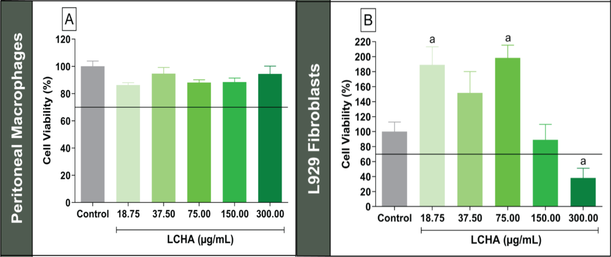
According to NBR ISO 10993-5, 2009, this assay should be one of the initial tests performed to assess the biocompatibility and toxicity of samples, establishing that in vitro cytotoxicity results must not fall below 70% for a sample to be considered biocompatible. As shown in Figure 1, cell viability remained above 80% for all evaluated concentrations of LCHA in peritoneal macrophages, indicating no cytotoxicity effects on this cell line. However, in L929 fibroblasts, viability remained above 70% only up to a concentration of 150 μg/mL, suggesting a concentration-dependent reduction in cell viability.

Evaluation of cell viability in peritoneal macrophages (A) and L929 fibroblast (B) cell lines treated with varying concentrations of the hydroalcoholic extract from the leaves of L. camara (LCHA). Control - Cells treated with vehicle (DMSO). The letter 'a' indicates a statistically significant difference (p< 0.05) when compared exclusively to the Control group. Statistical analysis was performed using ANOVA followed by the Bonferroni test.
While these findings align with prior reports in L. camara extracts, such as Pour and coauthors [43], who observed a concentration-dependent reduction in viability of Vero cells exposed to leaf extracts, the differential sensitivity between immune and mesenchymal cells observed here emphasizes that LCHA components may interact variably with distinct cellular pathways.
Anti-inflammatory activity
Nitric oxide (NO) is a reactive oxygen species, being liposoluble, it can permeate the cell membrane [44]. Involved in several diseases, its excessive production is associated with inflammatory processes [17]. During inflammation, pro-inflammatory cytokines induce an increase in NO synthesis in immune system cells, acting as a mediator of inflammation. The overproduction of NO (in cases of exacerbated inflammatory processes) can lead to various damages [44]. Thus, cellular NO production was measured by Griess test in peritoneal macrophages cultures treated with LCHA at the same concentrations used in the cell viability assay (MTT).
LCHA has significantly (p < 0.05) reduced NO levels at all concentrations tested, except for the lowest concentration of 18.75 μg/mL (Figure 2A), showing a IC50 value of 27.67 ± 2.18 μg/mL. The extract demonstrated a reduction in NO production of approximately 32.45 ± 4.91% at its lowest concentration (18.75 μg/mL) and a reduction of 61.04 ± 2.30% at 37.50 μg/mL, increasing to 96.09 ± 2.95% and 96.98 ± 4.04% at concentrations of 75 μg/mL and 150 μg/mL, respectively, reaching a reduction of approximately 100 ± 15.05% at its highest concentration evaluated (300 μg/mL). These results are consistent with the literature, in which Bairagi and coauthors [10] highlight the promising anti-inflammatory activity of the species. However, while the present study employed in vitro assays to evaluate this activity, the cited study of Bairagi and coauthors adopted an in vivo approach. Biological responses can vary substantially between a controlled in vitro system and the physiological complexity of a living organism, which may account for the differences observed in the results.

Anti-inflammatory activity of the hydroalcoholic extract from L. camara leaves (LCHA) in different concentrations: (A) Nitric oxide production by peritoneal macrophages; (B) lipid bodies accumulation by peritoneal macrophages. Control - Stimulated cells (LPS and IFN-γ) treated with vehicle (DMSO). Basal - Unstimulated cells treated with vehicle (DMSO). The letter 'a' indicates a statistically significant difference (p< 0.05) when compared exclusively to the Control group. Statistical analysis was performed using ANOVA followed by the Bonferroni test.
Lipid Bodies (LBs) are organelles present in the cytoplasm of various cells. In leukocytes, their accumulation is associated with their role as modulators of the immune response during an inflammatory process. Generally, LBs are linked to the initiation of the synthesis cascade of inflammatory mediators such as prostaglandins and leukotrienes [45]. Thus, the production and intracellular accumulation of LBs in peritoneal macrophages cultures treated with LCHA at 75 to 300 μg/mL was assessed. As shown in Figure 2B, LCHA significantly reduced LB accumulation (p < 0.05) at all concentrations tested. The extract reduced accumulation by approximately 68.23 ± 3.94% at the lowest concentration (75 μg/mL) and by 68.61 ± 3.26% at the highest concentration evaluated (300 μg/mL).
The results obtained highlight the anti-inflammatory potential of LCHA, a property already described in the literature for the species [46]. However, a detailed search of the literature revealed no prior studies investigating the effect of L. camara leaf extracts on nitric oxide (NO) production or lipid body accumulation. This highlights the originality of the present study, positioning it as possibly one of the first to evaluate the anti-inflammatory activity of this specific extract through these important cellular mechanisms.
The significant inhibition of NO production and lipid body accumulation observed in this study is strongly supported by the complex phytochemical profile of LCHA. The prior identification of high levels of terpenoids and saponins is particularly relevant, as these classes are known to target key inflammatory pathways [47]. Furthermore, the specific compounds identified by UFLC-QTOF-MS provide a mechanistic basis for the anti-inflammatory effects, as many of these constituents, such as lamiide [48], geniposide [49], and various flavonoids [50], possess well-documented anti-inflammatory potential. Specifically, the anti-inflammatory potential of pectolinarin [51] and the antioxidant/anti-inflammatory effects of luteolin-7-O-glucoside, including its reduction of reactive oxygen species (ROS) production [52], further support our findings.
The mechanisms underlying our results are likely multifactorial, aligning with previous studies on L. camara. Isolated triterpenoids from the species have been shown to inhibit the NF-κB-regulated expression of COX-2 and suppress NO release in LPS-induced cell models [53]. This documented ability provides a strong precedent for the nearly 100% dose-dependent reduction of NO we observed. The NF-κB pathway, which controls both iNOS (NO production) and lipid body formation machinery, is a central target, explaining the significant effects observed and aligning with the species' broader capacity to downregulate multiple inflammatory mediators, including NO, ROS, and COX-2 [53, 54].
The combined presence of terpenes, saponins, and phenolic compounds in LCHA reinforces its anti-inflammatory potential. Terpenes mitigate oxidative stress and inflammatory damage by reducing pro-inflammatory cytokines (TNF-α, IL-1, IL-6) and modulating MAPK signaling [40]. Saponins inhibit COX enzymes, reduce prostaglandin synthesis, block TNF-α production, and repress NF-κB activation [55]. Similarly, phenolic compounds modulate inflammatory pathways by inhibiting pro-inflammatory mediators and regulating key transcription factors such as NF-κB and Nrf2, suggesting a strong correlation between high phenolic compound levels and a reduced inflammatory response [56]. The combined action of these abundant bioactive classes in LCHA confirms its potent anti-inflammatory activity, consistent with both the literature and the present study's findings.
Antioxidant activity
Exacerbated inflammation can trigger the excessive production of reactive oxygen species (ROS), which oxidize essential macromolecules such as membrane lipids, structural proteins, enzymes, and nucleic acids. This oxidation leads to cellular damage, metabolic dysfunction, and even cell death [2,57]. Oxidative stress, characterized by an imbalance between ROS production and the antioxidant capacity of cells, plays a critical role in inflammatory processes and is associated with the development of various pathologies [4]. An antioxidant molecule is defined as one that can prevent or delay the oxidation of other molecules [58].
The application of diverse methodologies allows for a more comprehensive evaluation of complex samples, as their compounds may exert antioxidant effects through distinct mechanisms [59]. Therefore, the DPPH radical scavenging assay was used to evaluate the capacity of LCHA to scavenge free radicals by donating hydrogen atoms [58]. LCHA exhibited an IC% of 90.83 ± 0.61% at the highest concentration evaluated (250 μg/mL), presenting a IC50 value of 88.79 ± 17.64 μg/mL (Table 2), significantly different from the value obtained for the standard rutin (0.44 ± 0.22 μg/mL) (p < 0.05).
Evaluation of the antioxidant activity of the hydroalcoholic extract from L. camara leaves (LCHA) through DPPH, phosphomolybdenum, and β-carotene/linoleic acid system assays.
Several studies in the literature have evaluated different extracts from the leaves of L. camara using the DPPH assay, revealing a wide range of IC50 values. Various methanolic extracts have shown IC50 values starting from 16.02 ± 0.94 µg/mL [12], with reported values such as 34.01 ± 1.32 µg/mL [11] and 53.88 ± 6.40 µg/mL [60], reaching up to 114.63 ± 6.16 µg/mL [61]. Kumar and coauthors [29] evaluated four different variants of the species, reporting IC50 values ranging from 33.30 ± 2.39 μg/mL (CYV variant, yellow) to 927.16 ± 2.88 μg/mL (CPV variant, purple). This significant variability can be attributed to several critical factors. Such as the use of different extraction solvents, the phytochemical composition of L. camara (that is heavily influenced by regional, environmental, and genetic factors) and the existence of different chemotypes or variants, as shown in Kumar and coauthors study [29].
The phosphomolybdenum complex reduction assay provides data on the total antioxidant capacity of both hydrophilic and lipophilic substances, making it effective for analyzing complex mixtures (such as plant extracts) [20]. The RAA% values for rutin and quercetin obtained for LCHA were 73.63 ± 5.66 and 45.10 ± 3.46, respectively (Table 2). These results suggest that the compounds present in LCHA exhibit antioxidant potential more similar to that of rutin than to quercetin. A thorough literature search revealed a study by Kumar and coauthors [29] that also employed this assay on L. camara, however, that work utilized ascorbic acid as the reference standard. As the expression of total antioxidant capacity is relative to the standard used, a direct quantitative comparison with the rutin and quercetin equivalent results is not feasible. This highlights the originality of our approach and positions this work as potentially the first to benchmark the antioxidant capacity of this extract against these specific flavonoid standards, providing a new comparative perspective.
Another assay performed was the β-carotene/linoleic acid system, which evaluates the sample's ability to inhibit lipid peroxidation [58]. In this assay, LCHA exhibited an IC50 greater than 38.46 μg/mL (not determined) and a lipid peroxidation inhibition percentage of 41.94 ± 0.11% (Table 2), both values statistically different from the standard rutin (p < 0.05). In addition to these data, this assay also provides F1 and F2 values, which represent, respectively, the antioxidant effectiveness in blocking free radical chain reactions (initial oxidation stage, peroxide formation) and the effectiveness in the propagation phase (reactions that accelerate the oxidative process, associated with oxidative stress). For these values, results lower than 1 and closer to 0 are expected to indicate significant antioxidant potential [62]. LCHA showed an F1 value of 0.3656 ± 0.0540, which is in the ideal range for good antioxidant activity, indicating effectiveness in blocking reactions in the initial oxidation stage. However, its F2 value of 1.2029 ± 0.0447 is outside the ideal range for good antioxidant activity related to the propagation phase.
The evaluation of ROS production levels in peritoneal macrophage cultures treated with LCHA was also performed. The results obtained (Figure 3) show that the extract led to a statistically significant reduction in production (p < 0.05) at all concentrations tested, indicating its antioxidant potential in a cellular model. The reduction ranged from 29.75 ± 6.34% (lowest concentration of 18.75 μg/mL) up to 65.04 ± 6.58% (highest concentration of 300 μg/mL), showing an IC50 value of 79.06 ± 4.75 μg/mL.
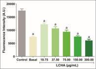
Antioxidant activity of the hydroalcoholic extract from L. camara leaves (LCHA). Reductions of reactive oxygen species (ROS) levels in peritoneal macrophages treated with varying concentrations of LCHA. Control - Stimulated cells (LPS and IFN-γ) treated with vehicle (DMSO). Basal - Unstimulated cells treated with vehicle (DMSO). The letter 'a' indicates a statistically significant difference (p< 0.05) when compared exclusively to the Control group. Statistical analysis was performed using ANOVA followed by the Bonferroni test.
A thorough review of the literature revealed no prior studies that have employed either the β-carotene/linoleic acid system or an intracellular ROS quantification assay, both specific methodologies to evaluate the antioxidant activity of L camara leaf extracts. This finding is significant, as it underscores the novelty of our study in providing the first evidence of this extract's protective effects against both lipid peroxidation and cellular oxidative stress, offering new insights into its potential cytoprotective mechanisms.
Based on the results obtained, LCHA demonstrates clear antioxidant potential, particularly in cellular models, with greater effectiveness observed during the early stages of oxidation. These findings suggest that LCHA may act as an antioxidant agent.
This antioxidant potential can be attributed to the bioactive compounds present in L. camara, particularly phenolic compounds and terpenes. The antioxidant activity of phenolic compounds is closely linked to their chemical structure, especially the number and position of hydroxyl groups. By donating hydrogen atoms or electrons, phenolic compounds neutralize free radicals, which significantly contributes to their overall antioxidant potential [25].
Similarly, the antioxidant potential of terpenes is related to their ability to modulate oxidative stress pathways. Terpenes reduce lipid peroxidation induced by H₂O₂, inhibit ROS formation, and enhance the activity of key antioxidant enzymes [40]. Together, these bioactive compounds present in LCHA reinforce its antioxidant potential.
CONCLUSION
The phytochemical analysis of the L. camara hydroalcoholic extract (LCHA) confirmed a rich profile of bioactive compounds, including lamiide, geniposide, pectolinarin, luteolin-7-O-glucoside, and rhamnocitrin-O-glucoside. The in vitro results demonstrated significant anti-inflammatory and antioxidant potential, with LCHA achieving up to 100% reduction in nitric oxide (NO) and a 68.61% decrease in lipid body (LB) accumulation. Furthermore, LCHA effectively inhibited lipid peroxidation and reduced intracellular ROS production. The demonstrated action of LCHA in mitigating both oxidative stress and key inflammatory mediators renders it an attractive candidate for topical formulations aimed at managing chronic inflammatory skin conditions, such as psoriasis or atopic dermatitis. While these findings are promising, the in vitro nature of the study highlights the need for subsequent in vivo validation to confirm its efficacy and safety. Also, this work reinforces the value of Brazilian biodiversity and traditional medicinal knowledge, providing a scientific foundation for its sustainable use.
Funding:
Institutional Review Board Statement:
Informed Consent Statement:
Acknowledgments:
The authors thank Delfino Antonio Campos of the Department of Biochemistry, Federal University of Juiz de Fora, for technical assistance and Analytical Methods Platform of Farmanguinhos/FIOCRUZ by UFLC-QTOF-MS analysis.
Use of Generative Artificial Intelligence
The authors declare that large language models and other generative artificial intelligence (AI) or AI-assisted technologies cannot be credited as authors and have not been listed as authors of this paper.
The author declare that did not use the artificial intelligence.
Data Availability Statement:
Data are available in the UFJF repository (https://repositorio.ufjf.br/jspui/handle/ufjf/18014).
REFERENCES
-
1 Barboza JN, Filho CSMB, Silva RO, Medeiros JVR, de Sousa DP. An Overview on the Anti-inflammatory Potential and Antioxidant Profile of Eugenol. Oxid Med Cell Longev. 2018;2018:1-9. https://doi.org/10.1155/2018/3957262
» https://doi.org/10.1155/2018/3957262 -
2 Lemos ASO, Campos LM, Souza TF, Paula PL, Granato JT, da Silva JVG, et al. Pharmacological investigation of antioxidant and anti-inflammatory activities of aqueous extract from Mitracarpus frigidus (Rubiaceae). J Pharm Pharmacol. 2022;74(5):750-60. https://doi.org/10.1093/jpp/rgac005
» https://doi.org/10.1093/jpp/rgac005 -
3 Speeckaert R, Speeckaert MM, Van Geel N. The Multifaceted Aspects of Oxidative Stress in the Skin and Other Tissues. Antioxidants (Basel). 2024;13(9):1-5. https://doi.org/10.3390/antiox13091081
» https://doi.org/10.3390/antiox13091081 -
4 Forman HJ, Zhang H. Targeting oxidative stress in disease: promise and limitations of antioxidant therapy. Nat Rev Drug Discov. 2021;20:689-709. https://doi.org/10.1038/s41573-021-00233-1
» https://doi.org/10.1038/s41573-021-00233-1 -
5 Ren Q, Qu L, Yuan Y, Wang F. Natural Modulators of Key Signaling Pathways in Skin Inflammageing. Clin Cosmet Investig Dermatol. 2024;17:2967-88. https://doi.org/10.2147/CCID.S502252
» https://doi.org/10.2147/CCID.S502252 -
6 Bharadvaja N, Gautam S, Singh H. Natural polyphenols: a promising bioactive compounds for skin care and cosmetics. Mol Biol Rep. 2023;50:1817-28. https://doi.org/10.1007/s11033-022-08156-9
» https://doi.org/10.1007/s11033-022-08156-9 -
7 Gushiken LFS, Beserra FP, Bastos JK, Jackson CJ, Pellizzon CH. Cutaneous Wound Healing: An Update from Physiopathology to Current Therapies. Life (Basel). 2021;11(7): 1-15. https://doi.org/10.3390/life11070665
» https://doi.org/10.3390/life11070665 - 8 Dange VN, Dinde SR, Doiphode AR, Dhavane SM, Dudhal BA, Shid SJ. Formulation and Evaluation Of Herbal Gel Containing Lantana Camara For Management Of Acne Vulgaris. J Univ Shanghai Sci Technol. 2020;22(11):799-809.
-
9 Nea F, Kambiré DA, Genva M, Tanoh EA, Wognin EL, Martin H, et al. Composition, Seasonal Variation, and Biological Activities of Lantana camara Essential Oils from Côte d’Ivoire. Molecules. 2020;25(10):1-22. https://doi.org/10.3390/molecules25102400
» https://doi.org/10.3390/molecules25102400 -
10 Bairagi S, Pathan DrI, Nema N. Analgesic and anti-inflammatory activity of crude leaf and bark extract of Lantana Camara. Marmara Pharm J. 2017;21(4):810-7. https://doi.org/10.12991/mpj.2017.7
» https://doi.org/10.12991/mpj.2017.7 -
11 El-Din MIG, Fahmy NM, Wu F, Salem MM, Khattab OM, El-Seedi HR, et al. Comparative LC-LTQ-MS-MS Analysis of the Leaf Extracts of Lantana camara and Lantana montevidensis Growing in Egypt with Insights into Their Antioxidant, Anti-Inflammatory, and Cytotoxic Activities. Plants (Basel). 2022;11(13):1-21. https://doi.org/10.3390/plants11131699
» https://doi.org/10.3390/plants11131699 -
12 Mahdi-Pour B, Jothy SL, Latha LY, Chen Y, Sasidharan S. Antioxidant activity of methanol extracts of different parts of Lantana camara. Asian Pac J Trop Biomed. 2012;2(12):960-5. https://doi.org/10.1016/S2221-1691(13)60007-6
» https://doi.org/10.1016/S2221-1691(13)60007-6 -
13 Lopes FB, Sarandy MM, Novaes RD, Valacchi G, Gonçalves RV. OxInflammatory responses in the wound healing process: a systematic review. Antioxidants (Basel). 2024;13(7):1-23. https://doi.org/10.3390/antiox13070823
» https://doi.org/10.3390/antiox13070823 -
14 Folin O, Ciocalteu V. On tyrosine and tryptophane determinations in proteins. J Biol Chem. 1927;73(2):627-50. https://doi.org/10.1016/S0021-9258(18)84277-6
» https://doi.org/10.1016/S0021-9258(18)84277-6 -
15 Pedrosa AM, de Castro WV, Castro AHF, Duarte-Almeida JM. Validated spectrophotometric method for quantification of total triterpenes in plant matrices. DARU J Pharm Sci. 2020;28:281-6. https://doi.org/10.1007/s40199-020-00342-z
» https://doi.org/10.1007/s40199-020-00342-z -
16 Mosmann T. Rapid colorimetric assay for cellular growth and survival: Application to proliferation and cytotoxicity assays. J Immunol Methods. 1983;65(1-2):55-63. https://doi.org/10.1016/0022-1759(83)90303-4
» https://doi.org/10.1016/0022-1759(83)90303-4 -
17 Sun J, Zhang X, Broderick M, Fein H. Measurement of Nitric Oxide Production in Biological Systems by Using Griess Reaction Assay. Sensors (Basel). 2003;3(8):276-84. https://doi.org/10.3390/s30800276
» https://doi.org/10.3390/s30800276 -
18 Basselin M, Robert-Gero M. Alterations in membrane fluidity, lipid metabolism, mitochondrial activity, and lipophosphoglycan expression in pentamidine-resistant Leishmania. Parasitol Res. 1997;84:78-83. https://doi.org/10.1007/s004360050361
» https://doi.org/10.1007/s004360050361 -
19 Brand-Williams W, Cuvelier ME, Berset C. Use of a free radical method to evaluate antioxidant activity. LWT. 1995;28(1):25-30. https://doi.org/10.1016/S0023-6438(95)80008-5
» https://doi.org/10.1016/S0023-6438(95)80008-5 -
20 Prieto P, Pineda M, Aguilar M. Spectrophotometric Quantitation of Antioxidant Capacity through the Formation of a Phosphomolybdenum Complex: Specific Application to the Determination of Vitamin E. Anal Biochem. 1999;269(2):337-41. https://doi.org/10.1006/abio.1999.4019
» https://doi.org/10.1006/abio.1999.4019 - 21 Melo MSOM, Mancini-Filho J. [Natural antioxidants from the oil palm fruit (elaeis guineensis, jacq)]. Rev Farm Bioquim Univ Sao Paulo. 1989;25(2):147-57.
-
22 Stroppa PHF, Antinarelli LMR, Carmo AML, Gameiro J, Coimbra ES, da Silva AD. Effect of 1,2,3-triazole salts, non-classical bioisosteres of miltefosine, on Leishmania amazonensis. Bioorg Med Chem. 2017;25(12):3034-45. https://doi.org/10.1016/j.bmc.2017.03.051
» https://doi.org/10.1016/j.bmc.2017.03.051 -
23 Yolbas I. Phenolic Compound Content and Antioxidant Activity of Ribes rubrum L. Shells. J Chem. 2024;2024:1-8. https://doi.org/10.1155/2024/9151180
» https://doi.org/10.1155/2024/9151180 -
24 Stafussa AP, Maciel GM, Bortolini DG, Maroldi WV, Ribeiro VR, Fachi MM, et al. Bioactivity and bioaccessibility of phenolic compounds from Brazilian fruit purees. Future Foods. 2021;4:1-7. https://doi.org/10.1016/j.fufo.2021.100066
» https://doi.org/10.1016/j.fufo.2021.100066 -
25 Vuolo MM, Lima VS, Maróstica Junior MR. Chapter 2 - Phenolic Compounds: Structure, Classification, and Antioxidant Power. Bioact Compd. 2019;2019:33-50. https://doi.org/10.1016/B978-0-12-814774-0.00002-5
» https://doi.org/10.1016/B978-0-12-814774-0.00002-5 -
26 Masyita A, Sari RM, Astuti AD, Yasir B, Rumata NR, Emran TB, et al. Terpenes and terpenoids as main bioactive compounds of essential oils, their roles in human health and potential application as natural food preservatives. Food Chem X. 2022;13:1-13. https://doi.org/10.1016/j.fochx.2022.100217
» https://doi.org/10.1016/j.fochx.2022.100217 -
27 Wijesekara T, Luo J, Xu B. Critical review on anti-inflammation effects of saponins and their molecular mechanisms. Phytother Res. 2024;38(4):2007-22. https://doi.org/10.1002/ptr.8164
» https://doi.org/10.1002/ptr.8164 -
28 Nguyen L, Fărcaş A, Socaci S, Tofana M, Diaconeasa Z, Pop OL, et al. An Overview of Saponins -A Bioactive Group. Bull Univ Agric Sci Vet Med Cluj-Napoca Food Sci Technol. 2020;77(1):25-36. https://doi.org/10.15835/buasvmcn-fst:2019.0036
» https://doi.org/10.15835/buasvmcn-fst:2019.0036 -
29 Kumar S, Sandhir R, Ojha S. Evaluation of antioxidant activity and total phenol in different varieties of Lantana camara leaves. BMC Res Notes. 2014;7:1-9. https://doi.org/10.1186/1756-0500-7-560
» https://doi.org/10.1186/1756-0500-7-560 -
30 Orji EA, Ejere VC, Orji CT, Anorue EC, Ossai NI, Ojua EO, et al. Phytochemical Profiling and GC-MS Analysis of Lantana camara Leaf Extract. Trop J Nat Prod Res. 2024;8(7):7920-7. https://doi.org/10.26538/tjnpr/v8i7.40
» https://doi.org/10.26538/tjnpr/v8i7.40 - 31 Ezebo RO, Okonkwo CC, Ozoh CN, Nwankwo CA, Nwafor EC, Esimai BG, et al. Phytochemical Screening and Antimicrobial Activity of Ethanol and Methanol Extracts of Lantana camara Leaf. World News Nat Sci. 2021;37:151-63.
- 32 Bhuvaneswari E, Giri RS. Physicochemical and phytochemical screening in Lantana camara leaves. J Pharmacogn Phytochem. 2018;7(6):1962-6.
-
33 El-Banna AA, Darwish RS, Ghareeb DA, Yassin AM, Abdulmalek SA, Dawood HM. Metabolic profiling of Lantana camara L. using UPLC-MS/MS and revealing its inflammation-related targets using network pharmacology-based and molecular docking analyses. Sci Rep. 2022;12:1-17. https://doi.org/10.1038/s41598-022-19137-0
» https://doi.org/10.1038/s41598-022-19137-0 -
34 Kostova M, Konaklieva M, Alipieva K, Popov S, Handjieva N. ESIMS study of some C 10 iridoid glycosides. Instrum Sci Technol. 2005;33(6):691-702. https://doi.org/10.1080/10739140500311288
» https://doi.org/10.1080/10739140500311288 -
35 Zhou H, Zhang S, Chen L, Liu Y, Shen L, Zhang J. Effective Therapeutic Verification of Crocin I, Geniposide, and Gardenia (Gardenia jasminoides Ellis) on Type 2 Diabetes Mellitus In Vivo and In Vitro. Foods. 2023;12(8):1-24. https://doi.org/10.3390/foods12081668
» https://doi.org/10.3390/foods12081668 -
36 Sousa EO, Costa JGM. Genus Lantana: chemical aspects and biological activities. Rev Bras Farmacogn. 2012;22(5):1155-80. https://doi.org/10.1590/S0102-695X2012005000058
» https://doi.org/10.1590/S0102-695X2012005000058 -
37 Othmen K, Maaloul N, Nhidi S, Cherif MM, Idoudi S, Elfalleh W. Phytochemical Profiles, in vitro Antioxidants, and Anti-inflammatory Activities of Flowers and Leaves of Lantana camara L. Grown in South of Tunisia. Period Polyt Chem Eng. 2024;68(1):72-84. https://doi.org/10.3311/PPch.22159
» https://doi.org/10.3311/PPch.22159 -
38 Es-Safi N-E, Kerhoas L, Einhorn J, Ducrot P-H. Application of ESI/MS, CID/MS and tandem MS/MS to the fragmentation study of eriodictyol 7-O-glucosyl-(1→2)-glucoside and luteolin 7-O-glucosyl-(1→2)-glucoside. Int J Mass Spectrom. 2005;247(1-3):93-100. https://doi.org/10.1016/j.ijms.2005.10.002
» https://doi.org/10.1016/j.ijms.2005.10.002 -
39 Barreto RSS, Albuquerque-Júnior RLC, Araújo AAS, Almeida JRGS, Santos MR V, Barreto AS, et al. A Systematic Review of the Wound-Healing Effects of Monoterpenes and Iridoid Derivatives. Molecules 2014;19(1):846-62. https://doi.org/10.3390/molecules19010846
» https://doi.org/10.3390/molecules19010846 -
40 Del Prado-Audelo ML, Cortés H, Caballero-Florán IH, González-Torres M, Escutia-Guadarrama L, Bernal-Chávez SA, et al. Therapeutic Applications of Terpenes on Inflammatory Diseases. Front Pharmacol. 2021;12:1-7. https://doi.org/10.3389/fphar.2021.704197
» https://doi.org/10.3389/fphar.2021.704197 -
41 Zulkefli N, Che Zahari CNM, Sayuti NH, Kamarudin AA, Saad N, Hamezah HS, et al. Flavonoids as Potential Wound-Healing Molecules: Emphasis on Pathways Perspective. Int J Mol Sci. 2023;24(5):1-29. https://doi.org/10.3390/ijms24054607
» https://doi.org/10.3390/ijms24054607 -
42 Kim ME, Lee JS. Advances in the Regulation of Inflammatory Mediators in Nitric Oxide Synthase: Implications for Disease Modulation and Therapeutic Approaches. Int J Mol Sci. 2025;26(3):1-19. https://doi.org/10.3390/ijms26031204
» https://doi.org/10.3390/ijms26031204 -
43 Pour BM, Latha LY, Sasidharan S. Cytotoxicity and Oral Acute Toxicity Studies of Lantana camara Leaf Extract. Molecules. 2011;16(5):3663-74. https://doi.org/10.3390/molecules16053663
» https://doi.org/10.3390/molecules16053663 -
44 Sharma JN, Al-Omran A, Parvathy SS. Role of nitric oxide in inflammatory diseases. Inflammopharmacology. 2007;15:252-9. https://doi.org/10.1007/s10787-007-0013-x
» https://doi.org/10.1007/s10787-007-0013-x -
45 Melo RCN, Weller PF. Lipid droplets in leukocytes: Organelles linked to inflammatory responses. Exp Cell Res. 2016;340(2):193-7. https://doi.org/10.1016/j.yexcr.2015.10.028
» https://doi.org/10.1016/j.yexcr.2015.10.028 -
46 Millycent SA, Mwonjoria KJ, Juma KK, Ngugi MP, Njagi ENM. Evaluation of analgesic, anti-inflammatory and toxic effects of lantana camara l. Int J Phytopharmacol. 2017;8(3):89-97. http://dx.doi.org/10.21276/ijp.2017.8.3.1
» http://dx.doi.org/10.21276/ijp.2017.8.3.1 -
47 Ono M, Hashimoto A, Miyajima M, Sakata A, Furusawa C, Shimode M, et al. Two new triterpenoids from the leaves and stems of Lantana camara. Nat Prod Res. 2021;35(21):3757-65. https://doi.org/10.1080/14786419.2020.1736063
» https://doi.org/10.1080/14786419.2020.1736063 -
48 Sobhy M, AbouZid SF, Kirollos FN, El-Shiekh RA, Abdel-Sattar E. Lamiide and Ipolamiide: A Comprehensive Review of Their Bioactive Properties and Therapeutic Potential. Chem Biodivers. 2024;21(12). https://doi.org/10.1002/cbdv.202401069
» https://doi.org/10.1002/cbdv.202401069 -
49 Shan M, Yu S, Yan H, Guo S, Xiao W, Wang Z, et al. A Review on the Phytochemistry, Pharmacology, Pharmacokinetics and Toxicology of Geniposide, a Natural Product. Molecules. 2017;22(10):1-29. https://doi.org/10.3390/molecules22101689
» https://doi.org/10.3390/molecules22101689 -
50 Wen K, Fang X, Yang J, Yao Y, Nandakumar KS, Salem ML, et al. Recent Research on Flavonoids and their Biomedical Applications. Curr Med Chem. 2021;28(5):1-25. https://doi.org/10.2174/0929867327666200713184138
» https://doi.org/10.2174/0929867327666200713184138 -
51 Patel K, Kumar Patel D. P-MD013. Importance of bioactive flavonoids “pectolinarin” in inflammation and pain: Validation through in-vivo, in-vitro and molecular study. Clin Neurophysiol. 2021;132(8). https://doi.org/10.1016/j.clinph.2021.02.232
» https://doi.org/10.1016/j.clinph.2021.02.232 -
52 De Stefano A, Caporali S, Di Daniele N, Rovella V, Cardillo C, Schinzari F, et al. Anti-Inflammatory and Proliferative Properties of Luteolin-7-O-Glucoside. Int J Mol Sci. 2021;22(3):1-18. https://doi.org/10.3390/ijms22031321
» https://doi.org/10.3390/ijms22031321 -
53 Wu P, Song Z, Wang X, Li Y, Li Y, Cui J, et al. Bioactive triterpenoids from Lantana camara showing anti-inflammatory activities in vitro and in vivo. Bioorg Chem. 2020;101. https://doi.org/10.1016/j.bioorg.2020.104004
» https://doi.org/10.1016/j.bioorg.2020.104004 - 54 Ashal TF, Ifora I, Oktavia S. Potential Anti-inflammatory Effects of Lantana camara L.: A Review. Int Res J Pharm Med Sci. 2020;3(6):1-4.
-
55 Moghimipour E, Handali S. Saponin: Properties, Methods of Evaluation and Applications. Annu Res Rev Biol. 2015;5(3):207-20. https://doi.org/10.9734/ARRB/2015/11674
» https://doi.org/10.9734/ARRB/2015/11674 -
56 Ambriz-Pérez DL, Leyva-López N, Gutiérrez-Grijalva EP, Heredia JB. Phenolic compounds: Natural alternative in inflammation treatment. A Review. Cogent Food Agric. 2016;2(1). https://doi.org/10.1080/23311932.2015.1131412
» https://doi.org/10.1080/23311932.2015.1131412 -
57 Tan BL, Norhaizan ME, Liew W-P-P, Sulaiman Rahman H. Antioxidant and Oxidative Stress: A Mutual Interplay in Age-Related Diseases. Front Pharmacol. 2018;9:1-28. https://doi.org/10.3389/fphar.2018.01162
» https://doi.org/10.3389/fphar.2018.01162 -
58 Moon J-K, Shibamoto T. Antioxidant Assays for Plant and Food Components. J Agric Food Chem. 2009;57(5):1655-66. https://doi.org/10.1021/jf803537k
» https://doi.org/10.1021/jf803537k -
59 Shahidi F, Zhong Y. Measurement of antioxidant activity. J Funct Foods. 2015;18:757-81. https://doi.org/10.1016/j.jff.2015.01.047
» https://doi.org/10.1016/j.jff.2015.01.047 -
60 Hoang TC, Nguyen MT, Nguyen TQ, Ho BTQ, Nguyen HT, Ngo TPD, et al. In vitro anti-leukemia, antioxidant, and anti-inflammatory properties of Lantana camara. Braz J Biol. 2024;84:1-11. https://doi.org/10.1590/1519-6984.279899
» https://doi.org/10.1590/1519-6984.279899 -
61 De Melo J, Araújo TAS, Castro VTNA, Cabral DLV, Rodrigues MD, do Nascimento SC, et al. Antiproliferative Activity, Antioxidant Capacity and Tannin Content in Plants of Semi-Arid Northeastern Brazil. Molecules. 2010;15(12):8534-42. https://doi.org/10.3390/molecules15128534
» https://doi.org/10.3390/molecules15128534 -
62 Borderes J, Costa A, Guedes A, Tavares LBB. Antioxidant activity of the extracts from Pycnoporus sanguineus mycelium. Braz Arch Biol Technol. 2011;54(6):1167-74. https://doi.org/10.1590/S1516-89132011000600012
» https://doi.org/10.1590/S1516-89132011000600012