 Grape Residue Extract Improves Mitochondrial Function and Promotes Sheep Oocyte Meiotic Resumption from In vitro-Grown Secondary Follicles
Abstract
Oxidative stress resulting from the inherent conditions of in vitro culture can compromise oocyte quality. In this regard, there is a growing interest in the use of natural products to prevent oxidative damage and to promote in vitro follicular development. This study was conducted to evaluate the effects of the addition of acidified extract of grape industrial residue (Vitis vinifera cv. Syrah) as a supplement to the base medium for in vitro culture of isolated sheep secondary follicles. Secondary follicles were isolated and cultured for 12 days in α-MEM+ (control medium) or in α-MEM+ supplemented with different grape residue extract concentrations (0.1, 0.2, or 0.4 mg/ml). Follicular morphology, antrum formation, follicular and oocyte diameter, glutathione (GSH) levels, mitochondrial activity, DNA fragmentation and meiotic resumption were evaluated. After 12 days of culture, there was no difference (P >0.05) among the treatments in relation to morphology, antral cavity formation, follicular diameter, DNA fragmentation and the percentage of fully grown oocytes (≥ 110 µm). Furthermore, the GSH levels were similar (P >0.05) among the α-MEM+, 0.2, and 0.4 mg/ml grape residue extract groups at the end of culture. Nevertheless, oocytes from secondary follicles cultured in 0.4 mg/ml grape residue extract showed higher (P <0.05) mitochondrial activity and greater meiotic resumption than oocytes cultured in the control medium. In conclusion, supplementation with 0.4 mg/ml grape residue extract maintained survival, improved mitochondrial activity, and promoted the meiotic resumption of oocytes from sheep secondary follicles cultured in vitro.
Keywords:
Antioxidant; In vitro culture; Ovine; Preantral follicle; Oxidative stress.
HIGHLIGHTS
Grape residue extract maintains survival, follicular development and GSH levels.
Grape residue extract (0.4 mg/mL) improves mitochondrial activity.
Grape residue extract (0.4 mg/mL) promotes meiotic resumption.
Grape extract can be an alternative supplement for the in vitro oocyte development.
INTRODUCTION
In vitro culture systems of preantral follicles are important to study early follicular biology [1], and to provide many oocytes that will be used for in vitro maturation (IVM) and fertilization [2]. However, oxidative stress caused by in vitro conditions may result in poor-quality oocytes [3,4]. In this context, there is a growing interest in the use of natural products to prevent oxidative damage and to promote follicular development in vitro. Herbal extracts with antioxidant properties have been successfully used as media or supplements for media to culture ovarian tissue and secondary follicles. Examples include Amburana cearensis [5,6], Morus nigra [7], and Justicia insularis [8].
Vitis vinifera, commonly known as grapevine, is a perennial woody climbing plant native to a broad region encompassing the Mediterranean Basin, Southern Europe, and Western Asia. It is currently cultivated throughout the world for the production of wine, juice and table grapes [9,10]. During industrial processing, large amounts of by-products, mainly skins and seeds, are generated and represent a rich source of structurally diverse phytochemicals. These residues contain protein, carbohydrates (fructose, glucose) lipids (monounsaturated fatty acids), minerals (iron, zinc), vitamins (vitamin C and E), and bioactive compounds (flavonoids, phenolic acids) with important biological properties, such as anti-inflammatory, anticancer, anti-aging, and antioxidant [9,11,12]. Grape residues can be applied in the pharmaceutical, food, and cosmetic industries to produce beneficial effects on human and animal health [13,14]. Supplementation of ewe diets with grape residue flour resulted in greater antioxidant and anti-inflammatory responses, improved milk production and quality, and reduced somatic cell count and lipid peroxidation compared with the control group [15,16]. In suckling lambs, supplementation with grape residue flour increased weight gain, enhanced glutathione S-transferase (GST) activity, and significantly reduced levels of reactive oxygen species (ROS). In addition, there was an improvement in immune function, evidenced by elevated serum immunoglobulins and reduced ceruloplasmin concentrations [57].
The health benefits of grape residues are mainly derived from its phenolic compounds with antioxidant properties, including resveratrol, rutin, procyanidins B1 and B2, gallic acid, and p-coumaric acid [17]. These compounds can scavenge reactive oxygen species (ROS), increase the activity of antioxidants, and attenuate mitochondrial damage, thus preventing cellular death from oxidative stress [18-20]. In vitro studies have demonstrated that some of these compounds induce the activation of primordial follicles (resveratrol: [21]; rutin: [22]); enhance the development of sheep secondary follicles (resveratrol: [23]; rutin: [24]; gallic acid: [25]); and improve IVM of porcine, murine, and human oocytes (resveratrol: [26]; procyanidin B1: [27]). However, supplementation of the culture medium of ovarian follicles with grape residue has not yet been studied. The aim of the present study was to evaluate the effect of the supplementation of base culture medium with acidified extract of industrial grape residue on the survival, development and meiotic resumption of oocytes from sheep secondary follicles cultured in vitro.
MATERIAL AND METHODS
Unless indicated otherwise, the media, supplements, and chemicals used in the present study were purchased from Sigma Chemical Co. (St. Louis, MO, USA).
Plant material and extract preparation
Industrial grape residues were collected from Santa Maria Winery, SA, Rural Zone, in the city of Lagoa Grande, Pernambuco (9°03'35.0"S, 40°11'32.1"W). To obtain the acidified ethanolic extract of grape residue (V. vinifera cv. Syrah), the plant material was dried at 42 ± 2 °C for 72 h. Then, the dry material was pulverized and subjected to fractional maceration with acid aqueous ethanol (ethanol:water:acetic acid: 70:30:1 [v/v/v]) with a solid:solvent ratio of 1:10. Five extractions were performed at 72-h intervals. The solution was filtered and concentrated on a SL-126 rotary evaporator (SOLAB Scientific, São Paulo, Brazil) under a vacuum at 50 ± 2 °C. After evaporation of the solvent, the grape residue extract was diluted in α-minimum essential medium (α-MEM), corresponding to concentrations of 0.1, 0.2, or 0.4 mg/ml. The extract was stored at 4ºC until further use. The characterization of the chemical constituents present in the grape residue extract was performed using HPLC-DAD-ESI-QTOF-MS/MS (supplementary material).
Source of ovarian tissue
Ovaries (n = 60) from 30 adult mixed-breed sheep (1-4 years old) were collected at a slaughterhouse and used for in vitro culture with different grape residue extract concentrations (n = 30 ovaries) and for in vitro maturation of oocytes from in vitro grown secondary follicles (n = 30 ovaries). Immediately after slaughter, the ovaries were removed and washed once in 70% alcohol and then twice in 0.9% saline solution supplemented with antibiotics (100 μg/ml penicillin and 100 μg/ml streptomycin). The ovaries were transported within 1 h to the laboratory in tubes containing 0.9% saline solution with antibiotics at 4°C [25].
Isolation, selection, and culture of secondary follicles
Isolation, selection, culture, and follicle evaluation were performed as described previously by [25]. Secondary follicles were obtained from ovarian cortical tissue. To this end, ovarian cortical slices (1-2 mm thick) were cut from the collected ovaries and subsequently placed in a holding medium consisting of α-MEM with HEPES and antibiotics (100 μg/mL penicillin and 100 μg/ml streptomycin). Secondary follicles (240-260 μm) were examined under a stereomicroscope (Nikon, Tokyo, Japan) and mechanically isolated by microdissection using 26-gauge (26G) needles. Approximately 15 secondary follicles were isolated from each pair of ovaries, resulting in a total of about 200 follicles used for in vitro culture. After isolation, all follicles were pooled and randomly distributed among the experimental groups, with around 50 follicles assigned to each of the four treatments. The follicles selected for the in vitro culture had an intact basement membrane, two or more layers of granulosa cells, and a visible and healthy oocyte that was round and centrally located within the follicle, without any dark cytoplasm.
The follicles were cultured individually in 100-μl droplets of culture medium with mineral oil in Petri dishes (60 × 15 mm, Corning, USA) at 39°C under 5% CO2 for 12 days. The base control medium consisted of α-MEM (pH 7.2-7.4) supplemented with 3.0 mg/ml bovine serum albumin (BSA), 10 ng/ml insulin, 2 mM glutamine, 2 mM hypoxanthine, 5.5 μg/ml transferrin, 5.0 ng/ml selenium, and 50 μg/ml ascorbic acid; this medium is referred to as α-MEM+. The selected follicles were cultured in α-MEM+ or α-MEM+ supplemented with different grape residue extract concentrations (0.1, 0.2, or 0.4 mg/ml). These grape residue extract concentrations were selected based on previous studies demonstrating that similar concentration ranges of plant-derived polyphenolic extracts support the development of secondary follicles in small ruminants (sheep: A. cearensis [6], M. oleifera [61]; goats: A. cearensis [5]) without inducing cytotoxic effects. In addition, grape seed extract has been shown to exert beneficial effects on sheep embryo development within comparable concentration ranges [28]. Every 2 days, 60 μl of the culture medium was replaced with fresh medium in each droplet.
Morphological evaluation of follicle development
Follicular morphology was assessed on days 0, 6, and 12 of culture. Only follicles showing an intact basement membrane, bright and homogeneous granulosa cells, and the absence of morphological indications of atresia were classified as morphologically normal follicles. Atretic follicles contained an oocyte with dark cytoplasm and/or surrounding granulosa cells or an abnormally shaped oocyte. The percentage of morphologically normal follicles was calculated as the number of normal follicles divided by the total number of cultured follicles (×100). Similarly, the percentage of atretic follicles was calculated as the number of atretic follicles relative to the total number of cultured follicles (×100). Furthermore, the following endpoints were assessed in the morphologically normal follicles: antral cavity formation, defined as the emergence of a visible translucent cavity within the granulosa cell layers, which was calculated as the number of follicles exhibiting an antral cavity divided by the total number of cultured follicles (×100); diameter, which was measured from the basement membrane using a pre-calibrated ocular micrometer attached to a stereomicroscope (Nikon) at 100× magnification; the daily growth rate, calculated as the diameter of normal follicles at day 12 minus the diameter of follicles at day 0 divided by the number of days of culture (12 days); and the general growth rate, calculated as the average diameter increase in normal follicles between days 0 and 12 of culture. At the end of the culture, the follicles with normal morphological features were carefully and mechanically opened with 26G needles using a stereomicroscope for oocyte recovery. The percentage of fully grown oocytes (i.e., oocyte ≥ 110 μm) was calculated as the number of acceptable quality oocytes (≥ 110 μm) recovered from the total number of cultured follicles (× 100).
Assessment of the glutathione (GSH) levels and mitochondrial activity
After 12 days of culture, from the 200 follicles cultured, approximately 40 oocytes per treatment were obtained. The recovered oocytes were then used to evaluate intracellular GSH levels and mitochondrial activity as described previously [25]. Briefly, oocytes were incubated in the dark for 30 min in phosphate-buffered saline (PBS) supplemented with 10 mM 4-chloromethyl-6.8-difluoro-7-hydroxycoumarin (CellTracker® Blue; Invitrogen Corporation, Carlsbad, CA, USA) and 100 nM MitoTracker Red (MitoTracker® Red, CMXRos, Molecular Probes, Melbourne, Australia) at 39 °C to detect the GSH level and mitochondrial activity as blue and red fluorescence, respectively. After incubation, the oocytes were washed with PBS, and the fluorescence was observed using an epifluorescence microscope with UV filters (370 nm for GSH and 579 nm for mitochondrial activity). The fluorescence intensities were analyzed using Image J software (National Institutes of Health, Bethesda, MD, USA) and normalized to oocytes cultured in α-MEM+.
Detection of DNA fragmentation with the terminal deoxynucleotidyl transferase-mediated dUTP nick end labeling (TUNEL) assay
Additional pairs of sheep ovaries (n = 30 ovaries) were collected, washed, transported to the laboratory, and the follicles were cultured (approximately 60 follicles per group) as described previously for 12 days in α-MEM+ or in α-MEM+ containing 0.4 mg/ml of grape residue extract (the concentration that produced the most desirable outcomes). At the end of culture, approximately 50 oocytes per treatment were recovered from the follicles and submitted to the TUNEL assay as described previously [29]. Briefly, after in vitro culture, oocytes were washed three times in 50-μl drops of polyvinylpyrrolidone (PVP)/PBS solution. Thereafter, they were fixed in 4% paraformaldehyde for 1 h at room temperature, washed three times in 50-μl drops of PVP/PBS, and incubated in 100-µl drops of permeabilizing solution (0.1% [v/v] Triton X-100 in 10 mM PBS) for 3 h at room temperature. Positive and negative controls were incubated in 100-µl drops containing DNase-free RNase (Invitrogen Corporation) at 37 °C for 1 h and washed three times in 50-µl drops of PVP/PBS. The TUNEL assay solution was prepared about 15 min prior to use and kept at 4 °C as recommended by the manufacturer (In Situ Cell Detection Kit, Fluorescein: Boehringer Mannheim/Roche Diagnostics). To this end, 12.5 µl terminal deoxynucleotidyl transferase enzyme and 112.5 µl of marker solution of 2-deoxyuridine triphosphate 5-FITC were combined. The experimental groups and the positive control were incubated with 15 µl of this solution for 1 h at 37°C in a moist chamber in the dark. The negative control was incubated with 15 µl of the marker solution alone. Oocytes were washed three times in 50-µl drops of PVP/PBS and incubated in drops containing 10 mM Hoechst 33342 for 15 min at room temperature in the dark. Oocytes were washed in PVP/PBS, and slides were prepared for evaluation using an epifluorescence microscope (Nikon E200, Tokyo, Japan) at 400× magnification. DNA fragmentation was observed as green fluorescence. The percentage of the TUNEL positive oocytes was calculated as the number of oocytes with fragmented DNA out of the total number of oocytes (× 100).
Maturation of oocytes from in vitro-grown secondary follicles
In vitro maturation (IVM) was performed in the oocytes derived from in vitro grown secondary follicles after 12 days of culture in α-MEM+ (control medium) or the treatment that yielded the best results (medium containing 0.4 mg/ml of grape residue extract). For IVM, additional pairs of ovine ovaries (n = 30 ovaries) were collected, washed, transported to the laboratory, and approximately 200 secondary follicles were cultured as described above. After 12 days of culture, the cumulus-oocyte complexes (COCs) were mechanically collected with 26G needles using a stereomicroscope. Only oocytes ≥ 110 μm of diameter with a homogeneous cytoplasm and surrounded by at least one compact layer of cumulus cells were selected for IVM. The COCs were transferred to 100-μl drops of maturation medium composed of tissue culture medium 199 (TCM 199) supplemented with 10% fetal calf serum (FCS), 1 μg/ml follicle-stimulating hormone (human recombinant FSH, Gonal-F, Serono Laboratórios, São Paulo, Brazil), and 1 μg/ml luteinizing hormone (LH, ovine pituitary), and incubated for 24 h at 39 °C in 5% CO2 in air [30]. After IVM, the oocytes were denuded and incubated in PBS drops containing 10 mM Hoechst 33342 for 15 min at room temperature in the dark and visualized using a fluorescence microscope (Nikon). The chromatin configuration was analyzed and classified as intact germinal vesicle (GV), meiotic resumption (including germinal vesicle breakdown [GVBD] and metaphase I [MI]) or nuclear maturation (metaphase II [MII]).
Determination of total phenolic content (TPC)
Total phenolic content was determined according to [31], with some modifications. Appropriate dilutions of the samples were prepared with water in test tubes, to an aliquot of 0.5 mL were added 2.5 mL of Folin-Ciocalteu reagent (diluted 1:10 with water) and 2 mL of Na2CO3 solution (7, 5%). The reaction occurred at 50 °C for 15 min and the absorbance was measured at 760 nm. The total phenolic content was measured using a gallic acid standard and expressed as mg of gallic acid/g (GAE/g).
Antioxidant activity by scavenging the DPPH (2,2-diphenyl-1-picrylhydrazyl) radical
The free radical scavenging activity was measured by the 2,2-diphenyl-1-picrylhydrazyl (DPPH) assay [32] with some modifications. 100 μL of samples were added to 3,900 μL of DPPH solution (60 μM in methanol). After incubation at 37°C for 10 min, the absorbance was measured at 515 nm. The Trolox was used as standard, and the results were expressed as mg Trolox/g of sample.
Statistical analysis
All statistical analyses were performed using BioEstat 5.3. The data for follicular survival, antrum formation, and fully grown oocytes after culture are expressed as percentages and were compared with the chi-square test. The data for follicular diameter, growth rates, GSH levels and mitochondrial activity were expressed as means and standard error of the mean (SEM) and then submitted to the D'Agostino test to verify the normal distribution, followed by the Kruskal-Wallis and Student-Newman-Keuls tests for comparisons. The data regarding meiotic resumption after IVM are expressed as percentages and were compared with Chi-squared test. P <0.05 was considered to indicate a statistically significant difference.
RESULTS
Follicular morphology and development after culture
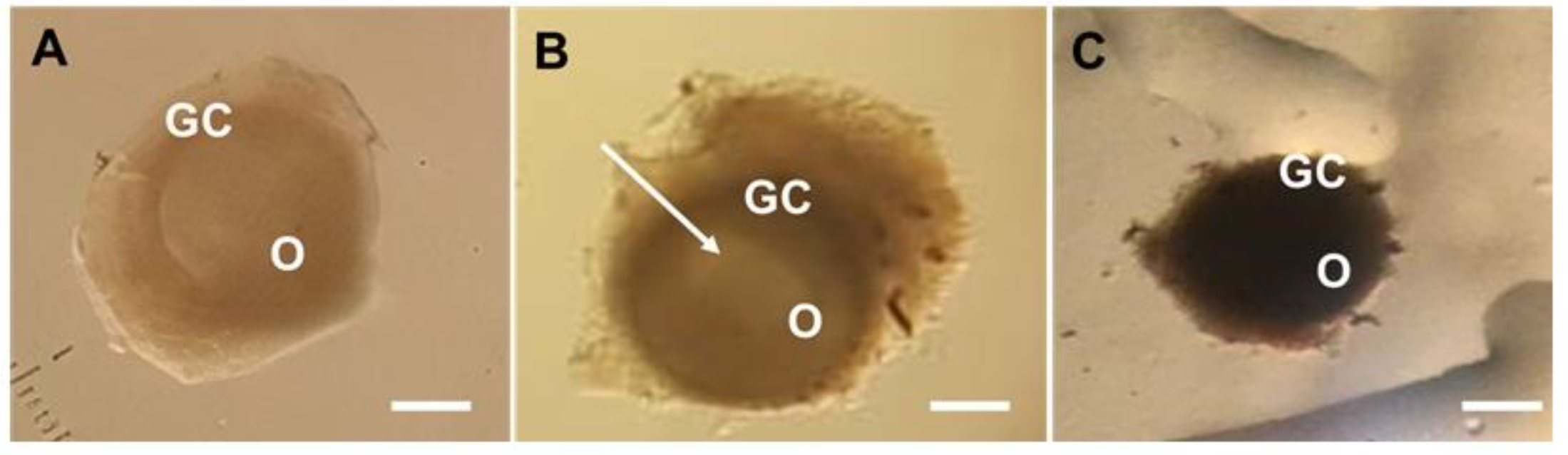
Follicular morphology was assessed throughout the culture period to verify the structural integrity of the follicles, a direct marker of their viability. The normal secondary follicles showed centrally located oocytes and granulosa cells with normal morphology (Figure 1A). At day 6 of the culture, antral (Figure 1B) and atretic (Figure 1C) follicles could be observed. At the end of the culture, the percentage of morphologically normal follicles did not differ (P >0.05) among the treatment groups (93.3%, 92.6%, 97.7%, and 92.6% for α-MEM+, 0.1, 0.2, and 0.4 mg/ml grape residue extract, respectively) (Figure 2).
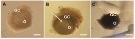
(A) Sheep secondary follicle with normal morphology at day 0 of culture. (B) antral follicle after 6 days of culture in 0.4 mg/ml acidified extract of industrial residue of grape and (C) atretic follicle after 6 days of culture in control medium. GC: granulosa cells; O: oocyte; Arrow: antral cavity; Scale bar: 100 μm (100x).
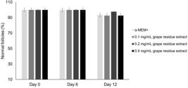
Percentages of morphologically normal follicles after culture of secondary follicles in control medium (α-MEM+) or in different concentrations of acidified extract of industrial residue of grape (0.1, 0.2 or 0.4 mg/ml). (a, b, c) Different letters indicate significant differences among culture periods in the same treatment group (P <0.05).
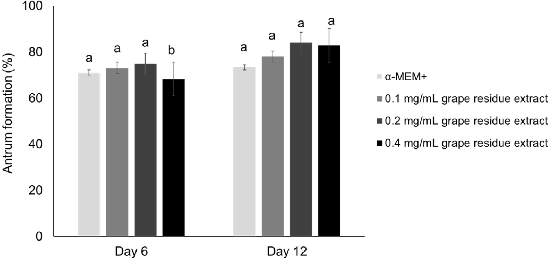
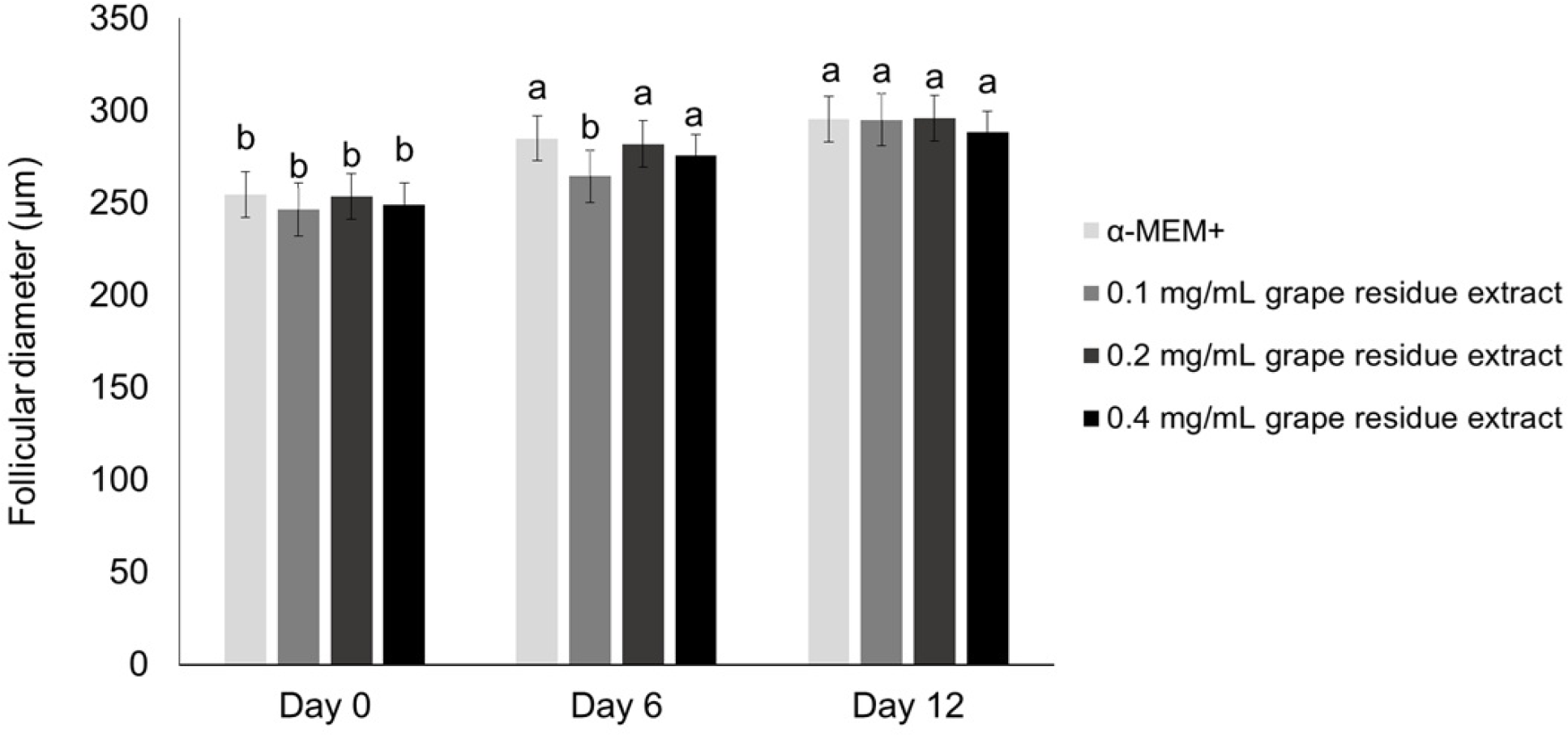
Follicular development was assessed through the formation of the antral cavity, follicular diameter, and growth rates, all morphological indicators representing the progression of follicles toward advanced stages of growth and differentiation across different follicular categories. After 6 days, all treatment groups showed a similar (P >0.05) percentage of antral cavity formation. However, the percentage of antrum formation increased significantly from day 6 to day 12 of culture only in the 0.4 mg/ml grape residue extract group (Figure 3). All treatments showed a significant increase in the follicular diameter from day 0 to day 12 of culture (Figure 4). However, at the end of the culture period, the antrum formation rate (Figure 3), follicular diameter (Figure 4), the daily growth rates (100%, 122%, 94.38%, and 91.54% for α-MEM+, 0.1, 0.2, and 0.4 mg/ml grape residue extract, respectively), general growth rates (100%, 119.19%, 96.68%, and 91.83% for α-MEM+, 0.1, 0.2, and 0.4 mg/ml grape residue extract, respectively) and the percentage of oocytes ≥ 110 μm (33.3%, 22.5%, 22.2%, and 21.9% for α-MEM+, 0.1, 0.2, and 0.4 mg/ml grape residue extract, respectively) were similar (P >0.05) among the treatments.

Percentages of antrum formation after culture of sheep secondary follicles in control medium (α-MEM +) or in different concentrations of acidified extract of industrial residue of grape (0.1, 0.2 or 0.4 mg/ml). (a, b) Different letters indicate significant differences among culture periods in the same treatment group (P<0.05).
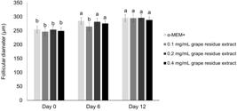
Follicular diameter (µm) after culture of sheep secondary follicles in control medium (α-MEM+) or in different concentrations of acidified extract of industrial residue of grape (0.1, 0.2 or 0.4 mg/ml). (a, b) Different letters indicate significant differences among culture periods in the same treatment group (P <0.05).
The GSH levels and mitochondrial activity after culture
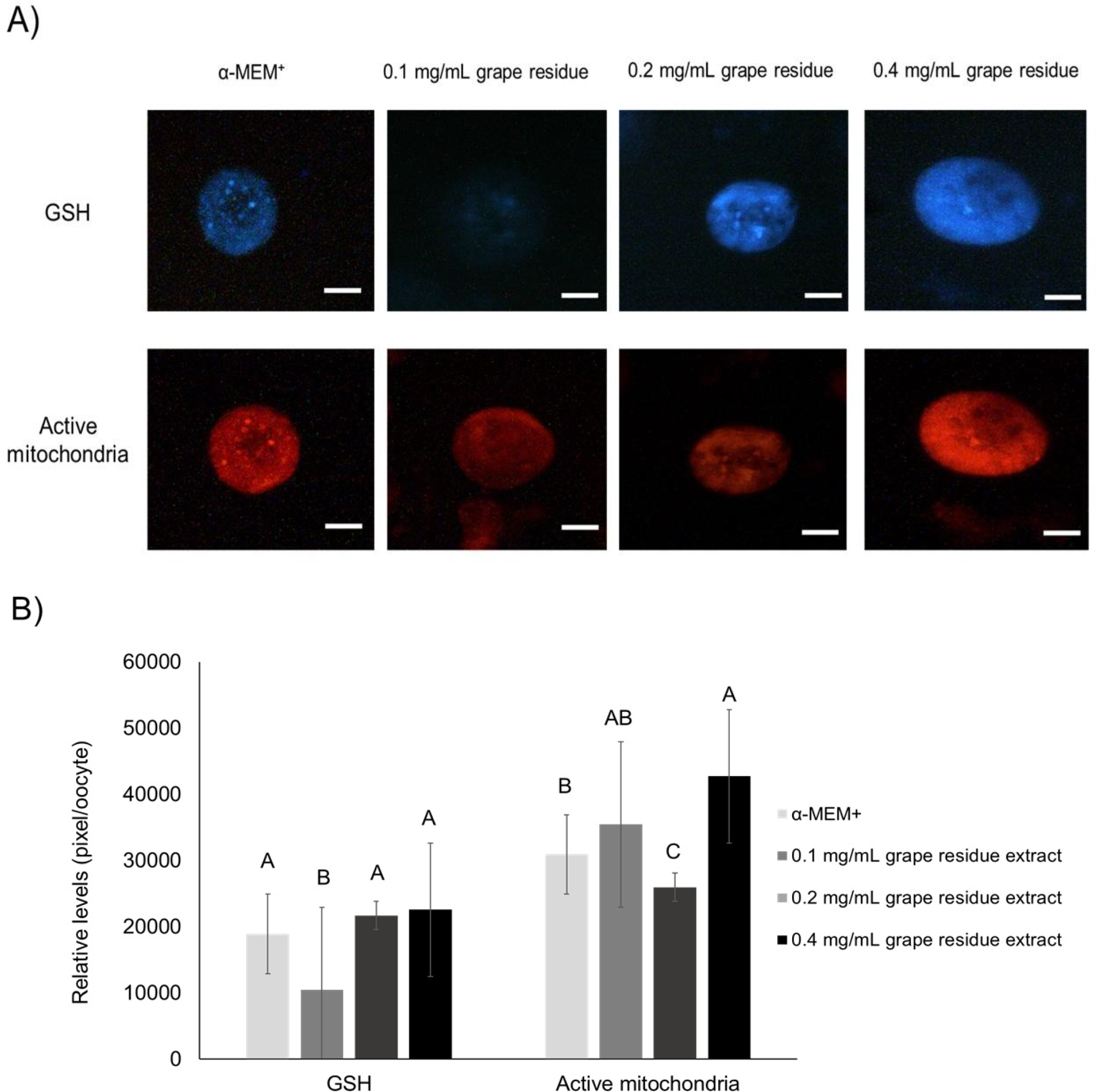
The metabolic health of oocytes was investigated through the assessment of their antioxidant defense, via glutathione (GSH) levels, and their bioenergetic capacity, via mitochondrial activity. After 12 days of culture, the GSH levels were similar (P >0.05) in the oocytes cultured with α-MEM+, 0.2, or 0.4 mg/ml grape residue extract. However, oocytes cultured with 0.4 mg/ml grape residue extract showed more (P <0.05) mitochondrial activity compared with the oocytes cultured with α-MEM+ or 0.2 mg/ml grape residue extract (Figure 5).

(A) Epifluorescent images of sheep oocytes stained with CellTracker Blue and MitoTracker Red to determine GSH concentrations and active mitochondria, respectively. Scale bars: 50 μm (100x). (B) Intracellular concentrations of GSH and active mitochondria in sheep oocytes from secondary follicles cultured in control medium (α-MEM+) or different concentrations of acidified extract of industrial residue of grape (0.1, 0.2 or 0.4 mg/ml). (A, B, C) Different letters indicate significant differences among treatments (P <0.05).
DNA fragmentation after culture
Nuclear integrity, an important indicator of oocyte quality and developmental competence, was evaluated by analyzing DNA fragmentation at the end of the culture period. All oocyte nuclei were stained by Hoechst 33342 (Figure 6). No DNA fragmentation was observed in either the control group or the 0.4 mg/ml grape residue extract group.
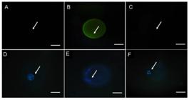
DNA fragmentation of sheep oocytes after in vitro culture. Normal oocyte cultured in medium containing 0.4 mg/ml grape residue extract (A and D), oocyte with DNA fragmentation in positive control (B and E), oocyte without DNA fragmentation in negative control (C and F). O: oocyte; Arrow: nuclear chromatin. Scale bars: 50 µm. Oocytes stained with TUNEL (A-C) and Hoechst 33342 (D-F).
Maturation of oocytes from in vitro-grown secondary follicles
Chromatin configuration analysis was performed to assess the stage of nuclear maturation achieved by oocytes after in vitro maturation, enabling the identification of effects of grape residue extract on meiotic progression. After IVM, the percentage of meiotic resumption was greater (P <0.05) in the 0.4 mg/ml grape residue extract group than in the α-MEM+ group (Table 1). No oocytes reached the MII stage.
Meiotic stages (%) after IVM of sheep oocytes from secondary follicles cultured in control medium (α-MEM+) or in medium containing 0.4 mg/mL grape residue.
Determination of total phenolic content (TPC) and antioxidant activity by scavenging the DPPH (2,2-diphenyl-1-picrylhydrazyl) radical
The antioxidant potential of the grape residue extract was evaluated by determining its total phenolic content (TPC) and radical scavenging activity using the DPPH assay, providing insight into its ability to counteract oxidative stress. TPC assay displayed values of 431.84, 10.41 and 15.55 GAE/g for pure grape residue extract, α-MEM+ and 0.4 mg/mL grape residue extract, respectively. In the DPPH analysis, the values obtained were 289.90 in 0.4 mg/mL grape residue extract and 1063.19 µM trolox/g for pure grape residue extract. No antioxidant activity was detected in α-MEM+, using the DPPH assay.
DISCUSSION
The present study is the first to evaluate the effects of acidified extract of industrial residue of grape (V. vinifera cv. Syrah) as a supplement of the medium used for the in vitro culture of ovarian follicles. After 12 days of culture, supplementation of the culture medium with 0.4 mg/ml grape residue extract had improved mitochondrial activity and oocyte meiotic resumption compared with the control medium (α-MEM+).
Mitochondria, the most abundant organelle in mammalian oocytes, play critical roles in cellular energetic metabolism and redox homeostasis; their functional integrity is essential for cellular survival and development [33,34]. This organelle is directly involved in several reproductive processes: It is crucial for the quality and functional competence of oocytes, fertilizing potential, embryo pre-implantation, and subsequent embryonic development [35-37]. Furthermore, mitochondrial dysfunction can cause excessive ROS production and induce oxidative stress and cell apoptosis, impairing the developmental competence of oocytes [38,39]. Similarly, to our results, increased mitochondrial activity has been observed in matured porcine oocytes and in endothelial cells cultured in vitro in medium supplemented with grape seed proanthocyanidin [40,41]. These beneficial effects are likely attributable to its phenolic compounds with antioxidant properties. These phenolic antioxidants contribute to improved mitochondrial activity by modulating cellular redox balance and limiting excessive reactive oxygen species (ROS) levels. Through their ability to donate electrons or hydrogen atoms and to stabilize unpaired electrons within their aromatic structures, phenolic compounds effectively neutralize free radicals and reduce oxidative damage to mitochondrial membranes. This attenuation of oxidative stress helps preserve mitochondrial integrity and functionality, thereby supporting mitochondrial metabolic activity and cellular homeostasis [62,63].
Chromatographic characterization associated with the use of mass spectrometry identified quercetin, luteolin, chrysoeriol, and isorhamnetin in the acidified extract of grape residue used in the present study (supplementary material). Furthermore, although there was no statistical difference the antioxidant capacity assessed by the TPC and DPPH methods in the medium that received 0.4 mg/ml of grape residue extract was higher compared to the control medium. These findings are in line with in vitro studies that have demonstrated that these compounds can improve the development of ovarian cells due to their antioxidant properties. In pigs, luteolin supplementation during IVM increased the rates of first polar body extrusion and blastocyst formation by reducing intracellular ROS and increasing the expression of genes that encode antioxidant enzymes (superoxide dismutase 1, superoxide dismutase 2, and catalase) [42]. In addition, IVM of goat oocytes with quercetin resulted in more MII oocytes by increasing mitochondrial activity [43]. Furthermore, quercetin, isorhamnetin, and luteolin increased sirtuin 1 (SIRT1) expression in different cells and tissues (muscle and brain: [44]; house ear institute-organ of Corti 1 cells: [45]). SIRT1 upregulates mitochondrial biogenesis through peroxisome proliferator-activated receptor γ coactivator 1-α [46] and modulates ROS production to promote resistance to oxidative stress [47].
It is noteworthy that most control oocytes remained at the GV stage. In contrast, treatment with grape residue extract increased meiotic resumption. It is likely that the quality of oocytes from sheep secondary follicles cultured with 0.4 mg/ml grape residue extract is related to the higher concentration of antioxidant components in its composition, which preserved or improved mitochondrial activity and, consequently, induced meiotic resumption. This is an important finding because in other studies, the use of antioxidant supplements during short-term culture (12 days) of sheep preantral follicles did not increase meiotic resumption compared with control medium (kaempferol: [48]; protocatechuic acid: [49]). However, it is important to note that no oocytes progressed to metaphase II (MII) stage, indicating that while the extract promotes initial maturation, it does not fully support the acquisition of developmental competence. The inability to reach MII suggests that the extract may be sufficient to overcome the initial block at the GV stage but lacks the necessary components, or fails to activate the specific signaling pathways, required for the complete nuclear and cytoplasmic maturation achieved with growth factors (IGF-1: [60]) or longer term hormonal supplementation (leptin: [51]; FSH: [50;60]). It is suggested that while antioxidants reduce oxidative stress, an important inhibitor of meiotic resumption, the complex cascade of events leading to MII, including adequate cytoplasmic maturation and cytoskeletal reorganization, depends on a broader set of signals. Therefore, our findings highlight the promising potential of grape residue extract as an additive to initiate maturation, but future studies should explore its combination with regulatory factors, such as growth factors and hormones, to support the oocyte through the final stages of meiotic maturation.
In this study, after 12 days of culture, oocytes treated with 0.4 mg/ml grape residue extract had similar percentages of survival and antrum formation, follicular and oocyte diameters, and GSH levels as oocytes cultured in α-MEM+. α-MEM has been routinely used as a base medium in the culture systems of preantral follicles; it is a rich source of electrolytes, non-essential amino acids, carbohydrates, vitamins, and DNA precursors, which help to maintain follicular viability and growth [48,52,53]. Furthermore, there was no DNA fragmentation in any treatment, which may be explained by the presence of antioxidants in the medium. Specifically, glutamine improved the DNA damage response in mouse neural cells [54], ascorbic acid reduced DNA fragmentation after culture of isolated rat secondary follicles [55], and supplementation of the IVM medium with selenium significantly reduced DNA damage in yak (Bos grunniens) cumulus cells [56]. Our findings are of significant importance because grape residue extract could be an alternative supplement to reduce the costs of the in vitro culture systems of ovarian follicles. Of note, an extended culture period may be necessary to induce significant changes in the viability and growth of secondary follicles conferred by grape residue extract. In addition, additional studies to evaluate whether higher grape residue extract concentrations could have greater effects on the in vitro follicle development are warranted.
Unexpectedly, supplementation with 0.1 or 0.2 mg/ml grape residue extract resulted in reduced GSH levels and mitochondrial activity, compared with α-MEM+, respectively. These results indicate a non-linear dose-response pattern, in which lower concentrations may be insufficient to maintain redox balance during in vitro culture. At these suboptimal doses, the antioxidant components of the extract may not reach the minimum level required for effective ROS neutralization, allowing oxidative stress to persist and impair mitochondrial function. Similar dose-dependent behavior has been described for other plant-derived antioxidants, where only specific concentrations are protective and lower doses produce inconsistent or adverse effects [58,59].
While this study provides promising evidence for the use of grape residue extract, some aspects merit consideration for future translation. The findings are based on a specific extract batch from a single cultivar and region. Although thoroughly characterized, natural variation between batches could influence outcomes. Future studies with standardized extracts and direct comparisons to pure antioxidants will be valuable to consolidate these initial findings and ensure reproducibility. Furthermore, the evaluated parameters are well-established indicators of oocyte health but represent intermediate endpoints, and their relationship with subsequent embryonic development was not directly assessed in the present study.
CONCLUSION
The treatment with 0.4 mg/ml grape residue extract can be used as a supplement of the base medium during the culture of sheep secondary follicles; it maintains follicular viability, increases mitochondrial activity, and oocyte meiotic resumption, and showed greater antioxidant capacity. Grape residue extract is inexpensive, has high biological value, and emerges as a promising and potentially cost-effective supplement for in vitro preantral follicle development. Furthermore, our results indicate that grape residue extract could be tested as a feed supplement to improve reproductive performance in sheep.
-
Funding:
I.M. Mota received a scholarship from the Fundação de Amparo à Ciência e Tecnologia do Estado de Pernambuco (FACEPE, Brazil, nº BIC-1560-5.05/20). M. H. T. Matos is supported by a grant from the Conselho Nacional de Desenvolvimento Científico e Tecnológico (CNPq, Brazil, nº 317582/2021-6).
-
Institutional Review Board Statement:
This study did not require ethical approval as it involved biological materials obtained from a slaughterhouse, and no live animals were used or subjected to experimental procedures for research purposes.
-
Informed Consent Statement:
Not applicable.
Acknowledgments:
The authors gratefully acknowledge the Regional Slaughterhouse of Petrolina (Pernambuco, Brazil) for providing the biological material (ovaries) used in this study.
Use of Generative Artificial Intelligence:
The authors declare that large language models and other generative artificial intelligence (AI) or AI-assisted technologies cannot be credited as authors and have not been listed as authors of this paper.
The author declare that did not use the artificial intelligence.
Data Availability Statement:
Research data are available in the body of the manuscript.
REFERENCES
- 1 Taghizabet N, Bahmanpour S, Zarei-Fard N, Rezaei-Tazangi F, Hassanpour A, Nejad EK, et al. In vitro growth of the ovarian follicle: taking stock of advances in research. J Bras Reprod Assist. 2022; 26(3):508-21.
- 2 Paulino LRFM, Assis EIT, Azevedo VAN, Silva BR, Cunha EV, Silva JRV. Why Is It So Difficult To Have Competent Oocytes from In vitro Cultured Preantral Follicles? Reprod Sci. 2022; 29(12):3321-34.
- 3 Agarwal A, Rosas IM, Anagnostopoulou C, Cannarella R, Boitrelle F, Munoz LV, et al. Oxidative Stress and Assisted Reproduction: A Comprehensive Review of Its Pathophysiological Role and Strategies for Optimizing Embryo Culture Environment. Antioxidants. 2022; 11(3):477.
- 4 Silva E, Almeida H, Castro JP. (In)Fertility and Oxidative Stress: New Insights into Novel Redox Mechanisms Controlling Fundamental Reproductive Processes. Oxid Med Cell Longev. 2020;2020:4674896.
- 5 Gouveia BB, Barros VRP, Gonçalves RJS, Barberino RS, Menezes VG, Lins TLB, et al. Effect of ovarian tissue transportation in Amburana cearensis extract on the morphology and apoptosis of goat preantral follicles. Anim Reprod 2015;12(2):316-23.
- 6 Barberino RS, Barros VRP, Menezes VG, Santos LP, Araújo VR, Queiroz MAA, et al. Amburana cearensis leaf extract maintains survival and promotes in vitro development of ovine secondary follicles. Zygote. 2015; 24(2):277-85.
- 7 Cavalcante AYP, Lins TLBG, Santos JMS, Barros VRP, Monte APO, Barberino RS, et al. Supplemented Morus nigra extract-based medium associated with FSH enables the survival and growth of isolated ovine secondary ovarian follicles. Reprod Domest Anim. 2018; 53(2):423-32.
- 8 Mbemya GT, Cadenas J, Sá NAR, Guerreiro DD, Donfack NJ, Vieira LA, et al. Supplementation of in vitro culture medium with FSH to grow follicles and mature oocytes can be replaced by extracts of Justicia insularis. PLoS One. 2018;13(12):e0208760.
- 9 Eyduran SP, Akin M, Ercisli S, Eyduran E, Maghradze D. Sugars, organic acids, and phenolic compounds of ancient grape cultivars (Vitis Vinifera L.) from igdir province of eastern Turkey. Biol Res. 2015; 48:2
- 10 Boso S, Gago P, Santiago JL, Teira-Brión A, Martín-Seijo M, Rey-Castiñeira J, et al. Morphometric comparison of current, Roman-era and medieval Vitis seeds from the north-west of Spain. Aust J Grape Wine Res. 2020; 26:300-8.
- 11 Sousa EC, Uchôa-Thomaz AMA, Carioca JOB, Morais SM, Lima A, Martins CG, et al. Chemical composition and bioactive compounds of grape pomace (Vitis vinifera L.), Benitaka variety, grown in the semiarid region of Northeast Brazil. Food Sci Technol. 2014;34(1):135-42.
- 12 Liu X, Lin X, Mi Y, Li J, Zhang C. Grape Seed Proanthocyanidin Extract Prevents Ovarian Aging by Inhibiting Oxidative Stress in the Hens. Oxid Med Cell Longev. 2018; 2018:9390810.
- 13 Martin ME, Grao-Cruces E, Millan-Linares MC, Montserrat-De la Paz S. Grape (vitis vinifera l.) seed oil: A functional food from the winemaking industry. Foods. 2020; 9(10):1360.
- 14 Labanca F, Faraone I, Nolè MR, Hornedo-Ortega R, Russo D, García-Parrilla MC, et al. New insights into the exploitation of vitis vinifera l. Cv. aglianico leaf extracts for nutraceutical purposes. Antioxidants. 2020; 9(8):708.
- 15 Mokni M, Amri M, Limam F, Aouani E. Effect of grape seed and skin supplement on milk yield and composition of dairy ewes. Trop Anim Health Prod. 2017; 49(1):131-7.
- 16 Alba DF, Campigotto G, Cazarotto CJ, Santos DS, Gebert RR, Reis JH, et al. Use of grape residue flour in lactating dairy sheep in heat stress: Effects on health, milk production and quality. J Therm Biol. 2019; 82(2019):197-205.
- 17 Padilha CVS, Biasoto ACT, Corrêa LC, Lima MS, Pereira GE. Phenolic compounds profile and antioxidant activity of commercial tropical red wines (Vitis vinifera L.) from São Francisco Valley, Brazil. J Food Biochem. 2017;41:e12346.
- 18 Ky I, Lorrain B, Kolbas N, Crozier A, Teissedre PL. Wine by-Products: Phenolic characterization and antioxidant activity evaluation of grapes and grape pomaces from six different French grape varieties. Molecules. 2014; 19(1):482-506.
- 19 Tabeshpour J, Mehri S, Behbahani FS, Hosseinzadeh H. Protective effects of Vitis vinifera (grapes) and one of its biologically active constituents, resveratrol, against natural and chemical toxicities: A comprehensive review. Phytother Res. 2018; 32(11):2164-90.
- 20 Pascoal GFL, Novaes GM, Sobrinho MP, Hirayama AB, Castro IA, Ong TP. Selenium Supplementation during Puberty and Young Adulthood Mitigates Obesity-Induced Metabolic, Cellular and Epigenetic Alterations in Male Rat Physiology. Antioxidants. 2022; 11(5):895.
- 21 Bezerra MÉS, Gouveia BB, Barberino RS, Menezes VG, Macedo TJS, Cavalcante AYP, et al. Resveratrol promotes in vitro activation of ovine primordial follicles by reducing DNA damage and enhancing granulosa cell proliferation via phosphatidylinositol 3-kinase pathway. Reprod Domest Anim. 2018; 53(6):1298-1305.
- 22 Lins TLBG, Barberino RS, Monte APO, Pinto JGC, Campinho DSP, Palheta RC, et al. Rutin promotes activation and reduces apoptosis of primordial follicles by regulating Akt phosphorylation after in vitro culture of ovine ovarian tissue. Theriogenology. 2021; 173(2021):64-72.
- 23 Macedo TJS, Barros VRP, Monte APO, Gouveia BB, Bezerra MES, Cavalcante AYP, et al. Resveratrol has dose-dependent effects on DNA fragmentation and mitochondrial activity of ovine secondary follicles cultured in vitro. Zygote. 2017;25(4):434-42.
- 24 Lins TLBG, Cavalcante AYP, Santos JMS, Menezes VG, Barros VRP, Barberino RS, et al. Rutin can replace the use of three other antioxidants in the culture medium, maintaining the viability of sheep isolated secondary follicles. Theriogenology. 2017; 89(2017):263-70.
- 25 Silva GAL, Araújo LB, Silva LCR, Gouveia BB, Barberino RS, Lins TLBG, et al. Gallic acid promotes the in vitro development of sheep secondary isolated follicles involving the phosphatidylinositol 3-kinase pathway. Anim Reprod Sci. 2021; 230(2021):106767.
- 26 Liu MJ, Sun AG, Zhao SG, Liu H, Ma SY, Li M, et al. Resveratrol improves in vitro maturation of oocytes in aged mice and humans. Fertil Steril. 2018;109(5):900-7.
- 27 Gao W, Jin Y, Hao J, Huang S, Wang D, Quan F, et al. Procyanidin B1 promotes in vitro maturation of pig oocytes by reducing oxidative stress. Mol Reprod Dev. 2021; 88(1):55-66.
- 28 Karimian M, Zandi M, Sanjabi MR, Masoumian M, Ofoghi H. Effects of grape seed extract, quercetin and vitamin C on ovine oocyte maturation and subsequent embryonic development. Cell Mol Biol (Noisy-le-grand) 2018 31;64(4):98-102.
- 29 Gouveia BB, Barberino RS, Menezes VG, Macedo TJS, Cavalcante AYP, Gonçalves RJS, et al. DNA damage and primordial follicle activation after in vitro culture of sheep ovarian cortex in Morus nigra leaf extract. Pesq Vet Bras. 2019;39(1):85-92.
- 30 Cecconi S, Barboni B, Coccia M, Mattioli M. In Vitro Development of Sheep Preantral Follicles. Biol Reprod. 1999; 60(3):594-601.
- 31 Singleton VL, Orthofer R, Lamuela-Raventós RM. Analysis of Total Phenols and Other Oxidation Substrates and Antioxidants by Means of Folin-Ciocalteu Reagent. Meth Enzymol. 1999; 299:152-78.
- 32 Brand-Williams W, Cuvelier ME, Berset C. Use of a free radical method to evaluate antioxidant activity. Food Sci Technol. 1995; 28(1):25-30.
- 33 Chiang JL, Shukla P, Pagidas K, Ahmed NS, Karri S, Gunn DD, et al. Mitochondria in ovarian aging and reproductive longevity. Ageing Res Rev. 2020; 63:101168.
- 34 Rodríguez-Varela C, Herraiz S, Labarta E. Mitochondrial enrichment in infertile patients: a review of different mitochondrial replacement therapies. Ther Adv Reprod Health. 2021; 15:26334941211023544.
- 35 Castaneda CA, Kaye P, Pantaleon M, Phillips N, Fry R, D’Occhio MJ. Circulating concentrations of leptin, ovarian follicle number, and oocyte lipid content and active mitochondria in Zebu crossbred cows maintained on standard or improved nutrition. Anim Reprod Sci. 2013; 140(1-2):7-13.
- 36 Reader KL, Stanton JAL, Juengel JL. The role of oocyte organelles in determining developmental competence. Biology (Basel). 2017; 6(3):35.
- 37 Gualtieri R, Kalthur G, Barbato V, Di Nardo M, Adiga SK, Talevi R. Mitochondrial dysfunction and oxidative stress caused by cryopreservation in reproductive cells. Antioxidants (Basel). 2021; 10(3):337.
- 38 Niu D, Chen KL, Wang Y, Li XQ, Liu L, Ma X, et al. Hexestrol deteriorates oocyte quality via perturbation of mitochondrial dynamics and function. Front Cell Dev Biol. 2021; 9:685102.
- 39 Richani D, Dunning KR, Thompson JG, Gilchrist RB. Metabolic co-dependence of the oocyte and cumulus cells: essential role in determining oocyte developmental competence. Hum Reprod Update. 2021;27(1):27-47.
- 40 Cerbaro AF, Rodrigues VSB, Rigotti M, Branco CS, Rech G, Oliveira DL, et al. Grape seed proanthocyanidins improve mitochondrial function and reduce oxidative stress through an increase in sirtuin 3 expression in EA.hy926 cells in high glucose condition. Mol Biol Rep. 2020; 47:3319-30.
- 41 Li W, He Y, Zhao H, Peng L, Li J, Rui R, et al. Grape seed proanthocyanidin ameliorates FB1-induced meiotic defects in porcine oocytes. Toxins (Basel). 2021; 13(12):841.
- 42 Park SH, Jeong PS, Joo YE, Kang HG, Kim MJ, Lee S, et al. Luteolin orchestrates porcine oocyte meiotic progression by maintaining organelle dynamics under oxidative stress. Front Cell Dev Biol. 2021; 9:687147.
- 43 Silva AAA, Silva MNP, Figueiredo LBF, Gonçalves JD, Silva MJS, Loiola MLG, et al. Quercetin influences in vitro maturation, apoptosis and metabolically active mitochondria of goat oocytes. Zygote. 2018; 26(6):465-70.
- 44 Davis JM, Murphy EA, Carmichael MD, Davis B. Quercetin increases brain and muscle mitochondrial biogenesis and exercise tolerance. Am J Physiol Regul Integr Comp Physiol. 2009;296(4):R1071-7.
- 45 Zhu RZ, Li BS, Gao SS, Seo JH, Choi BM. Luteolin inhibits H2O2-induced cellular senescence via modulation of SIRT1 and p53. Korean J Physiol Pharmacol. 2021;25(4):297-305.
- 46 Scarpulla RC. Metabolic control of mitochondrial biogenesis through the PGC-1 family regulatory network. Biochim Biophys Acta Mol Cell Res. 2011;1813(7):1269-78.
- 47 Salminen A, Kaarniranta K, Kauppinen A. Crosstalk between oxidative stress and SIRT1: impact on the aging process. Int J Mol Sci. 2013; 14(2):3834-59.
- 48 Santos JMS, Monte APO, Lins TLBG, Barberino RS, Menezes VG, Gouveia BB, et al. Kaempferol can be used as the single antioxidant in the in vitro culture medium, stimulating sheep secondary follicle development through the phosphatidylinositol 3-kinase signaling pathway. Theriogenology. 2019; 136:86-94.
- 49 Menezes VG, Santos JMS, Macedo TJS, Lins TLBG, Barberino RS, Gouveia BB, et al. Use of protocatechuic acid as the sole antioxidant in the base medium for in vitro culture of ovine isolated secondary follicles. Reprod Domest Anim. 2017; 52(5):890-8.
- 50 Barros VRP, Monte APO, Lins TLBG, Santos JM, Menezes VG, Cavalcante AYP, et al. In vitro survival, growth and maturation of sheep oocytes from secondary follicles cultured in serum-free conditions: impact of a constant or a sequential medium containing recombinant human FSH. Domest Anim Endocrinol. 2019; 67:71-9.
- 51 Macedo TJS, Santos JMS, Bezerra MÉS, Menezes VG, Gouveia BB, Barbosa LMR, et al. Immunolocalization of leptin and its receptor in the sheep ovary and in vitro effect of leptin on follicular development and oocyte maturation. Mol Cell Endocrinol. 2019; 495:110506.
- 52 Ferreira ACA, Cadenas J, Sá NAR, Correia HHV, Guerreiro DD, Lobo CH, et al. In vitro culture of isolated preantral and antral follicles of goats using human recombinant FSH: concentration-dependent and stage-specific effect. Anim Reprod Sci. 2018; 196:120-9.
- 53 Jachter SL, Simmons WP, Estill C, Xu J, Bishop CV. Matrix-free three-dimensional culture of bovine secondary follicles to antral stage: impact of media formulation and epidermal growth factor (EGF). Theriogenology. 2022; 181:89-94.
- 54 Chen J, Herrup K. Glutamine acts as a neuroprotectant against DNA damage, beta-amyloid and H2O2-induced stress. PLoS One. 2012;7(3):e33177.
- 55 Murray AA, Molinek MD, Baker SJ, Kojima FN, Smith MF, Hillier SG, et al. Role of ascorbic acid in promoting follicle integrity and survival in intact mouse ovarian follicles in vitro. Reproduction. 2001; 121(1):89-96.
- 56 Xiong X, Lan D, Li J, Lin Y, Li M. Selenium supplementation during in vitro maturation enhances meiosis and developmental capacity of yak oocytes. Anim Sci J. 2018; 89(2):298-306.
- 57 Molosse VL, Deolindo GL, Cécere BG, Marcon H, da Rosa G, Vedovatto M, et al. Effect of dietary supplementation with grape residue flour on weight gain, metabolic profile, leukogram, proteinogram and antioxidant response in suckling lambs. Res Vet Sci. 2021; 139:112-20.
- 58 Nowak M, Tryniszewski W, Sarniak A, Wlodarczyk A, Nowak PJ, Nowak D. Concentration Dependence of Antiand Pro-Oxidant Activity of Polyphenols as Evaluated with a Light-Emitting Fe2+-Egta-H2O2 System. Molecules. 2022; 27(11):3453.
- 59 Plauth A, Geikowski A, Cichon S, Wowro SJ, Liedgens L, Rousseau M, et al. Hormetic shifting of redox environment by pro-oxidative resveratrol protects cells against stress. Free Radic Biol Med. 2016; 99:608-22.
- 60 Monte APO, Barros VRP, Santos JM, Menezes VG, Cavalcante AYP, Gouveia BB, et al. Immunohistochemical localization of insulin-like growth factor-1 (IGF-1) in the sheep ovary and the synergistic effect of IGF-1 and FSH on follicular development in vitro and LH receptor immunostaining. Theriogenology. 2019; 129:61-9.
- 61 Guimarães VS, Silva RLS, Barberino RS, Mota IM, Pinto JGC, Torres MLGL, et al. Effect of the acidified extract of Moringa oleifera leaves as a supplement in the in vitro culture medium of sheep preantral follicles. Semina Cienc Agrar. 2023; 44(6):1991-2010.
- 62 Blokhina O, Virolainen E, Fagerstedt KV. Antioxidants, oxidative damage and oxygen deprivation stress: a review. Ann Bot. 2003; 91(2):179-94.
- 63 He L, He T, Farrar S, Ji L, Liu T, Ma X, et al. Antioxidants maintain cellular redox homeostasis by elimination of reactive oxygen species. Cell Physiol Biochem. 2017; 44(2):532-53.
-
Editor-in-Chief:
Bill Jorge Costa
-
Associate Editor:
Jane Manfron