 Photorealistic 3D Facial Meshes Obtained by Low-Cost Photogrammetry: Are They Precise Enough to Replace Colorless CT-scan Meshes in Virtual Surgical Planning?
Abstract
In Virtual Surgical Planning (VSP) for orthognathic procedures, 3D facial meshes are essential for soft tissue analysis and surgical simulation. Meshes obtained via CT segmentation are the standard, but they have the limitation of not incorporating skin texture. Facial 3D meshes obtained using optical devices overcome this limitation by incorporating skin texture, replacing texture-less CT-scan meshes with photorealistic and more patient-friendly 3D models. Since the high cost of these devices discourages widespread adoption, smartphone-based photogrammetry emerges as an accessible and eligible source of lifelike 3D images. However, photogrammetry accuracy in reproducing morphology similarly to CT-Scans may be a concern. This study evaluated the morphological accuracy of photogrammetry-derived texturized meshes to serve as replacement to texture-less CT-scan meshes for VSP. Photogrammetry meshes were generated from 40 facial photographs captured with a conventional smartphone and processed using proprietary (3DZephyr) and open-source (OrtogOnBlender) software. CloudCompare software was used to calculate surface-to-surface deviations of these meshes against the reference CT-based counterpart, determining tridimensional similarity. Individual and mean heatmaps were obtained. Statistical analyses were performed using the paired Wilcoxon test, with a significance level of 5%. Twenty-seven participants (14 males and 13 females) were included. Meshes processed by both software showed highly reliable accuracy, with mean RMSE (“3D error”) of 0.6734mm (±0.1274) for 3DZephyr and 0.6747mm (±0.1210) for OrtogOnBlender, with no statistically significant difference between them (p = 0.7366). These results support smartphone-based photogrammetry as a reliable low-cost source of 3D images, not only for VSP, but also for other specialties demanding precise tridimensional models.
Keywords:
Photogrammetry; Three-dimensional imaging; Orthognathic surgery; Open-source software; Data accuracy.
HIGHLIGHTS
Photogrammetry with photos captured by a smartphone is a low-cost modality of facial scanning.
We investigated if smartphone photogrammetry is accurate to replace colorless CT-scan meshes.
This photogrammetry provides lifelike facial meshes whose similar morphology can replace CT-scan.
Open-source photogrammetry algorithms perform as accurately as proprietary alternatives.
INTRODUCTION
Since the introduction of the concept of virtual surgical planning (VSP) and virtual osteotomies [1], surgical simulation has evolved to specialized programs that use mathematical models to predict the effects of changing parameters in hard and soft tissues [2]. This VSP process culminates in a tridimensional visualization of the treatment objectives (VTO), along with the tridimensional modeling and printing of surgical splints (or guides), which transfer to the surgical room what has been planned in the virtual environment [3].
The essential imaging set for VSP in orthognathic surgery includes helical or cone-beam facial computed tomography (CT) to obtain three-dimensional (3D) images of soft tissues and bony skeleton, and 3D images of the patient’s dentition [3]. Facial soft tissue meshes obtained by segmentation of a CT-scan reproduce precisely the patient’s anatomy. Besides, they are obviously acquired together with bony images for virtual osteotomies, making them widely used for accurate facial analysis and VTO. However, those images lack details on skin color and surface texture, which are important for a fully realistic virtual model.
This limitation of using a facial model without real details (such as color or nevus) can be resolved by adding a texturized 3D facial mesh to the VSP imaging set, which brings patient-friendly photorealistic images to the virtual environment.
In this scenario, the texture-less 3D mesh of soft tissues obtained from CT segmentation could be replaced by its texturized counterpart generated by different optical scanning devices, such as structured light scanning, laser scanning, stereophotogrammetry and photogrammetry [4].
While structured light scanning, laser scanning and stereophotogrammetry require specialized and often expensive equipment [4], monoscopic photogrammetry consists in capturing multiple two-dimensional (2D) photos with a conventional camera [5,6] and processing them with free-to-use or proprietary software to generate a 3D mesh [6,7].
Monoscopic photogrammetry shares with the previously mentioned optical devices the capability to provide tridimensional photorealistic images, but offers the distinct advantage of being feasible with relatively low-cost and widespread technology: a smartphone [6]. Besides, it doesn´t demand trained personnel to be perform, as demonstrated by previous publications [5]. Photo acquisition protocols are available in the literature [7], demonstrating the feasibility of the process.
Differently from CT-scan, though alike the other modalities, facial photogrammetry has the ability to capture the patient’s entire face in 3D without radiation exposure. Therefore, from the clinical point of view, they also allow the assessment of facial changes in growing children, analysis of facial changes in patients with pathologies, evaluation of asymmetries, besides the study of soft tissue in orthognathic surgery patients [8]. Regarding this latter advantage, the photorealistic image improves communication between professional and patients in the context of virtual surgical planning [9].
However, the central question of this study is whether a texturized facial mesh obtained using affordable monoscopic photogrammetry is morphologically accurate enough to replace a CT-based mesh in virtual surgical planning without loss of diagnostic information.
The importance of answering this question stems from the possibility of providing clinicians with an accessible and reliable tool to improve the quality and communicability of their virtual surgical planning, regardless of their economic reality. Besides, it may reinforce de applicability of photogrammetry as a tool to obtain reliable 3D morphological data which may be used by different medical specialties, such as orthopedics, dermatology and maxillofacial prosthodontics.
Thus, the objective of this study was to validate the morphological accuracy of low-cost photogrammetry-derived 3D facial meshes in comparison with CT-scan meshes, assessing their suitability as a replacement for CT-derived soft tissue models in virtual surgical planning and related clinical applications.
MATERIAL AND METHODS
This project was approved by the institutional ethics review board of Faculdade São Leopoldo Mandic (num.79377624.9.0000.5374). Informed consent was obtained from all participants.
Photogrammetry meshes were analyzed against CT-scan meshes using software capable of quantifying surface-to-surface deviations through a cloud point analysis approach, which computes the “3D error” between the point clouds of the two meshes. Different studies have applied a similar methodology [5, 10, 11]. To further assess affordability, open-source and proprietary photogrammetry software programs were used to generate two separate photogrammetry models.
This study included patients over 18 years who underwent VSP at the Armed Forces Hospital (Brasília, Brazil) between 2023 and 2024. The eligible patients had undergone facial helical CT and monoscopic photogrammetry within 7 days of each other.
Figure 1 presents the pipeline of this methodology, which is detailed in the following sections.

Data acquisition
All patients in this study underwent helical CT-scan encompassing the entire face, besides a multiple facial photography section.
CT-scan acquisition is essential for performing virtual surgical planning, as the bony mesh obtained by its segmentation forms the basis for virtual osteotomies. Consequently, all patients enrolled in this study underwent a CT-scan. The DICOM (Digital Imaging and Communications in Medicine) files resulting from the CT-scan were used to generate the reference 3D model in this study in the same way it was generated for facial diagnosis and simulation in the context of orthognathic surgery preparation.
Multiple facial photo acquisition, in turn, is necessary for the obtention of a texturized 3D model by photogrammetry, since this modality relies on the processing of multiple 2D images by a specific algorithm. Therefore, in order to generate texturized low-cost tridimensional facial images for this study, a total of 40 photos (20 per height) were captured with a Samsung Note 10 smartphone (2020) at 1908 × 4032 resolution, using the standard camera app available in the Android operational system.This 40 photo-protocol was based on Moraes and coauthors [7], who recommended 26 photos at two heights, and Nightingale and coauthors [5], who found that 40 photos are sufficient for monoscopic photogrammetry. The 40 photos were evenly distributed around the patient’s face (20 photos × 2 heights). For the first height, the smartphone camera was positioned at the same level as the subject’s eyes, while for the second height, the vertical position was right below the chin, to capture the submental area. No filtering or editing were applied to any photo. For both CT-scans and photo acquisition, the subjects were instructed to maintain a relaxed and neutral expression.
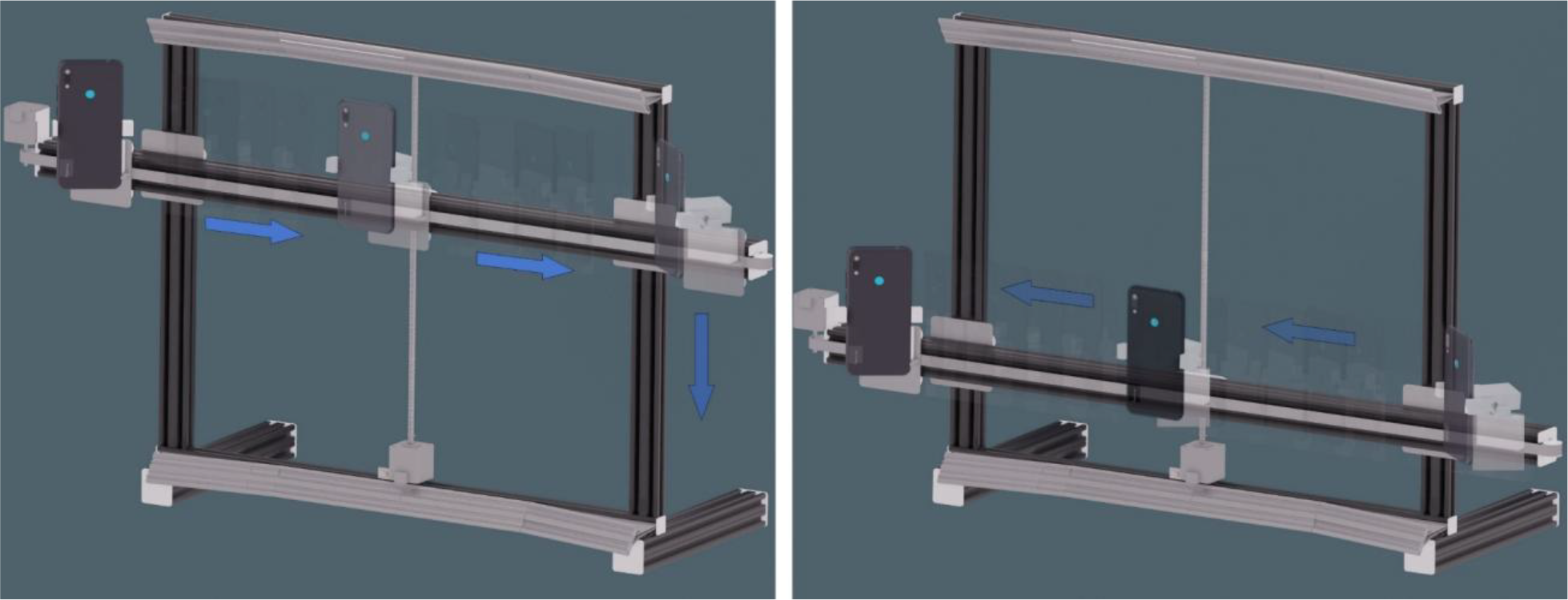
Photo acquisition was facilitated by a low-cost electromechanical device (patent BR 20 2023 010972 7), whose frame project and microcontroller code are shared for noncommercial use [12]. The device features motors, vertical and horizontal rails for precise linear and rotational movements of a smartphone that is attached to it (Figure 2), dimmable LED lighting, and a Bluetooth trigger to control the smartphone. Once the patient was positioned in front of the device, the attached and paired smartphone was moved through various angles at two heights, capturing photos of both sides of the face. Dimmable LED lighting ensured diffuse illumination, enhancing surface detail for photogrammetry. The automated photo capture took 32 seconds for each subject. A video illustrating the process is available at the project repository [12].

Electromechanical device for photogrammetry photo acquisition, illustrating the 40 different positions assumed by the smartphone. (a) Representation of the first height of scanning process; (b) Representation of the second height of the scanning process. While the smartphone slides laterally, the subject remains focused.
Mesh generation
For each subject in this study, three tridimensional facial models were generated:
-
1. M1: CT-scan mesh - This reference mesh was generated from CT segmentation using the Dolphin Imaging software (Dolphin & Management Solution, Chatsworth, CA, USA), which is designed and widely used for VSP. The resulting mesh was exported in the STL format (M1_original.stl). It must be emphasized that segmenting a CT-scan into a 3D mesh is the standard option to obtain soft tissue model for VSP [3], which explain the decision to use this mesh as the reference model for cloud point analysis.
-
2. M2P: proprietary photogrammetry software mesh - This model was generated from the 40 photos made with the Samsung Note 10 Smartphone. They were processed with the proprietary photogrammetry algorithm from the 3DZephyr software (3Dflow s.r.l., Verona, Italy), which has been used by different authors [13, 14]. The resulting mesh was exported as an OBJ file M2P_original.obj.
-
3. M2OS: open-source photogrammetry mesh - This mesh was created from the same 40 smartphone photographs, but processed using the open-source OpenMVG+OpenMVS algorithms within OrtogOnBlender, as performed by other authors [15, 16]. OrtogOnBlender is an open-source add-on that gives the software Blender3D (Blender Foundation, Amsterdam, the Netherlands) tools to perform VSP [17]. For the purpose of this article, the terms Blender and OrtogOnBlender are used interchangeably.
The .stl file from Dolphin (M1_original.stl) and the .obj file from 3DZephyr (M2P_original.obj) were imported into the same Blender environment where the open-source photogrammetry mesh was generated. From this point, the mesh preparation process proceeded with all the three subject´s models in the same Blender virtual environment, as depicted in Figure 1 - mesh.
Mesh preparation
The OrtogOnBlender tools were used for dimensioning, aligning, cutting, and exporting the final meshes for cloud point analysis. As for any photogrammetry, proprietary and open-source photogrammetry meshes were generated without specific spatial position or scale information. They were rescaled, marking the intercanthal points and inputting the intercanthal distance, which was measured directly from the patient’s face with a caliper. Alignment to the CT-scan mesh was done using 3-point alignment, followed by refinement with ICP (iterative closest point) algorithm, available in OrtogOnBlender. Fine alignment ensures the highest level of model superimposition, which is essential for the surface-to-surface analysis performed in the following section of the methodology.
After alignment, the three subject´s models were cropped to exclude irrelevant regions using a c-shaped uniform cleavage plane to separate the hair and ears from the facial areas. The same c-shaped cleavage plane was applied to every mesh of this study, with specific positional adjustments to ensure proper exclusion of areas not of interest while standardizing the resulting comparison meshes across all subjects. Additionally, the shaded area in the nostrils was excluded, along with the eyebrows and eyelids, because hair (including eyelashes) could also introduce errors (Figure 3).

Depiction of a subject’s resulting meshes from each software program: (a) M1 - CT-scan texture-less mesh; (b) M2P - proprietary photogrammetry software mesh; (c) M2OS - open-source photogrammetry mesh; (d) The three previous imagens cut and superimposed, ready for exportation as a STL file. As spatial position is saved and kept in the STL file, models remained superimposed when imported into CloudCompare software.
The cropped version of the models was exported in the STL format, generating the M1.stl, M2P.stl, and M2SO.stl files for each subject, respectively corresponding to CT-scan, 3DZephyr and OrtogOnBlender prepared models.
The cropped version of the models was exported in the STL format, generating the M1.stl, M2P.stl, and M2SO.stl files for each subject, respectively corresponding to CT-scan, 3DZephyr and OrtogOnBlender prepared models.
Mesh analysis
Photogrammetry meshes of a given subject were analyzed only against his/her own CT-scan mesh, since intersubject cloud point analysis would bring no meaningful data related to the precision of photogrammetry.
As CT-scan mesh (M1.stl) is the reference model against which photogrammetry meshes are tested, two comparisons were run for each participant:
-
1. 3DZephyr model (M2P.stl) versus CT-scan model (M1.stl)
-
2. OrtogOnBlender model (M2SO.stl) versus CT-scan model (M1.stl)
Both abovementioned analyses were performed separately by importing the pair of meshes into CloudCompare software. Once the pair of meshes were in CloudCompare environment, the following data were obtained:
-
• Mean distance: the average difference between corresponding points, providing a general measure of discrepancy;
-
• Standard deviation: indicates the dispersion of the differences relative to the mean. A higher standard deviation suggests greater variability;
-
• Root mean square error (RMSE): the square root of the average of squared differences, a measure for evaluating discrepancy magnitude, also referred to as “3D error.”
The data were analyzed to test the hypothesis that meshes generated through monoscopic photogrammetry are highly reliable compared with their CT-scan counterparts. The “3D error” value was evaluated against clinical criteria of similarity between facial patterns [18]. Additionally, data from the CloudCompare analysis of M1.stl versus M2P.stl and M1.stl versus M2OS.stl were evaluated to determine whether the open-source photogrammetry algorithm performs as effectively as the proprietary one.
Qualitatively, the meshes were analyzed using heatmaps, which visually depict the differences (point distances) between two overlaid 3D images. In the default CloudCompare color scale (used in this study), negative deviations (the analyzed point is deeper than the reference) are represented in blue, whereas positive deviations (the analyzed point overlies the reference) are represented in yellow to red. Areas of minimal discrepancy are represented in green. Furthermore, all the resulting heatmaps were fused using a Python algorithm [19] designed for this study to create an average image from the multiple facial heat maps. The algorithm computed the average by calculating the mean pixel values across all images. This final heatmap represented a visual summary of discrepancies and similarities across the maps from all samples.
RESULTS
Twenty-seven subjects (14 males and 13 females) met the inclusion criteria and were enrolled in this study, with a mean age of 33 years. A total of 81 three-dimensional facial models were generated: 27 CT-scan meshes (reference standard), 27 proprietary photogrammetry meshes (3DZephyr), and 27 open-source photogrammetry meshes (OrtogOnBlender).
Statistical analyses were performed using the R software [20], with a significance level set at 5%. Due to the non-parametric nature of the data, paired Wilcoxon tests were employed to compare the Root Mean Square Error (RMSE), mean distance, and standard deviation between the proprietary and open-source photogrammetry meshes. Effect sizes were calculated according to Cohen's criteria [21, 22].
The mean RMSE values for both photogrammetry approaches were below 0.7 mm (0.6734 mm for proprietary software and 0.6747 mm for open-source), with maximum RMSE values not exceeding 0.87 mm (Table 1). No statistically significant differences were observed between the two photogrammetry methods regarding RMSE (p = 0.7366), mean distance, or standard deviation (all p > 0.05), and effect sizes were consistently small (d_z = 0.01), indicating negligible differences in accuracy (Table 2).
The mean root mean square error (RMSE) and mean distance of equivalent points for the entire sample. The most and least accurate meshes are highlighted.
The mean (standard deviation), median (1st and 3rd quartile), and minimum and maximum root mean square error (RMSE), in millimeters, between the photogrammetry meshes and the computed tomography mesh (Dolphin Imaging).
Concerning cloud point distance, the mean and median were similar for 3DZephyr and OrtogOnBlender (Table 3), with comparable central tendencies. As with RMSE, there was no significant difference between the mean point distances and the SD for the meshes from 3DZephyr and OrtogOnBlender.
The mean (standard deviation), median (1st and 3rd quartiles), and minimum and maximum mean distance, in millimeters, between the photogrammetry meshes and the computed tomography mesh (Dolphin Imaging).
According to the clinical thresholds reported by Aung and coauthors [18], a deviation under 1 mm is considered highly reliable, as differences below this limit are generally imperceptible to the naked eye and unlikely to impact clinical evaluations. In practical terms, an observer would not be able to perceive morphological differences between the test meshes and the reference model.
Our results demonstrate that both photogrammetry methods produce 3D meshes with errors well within this clinically acceptable range, confirming their suitability for use in virtual surgical planning and other precision-dependent applications. These findings are further illustrated in the boxplots shown in Figure 4 (a) and (b), which demonstrate similar distributions of RMSE (Figure 4 - a) and mean distance (Figure 4 - b) between the two photogrammetry methods. The interquartile ranges and medians are nearly identical, and no relevant outliers were detected that might suggest inconsistency or bias. All values remained within submillimeter limits, reinforcing the suitability of both photogrammetry approaches for clinical use.

Boxplots comparing photogrammetry meshes generated with proprietary (3DZephyr) and open-source (OrtogOnBlender) software relative to the CT reference mesh: (a) root mean square error (RMSE, mm) and (b) mean point-to-point distance (mm).
The heat maps generated for each M1 versus MSOS cloud point analysis are depicted in Figure 5. Because there was no significant difference for any variable calculated by CloudCompare, the heatmaps presented for the M1 versus MSOS analysis are visually similar to those for the M1 versus M2P analysis, justifying the omission of the latter from this publication.

The signed distance heatmaps for the M1 versus MSOS comparison across the 27 subjects. The bluish areas indicate negative values (the analyzed points are deeper than the reference), while the greenish zones show minimal differences. The yellow to red regions represents positive values (the analyzed points overlie the reference). The scale reflects the distance in model units.
The average map created from the fusion of 27 facial heat maps is presented in Figure 6. Although superimposition of facial structures was not absolute due to different facial heights, this image clearly highlights areas where the photogrammetry mesh was often similar to the CT-scan mesh, and also indicates regions where imprecision was frequently detected.

DISCUSSION
By fusing 3D facial photographs with 3D bony skeleton from CT images, a photorealistic digital tridimensional model of the patient’s face is created (Figure 7). The enhanced 3D texture improves patient assessments and communication in the context of VSP [4], since the current facial condition and the surgical predictions are presented in a realistic fashion.

Photorealistic three-dimensional facial model integrated into a virtual surgical planning (VSP) workflow for orthognathic surgery. The soft tissue mesh was generated using smartphone-based photogrammetry, while hard tissues were segmented from the patient’s CT-scan: (a) Preoperative condition of a female patient with a Class II dentofacial deformity. (b) As bony movements are proposed, the planning software (OrtogOnBlender) generates a corresponding soft tissue prediction over the texturized mesh.
Furthermore, it is possible to generate postoperative serial tridimensional assessments of the patient´s face with radiation-free image acquisition [23]. However, replacing a CT-segmented soft tissue model with its photorealistic counterpart in the context of virtual planning for orthognathic procedures requires a discussion about accuracy and affordability.
Accuracy
Accurately capturing facial morphology significantly enhances the reliability of treatment planning, outcome predictability, and the quality of soft tissue analysis [24]. The most common methods for evaluating the accuracy of digital facial models include measuring inter-landmark linear distances and surface-to-surface deviations [25]. Surface-to-surface deviations provide a more comprehensive and objective evaluation of the capability of a 3D scanner by incorporating data from the entire or specific facial regions [10]. This method, as used in this study, is conducted using specialized software, such as CloudCompare, which generates data such as the RMSE and mean point distances with the SD, as well as a visual heatmap that illustrates discrepancies between the analyzed surfaces [11].
The mean distance between points of a mesh (Table 3) offers an intuitive indication of mesh congruency derived from cloud point analysis. In this study, the values were impressive: 0.0287 mm for M2P and 0.0435 mm for M2OS. However, this metric can mask discrepancies, as positive and negative differences may mutually cancel each other out. In contrast, RMSE (Table 2) is considered to be a more reliable to determine the “3D error”, because squaring the errors prevents cancellation and emphasizes larger deviations, making it more sensitive to significant differences between meshes [11]. In order to calculate RMSE, the software iterates through each point of the analyzed mesh, finding the closest corresponding point in the reference mesh. These distances are calculated for every point obtained from the cloud point analysis and applied to the formula:
where Xi represents the distance between a point Pi' n the test model, and N is the total number of point pairs. The RMSE then provides a measurement of how much the deviations between the two datasets vary from zero [10].
Concerning accuracy, a digital face scanner is classified as follows [18]:
-
Highly reliable if its mean accuracy is below 1.0 mm;
-
Reliable if its mean accuracy falls between 1.0 and 1.5 mm;
-
Moderately reliable if its mean accuracy ranges from 1.5 to 2.0 mm;
-
Unreliable if its mean accuracy exceeds 2.0 mm.
In a systematic review that analyzed facial scanning accuracy with stereophotogrammetry and smartphones, Quinzy and coauthors [25] concluded that smartphones show sufficiently reliable accuracy for clinical application, either using a conventional camera for multiple photo acquisition or the iPhone’s TrueDepth sensor. However, high-cost stationary stereophotogrammetry scanners, followed by portable ones, showed higher accuracy.
Abdelhakim and coauthors [16] compared nine facial scanning systems, including: monoscopic photogrammetry with both iPhone 13 Pro Max (1) and a Nikon Z 7II camera (2); stereophotogrammetry with Cloner (dOne 3D, Ribeirao Preto, SP, Brazil) (3) and Vectra H2 (Canfield Scientific, Inc., Parsippany, NJ, USA)(4); structured light scanning with Revopoint POP2 (Revopoint 3D Technologies Inc., Shenzhen, China) (5), Revopoint Mini (6), and Artec MHT (Artec 3D, Senningerberg, Luxembourg) (7); laser scanning with EXAscan (Creaform3D, Lévis, Québec, Canada) (8) and the Scaniverse App (Niantic Spatial Inc., San Francisco, CA, EUA) (9). Their reference model was generated by Vectra XT, a high-cost and precise stereophotogrammetry device, commonly used for this purpose. For monoscopic photogrammetry, both the iPhone 13 Pro Max and the Nikon Z 7II had their photos processed by the open-source algorithm implemented in OrtogOnBlender-the same employed in the present study-with no significant difference in mean RMSE (0.81 mm and 1.25 mm, respectively), when compared to their reference model. The most accurate system was Cloner, with the lowest mean RMSE of 0.60 ± 0.07 mm, while the poorest result was found with Vectra H2 (2.76 ± 0.04 mm). This study is noteworthy for evaluating nine different technologies under the same protocol, including two monoscopic photogrammetry approaches. In this context, the mean RMSE obtained with the iPhone 13 Pro Max (0.81 mm) was comparable to the 0.67 mm mean RMSE achieved by the meshes obtained from our Samsung Galaxy Note 10 photos. However, a limitation of their methodology is that all scans (a total of 10 per device) were performed on a single volunteer, which reduces morphological variability and may limit the generalizability of the findings. The present study, in turn, had a wider sample (27 subjects), which outnumbered this and other similar studies [5, 27]. Abdelhakim and colleagues also addressed cost considerations, as discussed in the following section.
Nightingale and coauthors [5] compared monoscopic photogrammetry (40 photos taken with an iPhone 8) to a 3D mesh obtained with the Artec Spider structured light scanner, enrolling 20 healthy volunteers. Their photo capture took less than 2 minutes, and processing with proprietary software (Agisoft Photoscan - Agisoft LLC, St. Petersbourg, Russia) yielded a mean ± SD RMSE of 1.3 ± 0.3 mm. The present study performed a cloud point analysis of a photogrammetry mesh (generated from 40 photographs) against a reference CT-scan mesh, as this is widely used by surgeons (it is replaced by texturized facial mesh only if this latter is available). The average accuracy of the photogrammetry meshes in this study was higher than the results reported by Nightingale and coauthors [5]. This can be attributed to the lack of human error in the photo capture [26], the use of diffuse lighting [7], and the more efficient capture time of 32 seconds due to automation of the photo-capture process.
Andrews and coauthors [27] evaluated the accuracy of the iPhone 11 Pro’s TrueDepth camera with the Bellus 3D app, using the 3dMD stereophotogrammetry system as a reference. They reported an overall accuracy of 0.86 ± 0.31 mm, with 20 of 29 participants achieving highly reliable results (RMSE < 1 mm). Although their use of a different reference model prevents a direct comparison with monoscopic photogrammetry, the present study placed all 27 samples within the highly reliable category.
Chong and coauthors [26] developed an electromechanical device to assist in facial scanning using a structured light 3D scanner. Similarly to the device used in the present study, their system featured motorized components that allowed the optical sensor to move laterally during image acquisition. To evaluate its accuracy, the authors compared 3D facial models from 15 participants with reference meshes generated by Vectra H1 stereophotogrammetry system (Canfield Scientific, Inc., Parsippany, NJ, USA). The author´s device showed a mean RMSE of 0.71 mm, a value very close to the accuracy observed in our study. They attributed this level of accuracy to the automation of the scanning process, which helped reduce operator variability. Even though their acquisition principle is different from ours (structured light vs. photogrammetry), this insight may also be extended to the present study, as both systems share the advantage of automation through movable components. It may be noted that, in both studies the capture time was reduced when compared with the manual method used by Nightingale and coauthors [5], likely explaining the lower average RMSE in comparison to this latter. However, the 32-second acquisition time of the electromechanical device is still much ‘longer than the milliseconds required by stereophotogrammetry systems, such as Vectra series or Cloner [16, 25].
As described in the methodology section, a rescaling step was required to match the photogrammetric 3D models to the actual dimensions of the subject. Although this procedure differs from stereophotogrammetry systems and the iPhone TrueDepth camera (both perform automatic scaling), photogrammetry itself is not inherently limited to manual rescaling. The use of standardized reference images based on ArUco markers was implemented in the current version of OrtogOnBlender [28], which allows automated scaling and spatial alignment of the reconstructed model. In this current scenario, if the natural head position is maintained during image acquisition, it will be also transferred to the virtual environment.
A limitation of the image acquisition of this present study is that the helical CT-scan was performed with the subject in the supine position, whereas in the photogrammetry session the subject assumed an upright position: differences in body posture are known to influence the distribution of facial soft tissues due to gravitational effects. Previous studies have reported position-dependent variations in soft-tissue thickness, particularly in the buccal, masseteric, and nasolabial regions [29]. The mean heat map shown in Figure 6 demonstrates that the inaccuracies observed in the present study were predominantly concentrated around the mouth and nose across the sample. This observation corresponds to the same facial regions reported to be most susceptible to gravity-induced deformation [29], which may be a reasonable explanation for the findings of the average image.
The use of this visualization with mean heat (Figure 6) is novel in the literature, and the Python code developed for this purpose has been made available [19] to permit replication and improvements.
Affordability
Stereophotogrammetry devices, such as the 3dMDtrio Stereophotogrammetry System (3dMD, Atlanta, GA, USA) and Vectra XT (Canfield Scientific, Inc., Parsippany, NJ, USA) have proven to be accurate for clinical purposes, allowing their resulting colorful facial 3D image to serve as a replacement for the CT-Scan mesh counterpart. However, they are constantly questioned for their cost-to-benefit ratio. As a natural response to such a question, low-cost alternatives have been tested, with a special mention to smartphone-based tridimensional facial scanning, either by monoscopic smartphone photogrammetry or the iPhone’s TrueDepth camera [25].
The TrueDepth technology is not available in Android smartphones, only in cutting-edge iPhone models. It utilizes a technology based on structured light and advanced depth sensing to capture detailed facial features, and has been proven to be accurate for clinical purposes in facial scanning [27]. However, being affordable may also mean taking advantage of a resource that is available on hand. In this sense, Android smartphones are more representative, because this operating system (OS) leads the global mobile market with a 71.84% share, followed by Apple’s iOS with 27.61% (September 2024) [30]. Moreover, monoscopic photogrammetry is feasible with any smartphone camera [7]. Thus, including Android smartphones as a resource for facial 3D scanning sounds appropriate, and may be considered when one intends to add colorful images in the VSP in a low-cost fashion.
In the abovementioned study by Abdelhakim and coauthors [16], the authors reported that the cost of the nine facial 3D scanning devices tested against the Vectra XT ranged from $615 (Revopoint POP2) to $27,000 (EXAscan) at the time of submission (2024). The most accurate device, Cloner, was priced at $10,000, while the least accurate result was obtained with the Vectra H2, which cost $8,000. In contrast, the iPhone 13 Pro Max, used in that study for monoscopic photogrammetry, was listed at $1,099. Although its classification as a low-cost option may vary depending on local socioeconomic conditions, smartphones-whether iOS or Android-are typically already owned for general use and fully suitable for photogrammetry. Therefore, they should not be regarded as dedicated equipment when adopted for facial scanning purposes, but rather as a readily available solution.
The automated device used in this study, developed by one of the authors (NSV), added only $350 to the overall cost of the methodology. It was assembled from electronic and metallic components widely used in open-source 3D printers, ensuring access to an affordable supply market. As cited above, the frame project and the microcontroller code that runs in the project were shared [12] for non-commercial use, as part of the author’s effort to promote accessible tools for facial 3D scanning.
In this study, the open-source OpenMVG+OpenMVS, within OrtogOnBlender, performed similarly to the proprietary algorithm (3DZephyr) for photogrammetry (Tables 2 and 3). Similar conclusions were reported in an in silico study in which a cloud point analysis was used to compare three proprietary photogrammetry solutions - 3DZephyr, MetaShape (Agisoft LLC, St. Petersburg, Russia), and RealityCapture (Capturing Reality, Bratislava, Slovakia)-with three open-source alternatives-OpenMVG+OpenMVS, SMVS, and Meshroom (AliceVision, open-source project)-using a three-dimensional skull model as reference [28]. In that study, 3DZephyr achieved the best overall performance, followed by OpenMVG+OpenMVS, whose results reached a level comparable to proprietary solutions. According to the authors, although paid software generally provided higher surface detail, open-source tools demonstrated similar global geometric compatibility while remaining free and cross-platform [28].
Depending on the software solution, version and included resources, license acquisition may negatively influence the overall cost of the process. Thus, the ability to use open-source software reinforces the value of photogrammetry as an accessible option for obtaining texturized 3D images when virtually planning orthognathic procedures.
Interdisciplinary Applications
The present authors apply photogrammetry within the context of virtual planning for orthognathic surgery, where photorealistic facial models are used for soft tissue analysis and simulation of skeletal movements (Figure 7). The clinical accuracy demonstrated in this present study supports its applicability not only in the field of maxillofacial surgery, but also across educational programs and other clinical specialties where photogrammetry has already shown interesting suitability [6, 31, 32, 33, 34].
In the field of maxillofacial prosthodontics, Salazar-Gamarra and coauthors [6] introduced in 2016 a practical and low-cost approach that combined smartphone photogrammetry with free software to create 3D models of patients with facial defects. The method proved effective in producing STL files that accurately replicated the patients’ facial anatomy, offering a viable basis for prosthesis fabrication. Beyond the technical success, this approach helped avoid the discomfort typically associated with conventional plaster impressions. However, at that time, the prosthesis fabrication process still relied heavily on manual work, although performed over a 3D printed facial defect model.
More recently, the same research group expanded this concept into a fully digital workflow for creating a right orbital prosthesis using full-color 3D printing [31]. They captured 39 facial photographs with a smartphone and generated a 3D model using an open-source photogrammetry algorithm available in OrtogOnBlender-the same software solution used in this present study. To reconstruct the missing orbital region, the unaffected left orbit was mirrored virtually, preserving not only the anatomical symmetry but also the skin texture and natural coloration acquired through the photogrammetric approach. The prosthesis was printed using a Stratasys J750 (Stratasys, Inc., Eden Prairie, MN, EUA), a printer capable of voxel-level color blending and multi-material printing. Importantly, the full-color 3D model retained the actual skin tones and surface patterns captured from the patient’s face, enabling the production of a lifelike facial prosthesis directly from the digital reconstruction. Remarkably, the entire process was completed without the need for physical impressions, direct molding, sculpture, or intrinsic pigmentation of silicone-representing a significant step toward a more efficient and patient-friendly prosthetic workflow.
From an educational perspective, human anatomy has traditionally been taught through cadaver-based instruction, but this method is not universally available due to financial, ethical, and safety constraints. Digital 3D anatomical models offer a more accessible alternative, allowing students to study anatomy anytime and anywhere [32]. Besides, a demand for these realistic cadaveric models has been sparked by the impact of COVID-19 pandemic, when students were prevented from attending traditional classes [33]. Petriceks and coauthors [32] digitalized cadaveric specimens of regional anatomy, including preserved and diseased organs, besides stepwise dissections. According to these authors, the 3D models were authentic to their original specimens, representing their irregular shapes and structures with nuanced accuracy, highlighting photogrammetry’s ability to capture unique pathologies with greater visuospatial engagement.
As mentioned above, the COVID-19 pandemic sparked the need to develop strategies to overcome the privation of social contact, and the necessity to provide healthcare under such constraints significantly boosted the use of telemedicine. However, according to Carpenter and coauthors [34], performing an appropriate physical exam in pediatric patients is challenging, as essential measurements for genetic and metabolic evaluations often lack standardized methods across households. Thus, a reliable clinical assessment remains elusive in virtual settings. In this context, Carpenter and colleagues suggested that image-based technologies may help overcome these limitations-particularly in pediatrics-highlighting 3D photogrammetry as a promising alternative. This approach enables more objective and reproducible assessments of growth and morphology over time. Such applications have the potential to enhance diagnostic accuracy, treatment planning, and communication in virtual clinical care [34].
CONCLUSION
This study showed that highly reliable 3D images (with a mean accuracy of < 1.0 mm) can be obtained using a conventional smartphone camera and either a proprietary or open-source photogrammetry algorithm. This level of accuracy means that any person couldn’t notice the measured difference from a clinical point of view, supporting their usage as a lifelike replacement for texture-less CT-scan meshes. The automated photo-acquisition process standardized the poses, lighting, and timing, contributing to the accuracy and consistency of the results.
Validating a method that is deemed affordable means that meaningful 3D images can be obtained with no substantial raising of the costs, enhancing communication between surgical staff and patients in a more accessible way.
In the field of maxillofacial surgery, these images can be valuable not only for virtual surgical planning (VSP) but also for serial postoperative assessments without radiation exposure, as well as for clinical research focused on 3D comparisons between preand postoperative appearances.
Different specialties have already taken advantage of 3D photogrammetry, such as maxillofacial prosthodontics [6, 31], anatomy education [32, 33] and telehealth in genetics/metabolism [34]. The results of this study on facial photogrammetry with a smartphone may encourage specialists to conduct similar accuracy studies in their respective fields. Furthermore, it may prompt specialties with clinical demands for 3D imaging [ 31, 32, 33, 34] to consider smartphone photogrammetry as a low-cost alternative to meet their needs.
Funding:
Institutional Review Board Statement:
Informed Consent Statement:
Use of Generative Artificial Intelligence:
Acknowledgments:
The authors thank Dr. Renato Ribeiro da Costa and Dr. Mauro Henrique Melo, who helped with CT segmentation with the Dolphin Imaging software.
Data Availability Statement:
Research data are available in the body of the manuscript.
REFERENCES
- 1 Xia J, Ip HH, Samman N, Wang D, Kot CS, Yeung RW, et al. Computer-assisted three-dimensional surgical planning and simulation: 3D virtual osteotomy. Int J Oral Maxillofac Surg. 2000 Feb;29(1):11-7.
-
2 Schendel SA, Jacobson R, Khalessi S. 3-dimensional facial simulation in orthognathic surgery: is it accurate? J Oral Maxillofac Surg. 2013 Aug;71(8):1406-14. doi: 10.1016/j.joms.2013.02.010.
» https://doi.org/10.1016/j.joms.2013.02.010. -
3 Rubio-Palau J, Prieto-Gundin A, Cazalla AA, Serrano MB, Fructuoso GG, Ferrandis FP, et al. Three-dimensional planning in craniomaxillofacial surgery. Ann Maxillofac Surg. 2016 Jul-Dec;6(2):281-6. doi: 10.4103/2231-0746.200322.
» https://doi.org/10.4103/2231-0746.200322. -
4 Cao R, Chen B, Xu H, Cai Y, Liu W. Accuracy of three-dimensional optical devices for facial soft-tissue measurement in clinical practice of stomatology: A PRISMA systematic review. Medicine (Baltimore). 2022 Nov 25;101(47):e31922. doi: 10.1097/MD.0000000000031922.
» https://doi.org/10.1097/MD.0000000000031922. -
5 Nightingale RC, Ross MT, Allenby MC, Woodruff MA, Powell SK. A Method for Economical Smartphone-Based Clinical 3D Facial Scanning. J Prosthodont. 2020 Dec;29(9):818-25. doi: 10.1111/jopr.13274.
» https://doi.org/10.1111/jopr.13274. -
6 Salazar-Gamarra R, Seelaus R, Silva JV, Silva AM, Dib LL. Monoscopic photogrammetry to obtain 3D models by a mobile device: a method for making facial prostheses. J Otolaryngol Head Neck Surg. 2016 May 25;45(1):33. doi: 10.1186/s40463-016-0145-3.
» https://doi.org/10.1186/s40463-016-0145-3. -
7 Moraes CAC, Dornelles RFV, Rosa ELS. [Ortogonblender - oficial documentation] [document]. figshare; 2020. doi:10.6084/m9.figshare.12871730.v1.
» https://doi.org/10.6084/m9.figshare.12871730.v1. -
8 Pellitteri F, Scisciola F, Cremonini F, Baciliero M, Lombardo L. Accuracy of 3D facial scans: a comparison of three different scanning system in an in vivo study. Prog Orthod. 2023 Dec 25;24(1):44. doi: 10.1186/s40510-023-00496-x.
» https://doi.org/10.1186/s40510-023-00496-x. -
9 Elnagar MH, Aronovich S, Kusnoto B. Digital Workflow for Combined Orthodontics and Orthognathic Surgery. Oral Maxillofac Surg Clin North Am. 2020 Feb;32(1):1-14. doi: 10.1016/j.coms.2019.08.004.
» https://doi.org/10.1016/j.coms.2019.08.004. -
10 Zhao YJ, Xiong YX, Wang Y. Three-Dimensional Accuracy of Facial Scan for Facial Deformities in Clinics: A New Evaluation Method for Facial Scanner Accuracy. PLoS One. 2017 Jan 5;12(1):e0169402. doi: 10.1371/journal.pone.0169402.
» https://doi.org/10.1371/journal.pone.0169402. -
11 Jayaratne YS, Zwahlen RA, Lo J, Cheung LK. Three-dimensional color maps: a novel tool for assessing craniofacial changes. Surg Innov. 2010 Sep;17(3):198-205. doi: 10.1177/1553350610370752.
» https://doi.org/10.1177/1553350610370752. -
12 Vitorino N. Eletromechanical device for photogrammetry photo-acquisition [dataset]. 2025 June 10. Mendeley Data. Available from https://data.mendeley.com/datasets/hszzkjy5m7/1
» https://data.mendeley.com/datasets/hszzkjy5m7/1 -
13 Chaves LB, Barbosa TL, Casagrande CPM, Alencar DS, Capelli Jr J, Carvalho FAR. Evaluation of two stereophotogrametry software for 3D reconstruction of virtual facial models. Dental Press J Orthod. 2022 Jul 4;27(3):e2220230. doi: 10.1590/2177-6709.27.3.e2220230.oar.
» https://doi.org/10.1590/2177-6709.27.3.e2220230.oar. -
14 Buzayan MM, Elkezza AH, Ahmad SF, Salleh NM, Sivakumar I. A comparative evaluation of photogrammetry software programs and conventional impression techniques for the fabrication of nasal maxillofacial prostheses. J Prosthet Dent. 2025 Jul;134(1):268-74. doi: 10.1016/j.prosdent.2023.08.027.
» https://doi.org/10.1016/j.prosdent.2023.08.027. -
15 Bertazzo TL, D'Ornellas MC. Protocol for capturing 3D facial meshes for rhinoseptoplasty planning. Braz J Otorhinolaryngol. 2023 Sep-Oct;89(5):101289. doi: 10.1016/j.bjorl.2023.101289.
» https://doi.org/10.1016/j.bjorl.2023.101289. -
16 Abdelhakim NA, Salazar-Gamarra R, Segaan LG, Soliman IS. Evaluation of different technologies used for extraoral surface data acquisition for 3D facial scanning. J Prosthodont. 2025 Feb 10. doi: 10.1111/jopr.14042.
» https://doi.org/10.1111/jopr.14042. -
17 Cunha HS, Moraes CAC, Dornelles RFV, Rosa ELS. Accuracy of three-dimensional virtual simulation of the soft tissues of the face in OrtogOnBlender for correction of class II dentofacial deformities: an uncontrolled experimental case-series study. Oral Maxillofac Surg. 2021 Sep;25(3):319-35. doi:10.1007/s10006-020-00920-0.
» https://doi.org/10.1007/s10006-020-00920-0. -
18 Aung SC, Ngim RC, Lee ST. Evaluation of the laser scanner as a surface measuring tool and its accuracy compared with direct facial anthropometric measurements. Br J Plast Surg. 1995 Dec;48(8):551-8. doi: 10.1016/0007-1226(95)90043-8.
» https://doi.org/10.1016/0007-1226(95)90043-8. -
19 Vitorino N. Python code for creation of an average image from multiple facial heat maps [dataset]. 2024 Dec 4. Mendeley Data. Available from https://data.mendeley.com/datasets/ngdbtzhftf/1
» https://data.mendeley.com/datasets/ngdbtzhftf/1 -
20 R Core Team. R: The R Project for Statistical Computing. Version 4.5.2 [software]. Available from https://www.r-project.org
» https://www.r-project.org - 21 Cohen J. Statistical power analysis for the behavioral sciences. 2nd ed. Hillsdale (NJ): Lawrence Erlbaum Associates; 1988.
-
22 Cohen J. A power primer. Psychol Bull. 1992 Jul;112(1):155-9. doi: 10.1037//0033-2909.112.1.155.
» https://doi.org/10.1037//0033-2909.112.1.155. -
23 Artopoulos A, Buytaert JA, Dirckx JJ, Coward TJ. Comparison of the accuracy of digital stereophotogrammetry and projection moiré profilometry for three-dimensional imaging of the face. Int J Oral Maxillofac Surg. 2014 May;43(5):654-62. doi: 10.1016/j.ijom.2013.10.005.
» https://doi.org/10.1016/j.ijom.2013.10.005. -
24 Mai HN, Kim J, Choi YH, Lee DH. Accuracy of Portable Face-Scanning Devices for Obtaining Three-Dimensional Face Models: A Systematic Review and Meta-Analysis. Int J Environ Res Public Health. 2020 Dec 25;18(1):94. doi: 10.3390/ijerph18010094.
» https://doi.org/10.3390/ijerph18010094. -
25 Quinzi V, Polizzi A, Ronsivalle V, Santonocito S, Conforte C, Manenti RJ, et al. Facial Scanning Accuracy with Stereophotogrammetry and Smartphone Technology in Children: A Systematic Review. Children (Basel). 2022 Sep 14;9(9):1390. doi: 10.3390/children9091390.
» https://doi.org/10.3390/children9091390. -
26 Chong Y, Xiao Y, Li Z, Huang J, Yu N, Ting W, et al. Introduction and validation of an automatic and low-cost three-dimensional facial imaging system: a comparison to direct anthropometry and Vectra H1. Quant Imaging Med Surg. 2023 May 1;13(5):2922-32. doi: 10.21037/qims-22-900.
» https://doi.org/10.21037/qims-22-900. -
27 Andrews J, Alwafi A, Bichu YM, Pliska BT, Mostafa N, Zou B. Validation of three-dimensional facial imaging captured with smartphone-based photogrammetry application in comparison to stereophotogrammetry system. Heliyon. 2023 Apr 28;9(5):e15834. doi: 10.1016/j.heliyon.2023.e15834.
» https://doi.org/10.1016/j.heliyon.2023.e15834. -
28 Moraes CAC, Moulon P, Bezzi A, Moura L, Rosa ELS, Dornelles RFV, et al. Photogrammetry in OrtogOnBlender XP: The Story of an (R)Evolution [document]. figshare; 2025. doi:10.6084/m9.figshare.30656390.
» https://doi.org/10.6084/m9.figshare.30656390. -
29 Bulut O, Liu CYJ, Koca F, Wilkinson C. Comparison of three-dimensional facial morphology between upright and supine positions employing three-dimensional scanner from live subjects. Leg Med (Tokyo). 2017 Jul;27:32-7. doi: 10.1016/j.legalmed.2017.06.002.
» https://doi.org/10.1016/j.legalmed.2017.06.002. -
30 StatCounter [internet]. Mobile Operating System Market Share Worldwide [cited 2024 Nov 1]. Available from: https://gs.statcounter.com/os-market-share/mobile/worldwide
» https://gs.statcounter.com/os-market-share/mobile/worldwide -
31 Salazar-Gamarra R, Cárdenas-Bocanegra A, Masch U, Moraes CAC, Seelaus R, Silva JVL, et al. Color translation from monoscopic photogrammetry +ID Methodology into a Polyjet final 3D printed facial prosthesis. F1000Res. 2022 May 27;11:582. doi: 10.12688/f1000research.111196.1.
» https://doi.org/10.12688/f1000research.111196.1. -
32 Petriceks AH, Peterson AS, Angeles M, Brown WP, Srivastava S. Photogrammetry of Human Specimens: An Innovation in Anatomy Education. J Med Educ Curric Dev. 2018 Sep 17;5:2382120518799356. doi: 10.1177/2382120518799356.
» https://doi.org/10.1177/2382120518799356. -
33 Gianotto I, Coutts A, Pérez-Pachón L, Gröning F. Evaluating a Photogrammetry-Based Video for Undergraduate Anatomy Education. Adv Exp Med Biol. 2023;1421:63-78. doi: 10.1007/978-3-031-30379-1_4.
» https://doi.org/10.1007/978-3-031-30379-1_4. -
34 Carpenter AB, Sheppard E, Atabaki S, Shur N, Tigranyan A, Benchoff T, et al. A Symposium on the Clinic of the Future and Telehealth: Highlights and Future Directions. Cureus. 2021 May 25;13(5):e15234. doi: 10.7759/cureus.15234.
» https://doi.org/10.7759/cureus.15234.