 25 Molecules Expressed in the AMPK Pathway After Exercise in Type 2 Diabetes: Evidence Map
ABSTRACT
This study investigates the expression of molecules in the AMPK pathway following physical exercise in people with type 2 diabetes, considering aerobic, resistance, and combined exercises. Although the literature indicates intracellular signaling in the AMP-activated protein kinase (AMPK) pathway occurs after exercise, it is essential to compile this information to support clinical decision-making. To summarize evidence on the expression of molecules in the AMPK pathway in people with type 2 diabetes following aerobic, resistance, and combined exercise. This integrative review included searches in MEDLINE/PubMed, SciELO, and Lilacs databases (2004-2024). After applying filters, duplicate evaluations, and screening titles and abstracts, nine studies met the eligibility criteria. An increase in the expression of 25 molecules, particularly GLUT4 and PGC1-α, was observed, with combined exercise proving more effective than aerobic and resistance exercises alone. Aerobic, resistance, and combined exercises can increase the expression of molecules in the AMPK pathway in people with type 2 diabetes, with combined exercise being the most effective. There are differences not only between exercise types but also in volume, intensity, and frequency.
Keywords:
Aerobic exercise; resistance exercise; signaling route; GLUT4; glycemic control.
HIGHLIGHTS
Combined exercise activates AMPK signaling more effectively than isolated exercises.
AMPK pathway shows a marked rise in key molecules, especially GLUT4 and PGC1-α.
Training volume, intensity, and frequency affect AMPK molecule expression levels.
INTRODUCTION
The Type 2 diabetes (T2D) is a metabolic syndrome characterized by hyperglycemia, hyperinsulinemia, and tissue insulin resistance [1]. This metabolic disorder affects 90-95% of the 425 million people with diabetes worldwide [2]. Data from the International Diabetes Federation revealed that in Brazil alone, the number of people with T2D reached nearly 17 million in 2019 [3].
The prevalence of T2D has been linked to genetic risk factors and social/environmental determinants, particularly lifestyle among the Brazilian population [4]. Given the complexity of T2D, various studies highlight the need for a multidisciplinary approach for its treatment and management [5]. Among interventions, physical exercise has been one of the most widely used non-pharmacological therapies due to its potential hypoglycemic effect on T2D [6], which can be explained by complex molecular mechanisms [7].
In this context, researchers have worked to understand which molecules and how they are involved in glucose uptake in T2D, in response to aerobic (AE), resistance (RT), and combined (CE) exercise. One of the most studied molecules today is AMP-activated protein kinase (AMPK) [8], which is activated by skeletal muscle contraction [6-9]. This activation induces a series of intracellular responses that aim to restore energy balance, including an increase in glucose uptake [10], improved insulin sensitivity in adipose and muscle tissue, and stimulation of fatty acid oxidation [11].
Although physical exercise is widely recognized for aiding in T2D management [10-12], an accurate understanding and comparison among different exercise modalities, such as aerobic, resistance, and combined exercise, on the AMPK pathway in people with T2D remains limited [13]. Better knowledge of its influence on the AMPK pathway will allow for optimized physical exercise prescriptions for T2D patients.
By identifying how these exercises affect AMPK pathway activation and its metabolic consequences, we aim to provide insights for developing more effective therapeutic strategies, particularly in the formulation and prescription of physical exercise. Thus, this study aimed to summarize evidence on the expression of molecules involved in the AMPK pathway in response to different exercise modalities in individuals with T2D.
MATERIAL AND METHODS
Characterization of Study Type and Guiding Question
This is an integrative literature review with a qualitative approach and descriptive nature [14]. Initially, the PICO strategy was used to answer the following research question: “Which molecules are expressed in the AMPK signaling pathway in people with type 2 diabetes after performing aerobic, resistance, and combined exercise?”
The PICO acronym stands for: 1) "P" for population (people with type 2 diabetes); 2) "I" for intervention (aerobic, resistance, and combined exercise); 3) "C" for comparison (control groups or other types of exercise); and 4) "O" for outcome (expression of molecules involved in the AMPK signaling pathway) [15].
Search Strategies
Articles were searched in the MEDLINE/PubMed (Medical Literature Analysis and Retrieval System Online/National Library of Medicine), SciELO (Scientific Electronic Library Online), and Lilacs (Latin American and Caribbean Health Sciences Literature) databases, with a timeframe of 2004 to 2024. The article analysis period concluded in January 2024. Additional searches were also conducted through reference lists.
The descriptors used were found in Medical Subject Headings (MESH) and Health Sciences Descriptors (DECS) from BIREME, including “type 2 diabetes,” “genes,” “expression,” “physical exercise,” and “signaling pathway,” in Portuguese, Spanish, and English. To narrow the search to the topic, Boolean operators “AND” and “OR” were used, along with filters for “the last 20 years,” “languages: English, Portuguese, and Spanish,” and “humans,” as shown in Table 1.
Eligibility Criteria
The inclusion criteria for this research were: primary and secondary studies that assessed the expression of molecules in type 2 diabetes through molecular techniques such as quantitative PCR (RT-PCR), microarray, or total RNA sequencing (RNA-Seq) and skeletal muscle biopsy; individuals of both sexes over 18 years of age, untrained, and diagnosed with type 2 diabetes; interventions that included combined, aerobic, and resistance exercises with acute or chronic analysis. Additionally, only studies with the following outcomes were included: 1) primary - expression of molecules in the AMPK pathway and 2) secondary - increased insulin sensitivity and blood glucose uptake.
In contrast, the exclusion criteria included studies that treated individuals with other types of interventions, unpublished study sources, or gray literature such as abstracts, theses, and dissertations, as well as basic research and studies that evaluated other outcomes.
Types of Studies Considered
This integrative review of the scientific literature considered observational analytical study designs, experimental and quasi-experimental research. Descriptive observational study designs were also included, such as case series, individual case reports, and cross-sectional descriptive studies, as well as systematic reviews that met the inclusion criteria.
Study Selection Process, Data Extraction, and Analysis
After conducting the search, all citations were grouped in the EndNote software, where duplicate studies were removed. Subsequently, titles and abstracts were screened by two independent reviewers who thoroughly evaluated the studies according to the inclusion criteria for the review. Potentially relevant sources were retrieved in full. The full text of the selected citations was evaluated in detail by two independent reviewers based on the inclusion criteria, with no discrepancies between them.
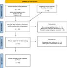
Reasons for excluding full-text sources that did not meet the inclusion criteria were recorded and reported in this review. The results of the search and study inclusion process were fully reported in the final integrative review and presented in a PRISMA (Preferred Reporting Items for Systematic Reviews and Meta-Analyses) flow diagram, shown in Figure 1.
Data were extracted from the articles included in the integrative review by two reviewers independently, using a data extraction tool developed by the reviewers themselves. The preliminary data extraction tool was revised as needed during the data extraction process for each included evidence source.
After data extraction, the publications were analyzed based on the following variables: article title; authors and year of publication; objectives; journal, database, and impact factor (IF); research method; study sample, and country (see Table 2). Table 3 includes the main molecules found. Subsequently, the interpretation of the obtained results was conducted through critical evaluation of the data (Table 4).
List of acronyms, full names, and categories of molecules involved in the AMPK signaling pathway in type 2 diabetes (T2DM)
RESULTS
Initially, 133 articles were identified from the databases and reference lists. After applying filters, evaluating duplicates, and reading titles and abstracts, 123 articles were excluded. After a complete reading of the 57 eligible studies, only 9 articles met the eligibility criteria for this study and were included. The others that were excluded contained: therapeutic treatments based on other types of intervention (n=30) and articles that assessed other outcomes (n=18).
Characteristics of the Included Studies
A total of 3 studies were conducted in Denmark, 1 study in the United Kingdom, 1 study in the United States, 1 study in Ireland, 2 studies in Australia, and 1 study in Spain. All included studies were published between 2004 and 2024. Out of the 7 articles, only 1 was conducted about 5 years ago. Sample sizes were generally small, and there was not a substantially large variation among the studies. A total of 8 studies had samples ranging from 15 to 28 participants, and only 1 systematic review had a more significant sample, with approximately 2,160 individuals investigated.
Research Methods and Bibliometric Indicators
Regarding the research methods of the included studies, it was observed that approximately 77.8% were clinical trials (randomized and non-randomized) and 22.2% were systematic reviews. As for bibliometric indicators, 100% of the studies were published and indexed in international journals that addressed molecular, physiological, and human metabolism aspects, particularly related to diabetes. Furthermore, all articles were published in journals with an impact factor (IF) > 1.5, in English.
Temporal Scope and Content Analysis of the Journals
Regarding the temporal scope, it was noted that about 84.6% of the articles were published more than 7 years ago, and only 15.4% were published in the last 5 years. In terms of topic, all studies (100%) addressed physiological, molecular, and morphological aspects involving different physical training in individuals with type 2 diabetes. Concerning the intervention duration of the studies, there was a large variation, with 1 article reporting a single acute session of physical training; 1 article up to one week; 3 articles up to six weeks; 1 article that did not report the intervention duration; 1 article up to eight weeks; 1 article up to ten weeks; and 1 article with up to fifty-two weeks.
DISCUSSION
The results of this study provided valuable insights into the expression of different molecules in the AMP-activated protein kinase (AMPK) pathway in individuals with type 2 diabetes (T2D) in response to aerobic, resistance, and combined exercises. Most studies indicated that regardless of the exercise modality practiced, activation of the AMPK pathway in people with diabetes can only occur through muscle contraction. Among the 25 molecules found in this pathway were cytokines/interleukins, proteins, enzymes, transcription factors, co-transcriptional activators, and protein hormones. Another significant finding in this study was the expression of the GLUT4 protein and the transcriptional co-activator PGC1-α in the majority of the included research [25-26].
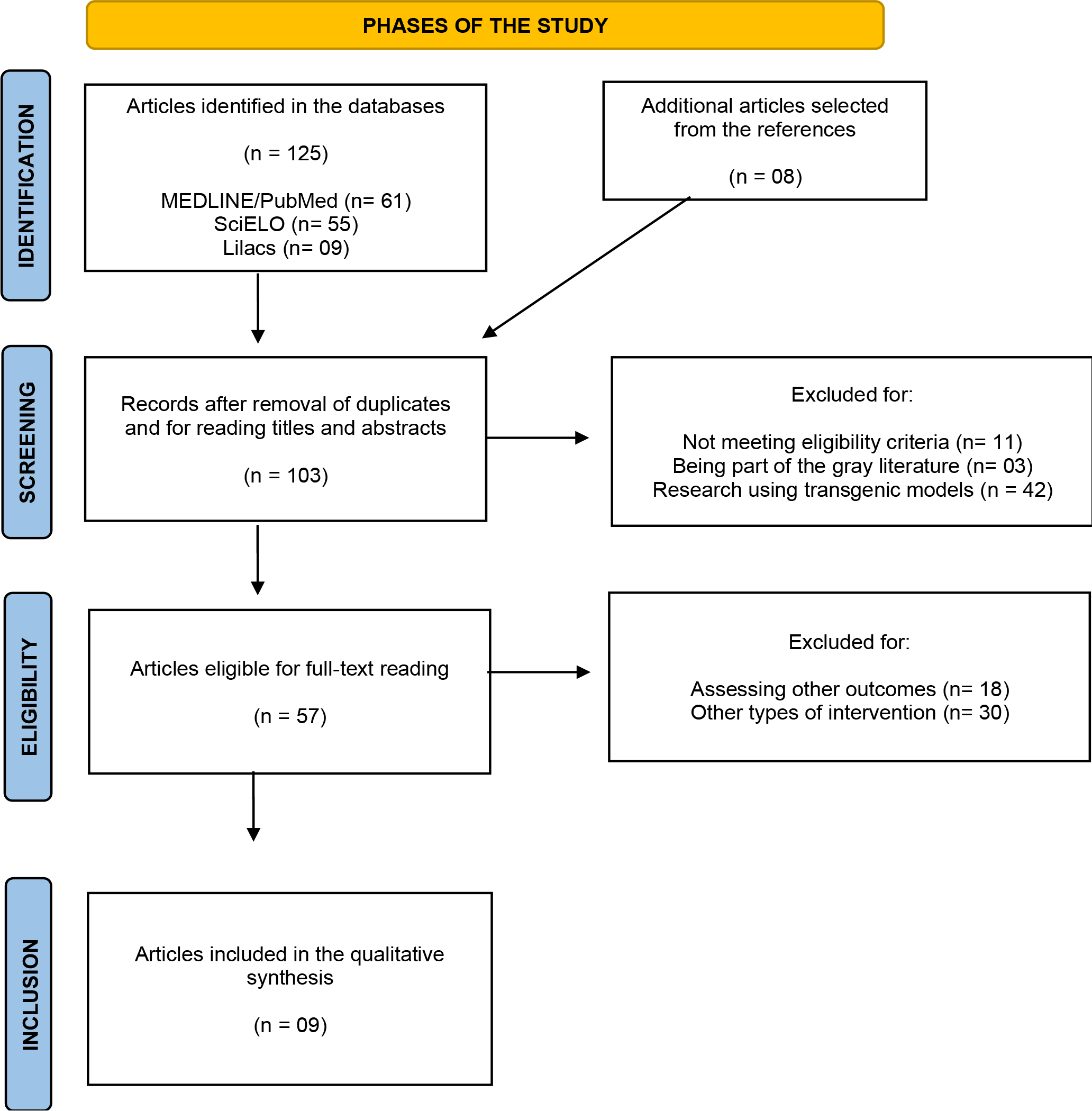
This supports the studies by Jeon [27] and Santos and coauthors [26], which have demonstrated that through physical exercise, skeletal muscles contract and alter cellular energy status, leading to increased metabolic demand in the body and changes in the AMP ratio. The phosphorylation of AMPK induced by different proteins activates PGC1-α and results in a peak in ATP production, facilitating the movement of vesicles containing GLUT4 to the plasma membrane [28-18]. Once GLUT4 is at the membrane, it can capture circulating glucose in the blood and transport it into the cells through facilitated diffusion [29]. Thus, blood glucose is reduced and transformed into ATP in the cytoplasm of the cell, and this entire process results in energy production for up to 48 hours, aiding in glycemic control of T2D [6-30].
Similarly, Wang, Simar, and Whang [20] highlighted that aerobic (AE), resistance (RE), and combined (CE) exercises are related to increases in muscle fiber area and capillary density, glycogen, glycogen synthase, and GLUT4 protein expressions in individuals with T2D. These same exercises can also induce changes in levels of adiponectin, fetuin-A, fibroblast growth factor-21, IL-6, IL-10, leptin, resistin, and TNF-α in T2D [31]. While AE appears to play a role more related to the expression of PGC1-α, mitochondrial adaptation, and oxidative capacity in skeletal muscles [21], RE is more associated with GLUT4 expression, muscle hypertrophy, and protein synthesis regulation [16].
Recent studies have supported the combination of these two exercise modalities, showing that combined exercise may be the best option for individuals with T2D aiming to maintain metabolic and cardiovascular control [32]. This was well elucidated in a meta-analysis by García-Hermoso and coauthors [24], where the authors compiled 40 randomized clinical trials and investigated 2,160 individuals with T2D in Spain. The authors highlighted that both exercise modalities led to significant positive changes in combined exercise, which were in turn related to changes in glycated hemoglobin (mean difference [MD] = 0.81%, 95% CI: 0.95% to 0.67%) and fasting blood glucose (MD = 23.43 mg/dL, 95% CI: 30.07 mg/dL to 16.80 mg/dL).
Beyond the training modality, this same study demonstrated that other variables that may be related to increased expression of molecules in the AMPK pathway and glycemic control in T2D include the volume and frequency of training per week, intensity, and total duration of intervention. Slightly stronger effects were observed with aerobic protocols, resistance, or high-intensity interval training (HIIT) at moderate to vigorous intensity, and with programs lasting over 24 weeks that included at least 3 sessions per week and more than 60 minutes per session.
The effects of molecular expression in the AMPK pathway can be readily observed after chronic exercise [33]. This was evidenced in a clinical trial conducted by Chris-Roberts and coauthors [17], where 8 weeks of AE increased GLUT4 expression by 38% and 22% in non-diabetic and diabetic individuals, respectively (p<0.05). The expression of serine protein kinase (AKT) increased in both groups after 8 weeks of AE (p<0.05). The total activity of glycogen synthase (GS) increased by 46% and 45% in non-diabetic and diabetic individuals, respectively, in response to AE (p<0.01). However, the high heterogeneity between the groups (control group with 16 individuals and intervention group with only 6 participants) may have influenced these results.
It appears that even a single session of physical exercise is capable of activating the AMPK pathway and increasing molecular expression [34]. In this regard, Hussey and coauthors [23] sought to analyze the expression of GLUT4 mRNA after a single session of aerobic exercise. Eighteen individuals were evaluated, nine of whom had T2D and nine with no previous history of comorbidities. The intervention consisted of AE lasting 60 minutes at 55% of VO2max. The results demonstrated that GLUT4 mRNA levels increased post-exercise in the T2D group (p<0.05), remaining elevated and only showing a decrease after 3 hours of the exercise intervention. However, when comparing the control and intervention groups, both increased GLUT4 expression, with no significant differences between them (p>0.05).
Not only does aerobic exercise have molecular effects, but resistance exercise has also demonstrated significant molecular effects in the AMPK pathway (18). In Holten and coauthors [16], a 6-week unilateral resistance exercise intervention was able to increase insulin action in the T2D group, elevate PKB and glycogen synthase levels, and induce significant increases in insulin receptor protein content (p<0.05). This same study showed that RE increased GLUT4 density by 40% in individuals with T2D (p<0.05). However, there was no difference in GLUT4 protein thickness between the control and intervention groups (p>0.05).
On the other hand, the authors showed that despite no differences in the expression of molecules like GLUT4 when comparing the intervention and control groups [16], discrepancies may be observed regarding increased insulin sensitivity in adipose and muscle tissues, as well as greater glucose disposal in individuals with T2D [19].
This exercise-induced increase in insulin sensitivity is regulated by a signaling mechanism known as AMPK-TBC1D4 [34]. This relationship was recently confirmed by several authors [31], who demonstrated that the phosphorylation of Rac1 protein and TBC1 domain family member 4 (TBC1D4) may provide a mechanism for exercise-induced improvements in muscle insulin sensitivity in individuals with T2D, particularly in those with higher insulin resistance, specifically the obese.
It is known that individuals with obesity tend to have greater insulin resistance [7]. Therefore, it is important to determine whether individuals with T2D and this clinical condition exhibit impaired AMPK signaling [21]. In view of this, a recent study [22] evaluated 26 male individuals with obesity divided into two groups: the intervention group (individuals with obesity and T2D) and the control group (obese individuals without diabetes).
The intervention consisted of 10 weeks of AE, comprising 4 to 5 days per week with a duration of 20 to 35 minutes at 65% of VO2 max. The findings demonstrated that phosphorylation of TBC1D4 showed no differences between the studied groups (p>0.05), but the study revealed an increase in TBC1D4 of approximately 20% within groups (p<0.05). In this same study, AKT1 and AKT2 proteins exhibited significant differences between groups. While AKT1 expression increased by 20% in the control group after AE, the group with individuals with T2D increased the expression of this molecule by up to 50% (p<0.05). Meanwhile, AKT2 increased in both groups (control and intervention), with values ranging between 30% and 40%, respectively.
Similar to this study, a clinical trial conducted in the UK [21] investigated 28 individuals (control group = 8 obese non-diabetic individuals and 8 lean non-diabetic individuals; intervention group = 12 obese individuals with T2D). After 10 weeks of low to moderate intensity aerobic exercise, the expression of the PGC1-α gene was acutely stimulated, peaking after a 150-minute rest period (p<0.05). PGC1-α, after being activated by AMPK and calcium/calmodulin-dependent kinase (CaMK), is responsible for increasing GLUT4 expression in individuals with T2D [26]. Furthermore, it regulates other genes (such as SLC2A4 itself), activates mitochondrial biogenesis, maintains glycemic homeostasis, and promotes the increase/remodeling of type I fibers (red and slow fibers) [35].
Different intervention protocols and the biochemical responses observed
Intervention protocols, especially resistance (RT) and aerobic (AE) exercises, play a crucial role in the biochemical responses observed in the muscles of individuals with type 2 diabetes. In studies such as those by Holten and coauthors [16] and Wojtaszewski and coauthors [18] participants performed RT three times a week with varying intensities and volumes over 6 weeks. The results showed improvements in insulin-mediated glucose uptake and the expression of glucose transporter-related proteins, such as GLUT4. Another interesting aspect was the relationship between exercise intensity and biochemical responses, which was observed with higher intensity and training volume in the final weeks of the intervention protocol. These variables contribute to favorable adaptations in insulin signaling, as evidenced by improvements in protein phosphorylation and the isoenzymatic composition of the AMPK pathway.
Moreover, AE protocols also produced significant responses, as seen in the studies by O'Gorman and coauthors [19] and Vind and coauthors [22] which used different aerobic exercise intensities in their interventions. By combining intensity with duration of AE, associations were found with improvements in insulin-mediated glucose disposal and various aspects of cellular signaling related to glucose metabolism. Exercise intensity was a critical factor in determining favorable effects on insulin signaling pathways, with programs that included progressive increases in exercise intensity and duration showing more pronounced responses. An example of this was the response in groups performing exercises at 60% to 80% of VO2peak.
Finally, the difference in sample collection times [36] and methods has also been highlighted [37, 38] as a factor that may influence biochemical responses in humans. In the studies by Sriwijitkamol and coauthors. (2007) [21] and García-Hermoso and coauthors (2023) [24], more specifically, muscle tissue samples were collected at different time intervals post-exercise. The analysis of acute and recovery biochemical responses revealed that the time elapsed after the exercise session could be crucial for understanding the adaptations of glucose metabolism and proteins involved in the insulin signaling process. Sample collection between 16 and 96 hours after exercise demonstrated that the time window for biochemical response measurements could be an important aspect to consider in the specific muscle adaptations in individuals with type 2 diabetes. Therefore, intervention protocols, exercise intensity, volume, and sample collection timing play critical roles in biochemical responses and training adaptations.
Overall, the results of this review suggest that the expression of molecules in the AMPK pathway induced by aerobic, resistance, and combined exercises may be important for metabolic control of T2D and provide multisystem benefits. However, much of the regulation of these molecules by exercise in individuals with T2D remains unknown and presents several challenges for its validation in clinical/therapeutic practice. Therefore, in treating patients with T2D, it is necessary to clarify these aspects and determine whether the expression of molecules in the AMPK pathway can directly impact glycemic control in this target population.
Strengths and Limitations of the Study
Most studies analyzing the expression of molecules present in the AMPK pathway do not examine these effects in individuals with type 2 diabetes and impaired glucose tolerance after different modalities of physical exercise. As strengths of our study, we highlight the fact that we conducted this analysis and avoided comparing research involving humans and transgenic models, aiming to increase the homogeneity of the sample in our research. Furthermore, we emphasize that most of the studies were clinical trials, and all articles included in the research were published in high-impact journals (>1.5). Thus, we believe that the results of this study are of great utility, as they provide initial data that have not yet been substantially studied.
However, we recognize that our study also has limitations. First, significant heterogeneity was found in the results for the molecules present in the AMPK pathway, which may be explained by the differences between studies in terms of variations in employed methods, exercise protocols, and studied samples. Despite the positive effects of physical exercise on molecular expression in the AMPK pathway being demonstrated in our study, our data cannot be generalized, since the sample from 8 of the 9 included studies was relatively small and not representative of the studied population, which diminishes its external validity.
Other limiting factors included the temporality of the studies and the lack of transparency in some of them. Only 1 study was published in the last five years. In general, the clinical trials did not specify whether randomization was performed or if participants or researchers were blinded during the interventions to reduce the risk of bias in the research. Only one systematic review included did this blinding. Nevertheless, most studies used a short intervention time, which may have prevented the observation of the long-term effects of physical exercise.
Therefore, additional research is suggested, especially randomized clinical trials, to assess the expression of molecules in the AMPK pathway in individuals with type 2 diabetes and their relationship with glycemic variables after different modalities of exercise, aiming to identify whether both are associated with better glycemic control in this target population.
CONCLUSION
It is concluded that physical exercise can increase the expression of molecules involved in the AMPK pathway in individuals with type 2 diabetes. However, when comparing exercise modalities, the studies indicated that combined exercise appears to be more effective than either resistance or aerobic exercise performed in isolation. Furthermore, there seems to be a difference not only between the types of exercises but also among the volume, intensity, and frequency of training
-
Funding:
This research received no external funding.
Acknowledgments:
None.
Data Availability Statement:
Research data are only available upon request for corresponding author.
REFERENCES
-
1 Camargo N, Bernardi DM, Lordani CRF. TCF7L2, a genetic risk factor for type 2 diabetes and dietary indications. Fag Journal of Health (FJH). 2020;2(4):470-3. Available from: doi: 10.35984/fjh.v2i4.262.
» https://doi.org/10.35984/fjh.v2i4.262. -
2 Laakso M. Biomarkers for type 2 diabetes. Mol Metab. 2019;27(Suppl). Available from: doi:10.1016/j.molmet.2019.06.016.
» https://doi.org/10.1016/j.molmet.2019.06.016. -
3 Brazilian Diabetes Society. Guidelines of the Brazilian Diabetes Society 2019-2020. Clannad Scientific Publisher; 2019. Available from: https://portaldeboaspraticas.iff.fiocruz.br/wp-content/uploads/2021/08/Diretrizes-Sociedade-Brasileira-de-Diabetes-2019-20201.pdf Accessed 22 Jan 2024.
» https://portaldeboaspraticas.iff.fiocruz.br/wp-content/uploads/2021/08/Diretrizes-Sociedade-Brasileira-de-Diabetes-2019-20201.pdf -
4 Tinajero MG, Malik VS. An update on the epidemiology of type 2 diabetes: a global perspective. Endocrinol Metab Clin. 2021;50(3):337-55. Available from: doi:10.1016/j.ecl.2021.05.013.
» https://doi.org/10.1016/j.ecl.2021.05.013. -
5 Aguiar C, Duarte R, Carvalho D. New approach to diabetes treatment: from glycemia to cardiovascular disease. Rev Port Cardiol. 2019;38(1):53-63. Available from: doi: https://doi.org/10.1016/j.repc.2018.03.013
» https://doi.org/10.1016/j.repc.2018.03.013 -
6 Ranasinghe C, Devage S, Constantino GR, Katulanda P, Hiils A, Rei NU. Glycemic and cardiometabolic effects of exercise in South Asian Sri Lankans with type 2 diabetes mellitus: A randomized controlled trial Sri Lanka diabetes aerobic and resistance training study (SL-DARTS). Diabetes Metab Syndr Clin Res Rev. 2021;15(1):77-85. Available from: doi:10.1016/j.dsx.2020.12.011.
» https://doi.org/10.1016/j.dsx.2020.12.011. -
7 Ferrari F, Motta MT, Helal L. Biochemical and molecular mechanisms of glucose uptake stimulated by physical exercise in the state of insulin resistance: role of inflammation. Arq Bras Cardiol. 2019;113:1139-48. Available from: doi:10.5935/abc.20190224.
» https://doi.org/10.5935/abc.20190224. -
8 Wang Z, Zhao C, Wang Y, Tian X, Lin J, Zhu B, et al. Exercise ameliorating myocardial injury in type 2 diabetic rats by inhibiting excessive mitochondrial fission involving increased irisin expression and AMP-activated protein kinase phosphorylation. J Diabetes. 2023;1-12. Available from: doi:10.1111/1753-0407.13475.
» https://doi.org/10.1111/1753-0407.13475. -
9 Franca R. Physical exercise and molecular mechanisms of glucose uptake in type 2 diabetes: integrative review. Disciplinarum Sci Saúde. 2021;22(2):1-15. Available from: doi:10.37777/dscs.v21n2-001.
» https://doi.org/10.37777/dscs.v21n2-001. -
10 Savikj M, Zierath JR. Train like an athlete: applying exercise interventions to manage type 2 diabetes. Diabetologia. 2020;63:1491-9. Available from: doi:10.1007/s00125-020-05166-9.
» https://doi.org/10.1007/s00125-020-05166-9. -
11 Spaulding HR, Yan Z. AMPK and the adaptation to exercise. Annu Rev Physiol. 2022;84:209-27. Available from: doi:10.1146/annurev-physiol-060721-095517.
» https://doi.org/10.1146/annurev-physiol-060721-095517. -
12 Liu F, Deng Y, Zhao Y, Li Z, Gao J, Zhang Y, et al. Time series RNA-seq analysis identifies MAPK10 as a critical gene in diabetes mellitus-induced atrial fibrillation in mice. J Mol Cell Cardiol. 2022;168:70-82. Available from: doi:10.1016/j.yjmcc.2022.04.013.
» https://doi.org/10.1016/j.yjmcc.2022.04.013. -
13 Blazev R, Carl CS, Ng Y, Molndjik J, Voldstedlund CT, Zhao Y, et al. Phosphoproteomics of three exercise modalities identifies canonical signaling and C18ORF25 as an AMPK substrate regulating skeletal muscle function. Cell Metab. 2022;34(10):1561-77.e9. Available from: doi:10.1016/j.cmet.2022.07.003.
» https://doi.org/10.1016/j.cmet.2022.07.003. - 14 Mendes KS, Silveira RCCP, Galvão CM. Integrative review: research method for incorporating evidence in health and nursing. Texto Contexto Enferm. 2008;17:758-64.
- 15 Alarcón-Gil MT, Osorio Toro S, Baena-Caldas GP. The evidence-based medicine PICO strategy applied to dentistry using MeSH, Emtree and DeCS. Rev Fac Odontol Univ Antioq. 2019;31(1):91-101.
-
16 Holten MK, Zacho MT, Gaster M, Juel C, Wojtaszewski JFP, Dela F. Strength training increases insulin-mediated glucose uptake, GLUT4 content, and insulin signaling in skeletal muscle in patients with type 2 diabetes. Diabetes. 2004;53(2):294-305. Available from: doi:10.2337/diabetes.53.2.294.
» https://doi.org/10.2337/diabetes.53.2.294. -
17 Christ-Roberts CY, Pratipanawatr T, Pratipanawatr W, Berria R, Belfort R, Kashyap S, et al. Exercise training increases glycogen synthase activity and GLUT4 expression but not insulin signaling in overweight nondiabetic and type 2 diabetic subjects. Metabolism. 2004;53(9):1233-42. Available from: doi:10.1016/j.metabol.2004.03.022.
» https://doi.org/10.1016/j.metabol.2004.03.022. -
18 Wojtaszewski JFP, Birk JB, Fosing C, Holten M, Pilegaard H, Dela F. 5′ AMP activated protein kinase expression in human skeletal muscle: effects of strength training and type 2 diabetes. J. Physiol. 2005;564(2):563-73. Available from: doi:10.1113/jphysiol.2005.082669.
» https://doi.org/10.1113/jphysiol.2005.082669. -
19 O’Gorman DJ, Karlsson HKR, McQuaid S, Yousif O, Rahman Y, Gasparro D, et al. Exercise training increases insulin-stimulated glucose disposal and GLUT4 (SLC2A4) protein content in patients with type 2 diabetes. Diabetologia. 2006;49:2983-92. Available from: doi:10.1007/s00125-006-0457-3.
» https://doi.org/10.1007/s00125-006-0457-3. -
20 Wang Y, Simar D, Singh MAF. Adaptations to exercise training within skeletal muscle in adults with type 2 diabetes or impaired glucose tolerance: a systematic review. Diabetes Metab Res Rev. 2009;25(1):13-40. Available from: doi:10.1002/dmrr.928.
» https://doi.org/10.1002/dmrr.928. -
21 Sriwijitkamol AS, Colleta AK, Wajcberg E, Balbontin GB, Reyna SM, Barrientes J, et al. Effect of acute exercise on AMPK signaling in skeletal muscle of subjects with type 2 diabetes: a time-course and dose-response study. Diabetes. 2007;56(3):836-48. Available from: doi:10.2337/db06-1119.
» https://doi.org/10.2337/db06-1119. -
22 Vind BF, Treebak JT, Birk J, Hey-Mogensen M, Beck-Nielsen H, Zierath J, et al. Impaired insulin-induced site-specific phosphorylation of TBC1 domain family, member 4 (TBC1D4) in skeletal muscle of type 2 diabetes patients is restored by endurance exercise-training. Diabetologia. 2011;54(1):157-67. Available from: doi: 10.1007/s00125-010-1924-4.
» https://doi.org/10.1007/s00125-010-1924-4. -
23 Hussey SE, McGee SL, Garnham U, McConell GK, Hargreaves M. Exercise increases skeletal muscle GLUT4 gene expression in patients with type 2 diabetes. Diabetes Obes Metab. 2012;14(8):768-71. Available from: doi:10.1111/j.1463-1326.2012.01585.x.
» https://doi.org/10.1111/j.1463-1326.2012.01585.x. -
24 García-Hermoso A, Ramírez-Vélez R, Díez J, González A, Izquierdo M. Exercise training-induced changes in exerkine concentrations may be relevant to the metabolic control of type 2 diabetes mellitus patients: A systematic review and meta-analysis of randomized controlled trials. J. Sport Health Sci. 2023;12(1):47-58. Available from: doi:10.1016/j.jshs.2021.12.003.
» https://doi.org/10.1016/j.jshs.2021.12.003. -
25 Stanford KI, Goodyear LJ. Exercise and type 2 diabetes: molecular mechanisms regulating glucose uptake in skeletal muscle. Adv Physiol Educ. 2014;38(4):308-14. Available from: doi:10.1152/advan.00080.2014
» https://doi.org/10.1152/advan.00080.2014 -
26 Santos JM, Moreli ML, Tewai S, Benite-Ribeiro SA. The effect of exercise on skeletal muscle glucose uptake in type 2 diabetes: An epigenetic perspective. Metabolism. 2015;64(12):1619-28. Available from: doi:10.1016/j.metabol.2015.09.013.
» https://doi.org/10.1016/j.metabol.2015.09.013. -
27 Jeon SM. Regulation and function of AMPK in physiology and diseases. Exp Mol Med. 2016;48(7). Available from: doi:10.1038/emm.2016.81.
» https://doi.org/10.1038/emm.2016.81. -
28 Steinberg GR, Hardie DG. New insights into activation and function of the AMPK. Nat Rev Mol Cell Biol. 2023;24(4):255-72. Available from: doi:10.1038/S41580-022-00547-X.
» https://doi.org/10.1038/S41580-022-00547-X. -
29 Koester AM, Geiser A, Bowman PRT, Van De Linde S, Gadegaard N, Bryant NJ, et al. GLUT4 translocation and dispersal operate in multiple cell types and are negatively correlated with cell size in adipocytes. Sci Rep. 2022;12(1):20535. Available from: doi:10.1038/s41598-022-24736-y.
» https://doi.org/10.1038/s41598-022-24736-y. -
30 Sylow L, Kleinert M, Richter EA, Jhensen TE. Exercise-stimulated glucose uptake-regulation and implications for glycaemic control. Nat Rev Endocrinol. 2017;13(3):133-48. Available from: doi:10.1038/nrendo.2016.162.
» https://doi.org/10.1038/nrendo.2016.162. -
31 Kjobsted R, Hingst JR, Fentz J, Foretz M, Sanz M, Pehmoller C, et al. AMPK in skeletal muscle function and metabolism. FASEB J. 2018;32(4):1741-77. Available from: doi:10.1096/fj.201700442R.
» https://doi.org/10.1096/fj.201700442R. -
32 Su X, He J, Cui J, Li H, Homens J. The effects of aerobic exercise combined with resistance training on inflammatory factors and heart rate variability in middle-aged and elderly women with type 2 diabetes mellitus. Ann Noninvasive Electrocardiol. 2022;27(6). Available from: doi:10.1111/anec.12996.
» https://doi.org/10.1111/anec.12996. -
33 Steenberg DE, Jørgensen NB, Birk JB, Sjøberg KA, Kiens B, Richter EA, et al. Exercise training reduces the insulin-sensitizing effect of a single bout of exercise in human skeletal muscle. J. Physiol. 2019;597(1):89-103. Available from: doi:10.1113/JP276735.
» https://doi.org/10.1113/JP276735. -
34 Cartee GD. AMPK-TBC1D4-Dependent Mechanism for Increasing Insulin Sensitivity of Skeletal Muscle. Diabetes. 2015;64(6):1901. Available from: doi:10.2337/db15-0010.
» https://doi.org/10.2337/db15-0010. -
35 Abreu P, Leal-Cardoso JH, Ceccatto VM. Skeletal Muscle Adaptation to Physical Exercise: Molecular and Energetic Considerations. Rev Bras Med Esporte. 2017;23:60-5. Available from: doi:10.1590/1517-869220172301167371.
» https://doi.org/10.1590/1517-869220172301167371. -
36 Edman S, Jones III RG, Jannig PR, Fernandez-Gonzalo R, Norrbom J, Thomas NT, et al. The 24-hour molecular landscape after exercise in humans reveals MYC is sufficient for muscle growth. EMBO Rep. 2024;1-28. Available from: doi: https://doi.org/10.1038/s44319-024-00299-z
» https://doi.org/10.1038/s44319-024-00299-z -
37 Newmire DE, Willoughby DS. The skeletal muscle microbiopsy method in exercise and sports science research: A narrative and methodological review. Scand J Med Sci Sports. 2022;32(11):1550-68. Available from: doi: https://doi.org/10.1111/sms.14215
» https://doi.org/10.1111/sms.14215 -
38 Dreher SI, Irmler M, Pivovarova-Ramich O, Kessler K, Jürchott K, Sticht C, et al. Acute and long-term exercise adaptation of adipose tissue and skeletal muscle in humans: a matched transcriptomics approach after 8-week training-intervention. Int J Obes. 2023;47(4):313-24. Available from: doi: https://doi.org/10.1038/s41366-023-01271-y
» https://doi.org/10.1038/s41366-023-01271-y
-
Editor-in-Chief:
Paulo Vitor Farago
-
Associate Editor:
Paulo Vitor Farago