 Cymbopogon citratus Essential Oil Protects Tubular Renal Cells against Ischemia/Reoxygenation Injury - Involvement Nrf2/Keap1 Pathway
Abstract
Ischemia and reoxygenation (I/R) cause acute kidney injury with progression related to oxidative damage, mitochondrial dysfunction and cell death. Nrf2-Keap1 pathway is involved in this process. Cymbopogon citratus, used in popular medicine, is a promising source of bioactive antioxidant compounds. This study investigated the cytoprotective effect of C. citratus essential oil (CCEO) in an I/R model, and the interaction of CCEO major components with the Nrf2-Keap1 pathway in silico. CCEO was characterized by GC-MS, presenting citral isomers trans-geranial (55.48%) and cis-neral (35.40%). I/R decreased LLC-MK2 kidney cells viability by around 50% (MTT assay); CCEO increased cell viability and decreased loss of membrane integrity (Flow cytometry with 7-AAD). CCEO decreased ROS and mitochondrial depolarization and reduced the release of KIM-1, a marker of tubular injury (ELISA). CCEO ameliorated ultrastructural (DCF and Rho123 staining) changes induced by I/R saw in scanning electron microscopy, such as volume retraction, formation of apoptotic bodies, and adhesion to extracellular matrix decrease. The interactions of citral isomers with Keap1 were made through molecular docking. Geranial and neral demonstrated a ∆G around -5.0 kcal/mol with Keap1, binding to some amino acids common to co-crystallized ligand. So, the cytoprotective effect in renal cells may be related to the interaction of citral (neral and geranial) with the Nrf2/Keap1 pathway.
Keywords:
citral; KEAP-1; acute kidney injury; antioxidants.
HIGHLIGHTS
CCEO citral isomers increase cell viability and reduce membrane injury.
CCEO citral isomers decreases ROS and mitochondrial depolarization.
CCEO citral isomers reduces the release a marker of tubular injury (KIM-1).

INTRODUCTION
Ischemia and reperfusion (I/R) are causes of acute kidney injury (AKI) seen in clinical practice [1], associated with high morbidity and mortality. This condition can occur in a variety of clinical disorders that lead to poor tissue perfusion, such as transplantation, shock, trauma and major surgeries [2,3]. It is estimated that 50% of critically ill patients die because there is no effective therapeutic strategy. Although reperfusion is necessary, it is known that it enhances oxidative stress, leading to reactive oxygen species (ROS) formation and an inflammatory response [4]. The excessive ROS generation during reperfusion modifies proteins, causes damage to the plasmatic membrane, and damages DNA, triggering cell death pathways [5,6].
It has been demonstrated that natural substances could regulate intracellular signaling cascades that control responses to stress, inflammation and cellular repair. The MAPK pathway responds to diverse stimuli, including oxidative stress, and has been implicated in nuclear factor erythroid 2-related factor 2 (Nrf2) inductions. Nrf2 is a regulator of cellular antioxidant responses, regulated by a protein called Keap1 (protein 1 similar to Kelly and associated with ECH), acting as a redox sensor and associated with the pathogenesis of a variety of diseases [7-10].
Cymbopogon citratus, known as lemon grass, is distributed worldwide in tropical and subtropical areas and grown mainly to obtain its essential oil [11]. C. citratus leaves are commonly used in popular medicine due to their variety of therapeutic properties [12] such antitumor [13], anti-inflammatory and antioxidant effects [11,14]. Citral, the major component of C. citratus essential oil (CCEO), is formed by a mixture of geranial (trans-citral) and neral (cis-citral) [15]. It was demonstrated that citral has a nephroprotective effect in segmental and focal glomerulosclerosis models [16] and lupus nephritis [17] by activating the Nfr2 factor and inhibiting MAPK phosphorylation.
This study investigated the cytoprotective effect of CCEO on LLC-MK2 renal cells exposed to I/R. The interactions between citral isomers and Nrf2-Keap1 were evaluated by molecular docking.
MATERIAL AND METHODS
Essential oil Collection and Extraction by steam-dragging
The C. citratus specimens (exsiccate registration #54263) were collected at the Francisco José de Abreu Matos Medicinal Plants Garden (Geographical coordinates: -3.7456214227893363, -38.57757958465461; at 8:00 a.m. in September 2019) and CCEO was extracted at the Garden Natural Products Laboratory from the Federal University of Ceará. The extraction took place through the use of 1000 g of dried leaves, crushed, and placed separately in a vial coupled to a condenser and a source of water vapor to extract the essential oil by steam-dragging. The extraction lasted for 2 hours, under a controlled temperature of approximately 100 °C, consistent with the boiling point of water under atmospheric pressure. After, CCEO was treated with sodium sulfate anhydrous, in order to remove moisture, and analyzed by Gas Chromatography coupled to Mass Spectrometer (GC-MS).
A CCEO aliquot was dissolved in dichloromethane/hexane 1:1-Chromatographic grade (1:100). A GC-MS detector Shimadzu QP-2010 plus was used, applying helium-dragging gas. The column used was Equit-5 (30 cm x 25 mm x 25 µm). The temperature programming started with 60 °C to 246 °C at a rate of 3 °C/min, totaling 62 min. The detector voltage used was 70 eV, with a mass range ranging from 30 to 500 m/z. The ionization source and interface temperature was 240 ºC. The injector temperature was 220 °C with a 1:10 split injection mode. The injection volume was 1μL. The compounds were identified by similarity with the NIST (National Institute of Standards and Technology) database mass spectra.
LLC-MK2 cell culture
LLC-MK2 kidney cells (ATCC CCL-7) [18], were cultured in Dulbecco’s modified Eagle’s medium (DMEM, Invitrogen, USA) with antibiotics and 10% fetal bovine serum (FBS) at 37 °C and 5% CO2.
In vitro I/R model
For ischemia induction, the culture medium was replaced by DMEM without glucose, pyruvate and FBS, so the plates were incubated in an anaerobic chamber for 24 hours. After that, the reoxygenation was performed by adding a complete DMEM and returning the cells to the 5% CO2 atmosphere for 3 hours [19]. Then, cells were treated with CCEO (250; 125; 62.5; 31.25; 15.6; 7.8; and 3.9 µg/ml) for 24 hours after reoxygenation.
Cell viabily evaluation
Cell viability was assessed by the MTT reduction assay. After treatment with CCEO for 24 hours, MTT (0.25 mg/mL) was added and incubated at 37 °C for 4 hours. In the presence of living cells, the yellow, insoluble MTT salt is converted to purple formazan, read at 570 nm [20].
The 7-AAD marker, a fluorescent DNA intercalator able to assess cell membrane fragmentation, was used by flow cytometry (FACSCalibur, BD Biosciences). Treated LLC-MK2 cells were labeled with 7-AAD and analyzed considering the percentage of labeled cells [21].
Proximal tubular injury marker determination (KIM-1)
KIM-1 levels were determined in the LLC-MK2 cells supernatant through a commercial enzyme-linked immunosorbent assay (ELISA) (R & D Systems, Inc - Cat. DY1750, Minneapolis, MN) [22].
Scanning electron microscopy (SEM)
LLC-MK2 cells were cultured on sterile circular glass slides. The samples were fixed with 2.5% glutaraldehyde 0.1 M (pH 7.2 at room temperature, for 2 hours). For SEM analysis, the fixed samples were dehydrated with crescent concentrations of ethanol, coated with a 20 nm gold layer in a QT150 ES-Quorum ion spray device, and evaluated in Quanta 450 FEG-FEI equipment (Thermo Fisher Scientific, Waltham, USA) [23].
CCEO redox potential analysis by flow cytometry
For assessment of intracellular ROS, the dye DCFH2-DA (2’,7’-dichlorofluorescein diacetate) was used. Cytosolic ROS oxidize DCFH, forming the fluorescent molecule DCFH-Ox, which exhibits a green fluorescence (FL1). LLC-MK2 cells were labeled with DCFH-DA (100 µM), as described elsewhere [24].
Mitochondrial depolarization was assessed with rhodamine 123 (Rho123) (Sigma-Aldrich, St. Louis, MO, USA). Cells were washed with PBS and stained with Rho123 (10 μg/mL) for thirty minutes. Then, the cells were analyzed by flow cytometry to measure the accumulation of Rho123 in viable mitochondria (FL2). Results were determined considering the change in the fold signal intensity geometric mean (relative fluorescence intensity) [25].
Molecular docking study
To determine the citral's isomers potential binding with the Keap1 protein, molecular docking studies were carried out with the AutodockVina software. Keap1 (PDB:4IQK) crystal structure with their respective ligands was recovered from the RSCB Protein Data Bank. Geranial (ICD: 638011) and Neral (ICD: 643779) ligands were obtained from the PUBCHEM database.
For the fitting studies, water molecules and the protein co-crystallized ligand were removed, and polar hydrogen atoms were added to the protein. Gasteiger charges were added to each atom, the non-polar hydrogen atoms were dissolved with the protein structure, and the grid box was settered (90 × 80 × 80 Å along the x, y, z and grid center -35.687, 0.38 and -16.998 Å). Results (free binding energy - ∆G) were validated by a redocking protocol with the co-crystallized protein ligand. Visualization and analysis of simulations were performed by PyMol and Discovery Studio Visualizer 2020.
Statistical analysis
All data were expressed as the mean ± standard error of the mean. Statistical comparisons were performed by One-way ANOVA with Bonferroni post-test and p <0.05 as significant criteria. Statistical analyzes were performed using the GraphPad Prism5.0 software (USA).
RESULTS
CCEO chemical composition
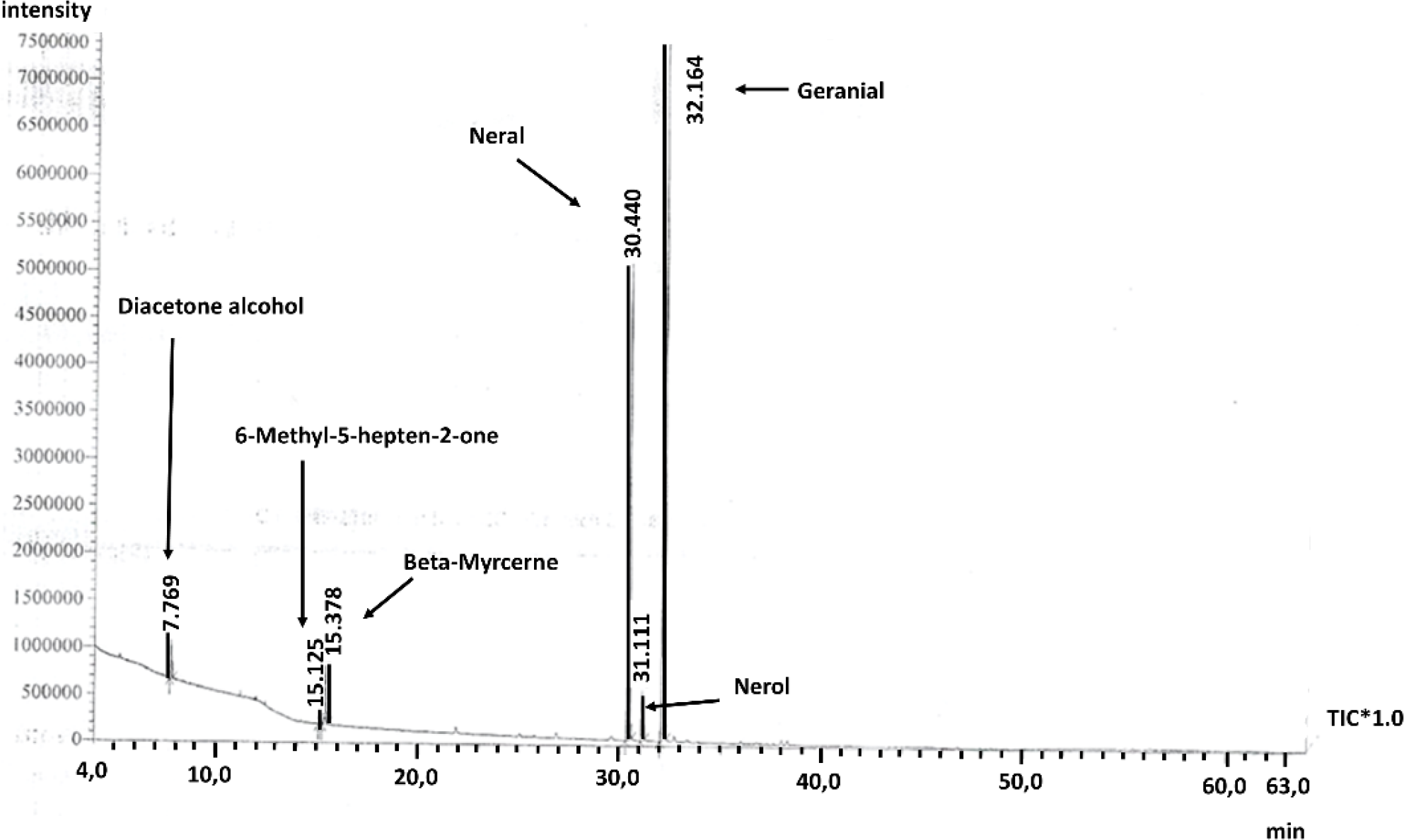
CCEO extraction resulted in 1.98% (volume/weight) of yield. CCEO is mainly constituted (90.88%) by the monoterpenes citral aldehyde, formed by geranial (55.48%) and neral (35.40%). Additionaly, it has chemical composition: nerol (3.49%), beta myrcene (3.49%), 6-methyl-5-hepten-2-one (0.46%) and diacetone alcohol (1.68%) demonstrated in Figure 1.

Cell viability assays
The MTT assay showed that the two higher concentrations of CCEO decreased the cell viability. I/R protocol reduced cell viability by around 50% when compared to the control. Cells treated with CCEO at 125 µg/mL and 250 µg/mL after I/R showed an increase in cell viability of 20% (Figure 2).

LLC-MK2 cell viability by MTT assay. (A) Cytotoxicity; CT: = Negative control; *p<0.05; regarding the C. citratus EO concentrations. (B) Cell viability of cells subjected to ischemia and reperfusion (I/R). The results are shown as mean ± SEM. p <0.05 vs. control group (CT); #p< 0.05 vs. I/R group.
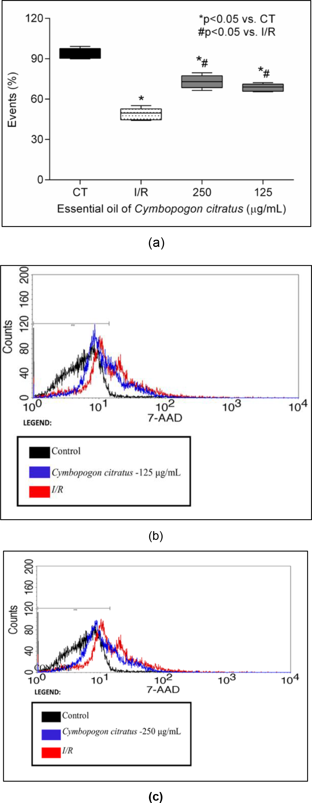
The cell membrane fragmentation assay (Figure 3) showed a decrease in cell viability in the I/R group, 48.88% when compared to the control, while treatment with CCEO (125 and 250 µg/mL) decreased the damage in LLC-MK2 caused by I/R.
Membrane fragmentation assay. (A) Statistical analysis and event distribution percent (cells). B and C histogram analysis - The population highlighted by the black line represents the control; the cells in red are those subjected to ischemia/reperfusion; the cells in blue are those treated with C. citratus essential oil at 125 μg/mL and 250 μg/mL concentrations, respectively. The data are expressed as fluorescence ratio when compared to control ± SEM. p <0.05 vs. control group (CT); #p <0.05 vs. I/R group.
Proximal tubular injury marker KIM-1
In I/R group supernatant (68 ng/mg ± 2.73), KIM-1 levels were more than doubled when compared to the control group (29.19 ng/mg ± 3.52). The CCEO 125 μg/mL treatment was able to decrease KIM-1 levels (22.24 ng/mg ± 2.59).
Scanning electron microscopy
Cells subjected to the I/R had ultrastructural changes, such as cell volume retraction, formation of apoptotic bodies by cytoplasmic fragmentation, and a decrease in adhesion to the extracellular matrix (Figure 4). The CCEO treatments at 125 μg/mL and 250 μg/mL were able to partially reverse the changes observed.

Scanning electron microscopy in in vitro I/R (Ischemia/Reperfusion) model using LLC-MK2 cells. (A) Control group; (B) I/R group; (C and D) I/R after treatment with C. citratus EO at 125 μg/mL and 250 μ/mL concentrations, respectively.
ROS production
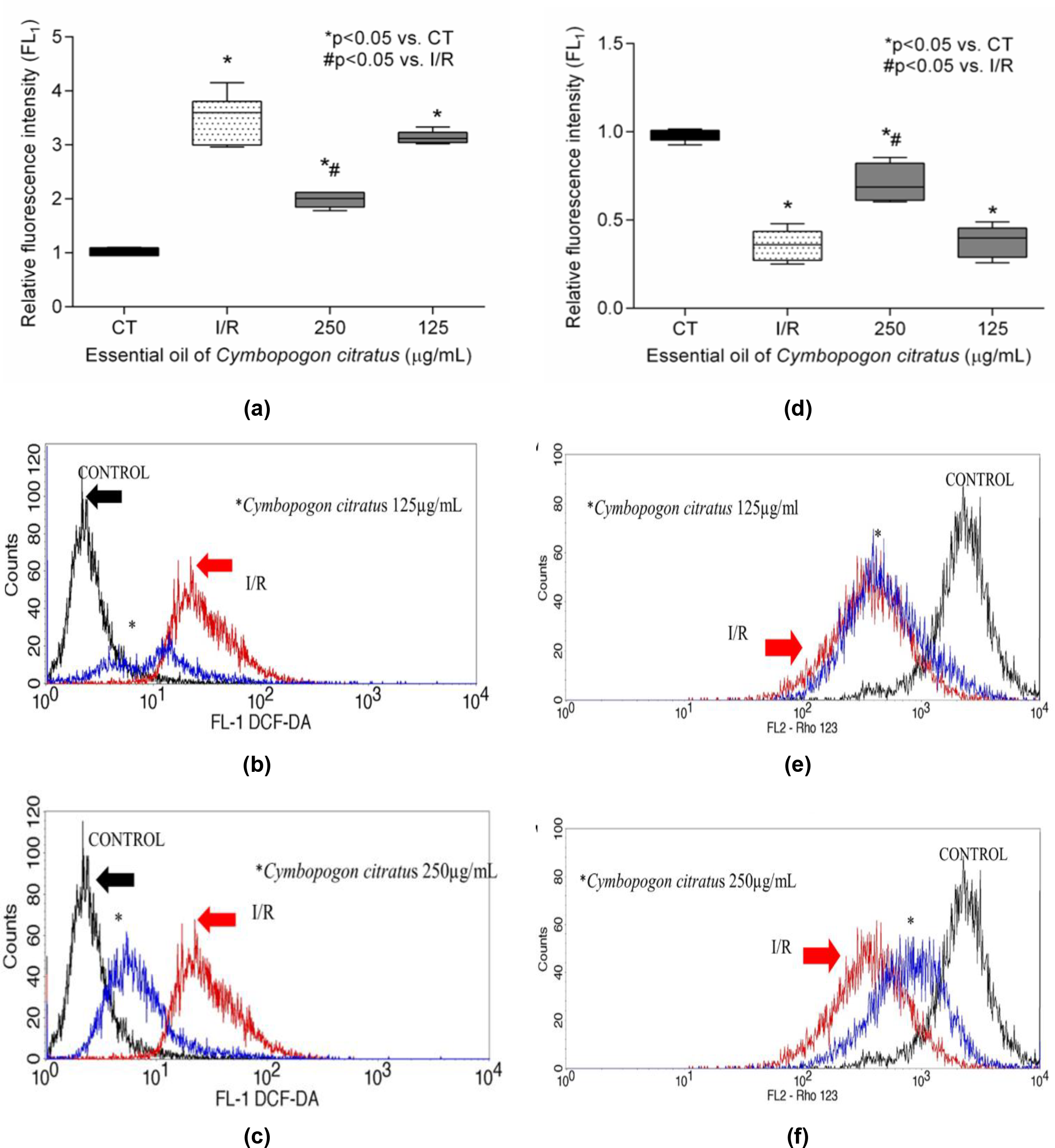
Figure 5 (A-C) shows that I/R increased ROS levels. Only CCEO 250 μg/mL was able to attenuate the fluorescence intensity by 43.3% compared to the I/R group. Figure 5 (D-F) showed a decrease in Rho123 fluorescence emission in I/R when compared to the control (64%). CCEO (250 μg/mL) improved transmembrane potential when compared to the I/R group.

Oxidative stress analysis by flow cytometry. (A) Analysis of reactive oxygen species production by DCFH-DA. (B and C). Histogram analysis of representative reactive oxygen species in the population of cells treated with C. citratus essential oil at 125 and 250μg/mL, respectively. D analysis of mitochondrial transmembrane potential by rhodamine 123 labeling. (E and F) Histogram analysis of the representative mitochondrial transmembrane potential in the cell population treated with essential oil at 125 and 250μg/mL, respectively. The data are expressed as fluorescence ratio when compared to control ± SEM * P <0.05 vs . control; #P <0.05 vs. I/R Group.
Interaction with Keap1 protein
Table 1 shows the scoring parameters of ligands and their intermolecular interactions with the Keap1 protein. Geranial and neral showed similar ∆G values, -5.5 and -5.2 kcal/mol, respectively. The geranial isomer showed more hydrophobic bonds than neral. It is noteworthy that the geranial isomers have bound to some amino acids common to the co-crystallized ligand (TYR 572 and TYR 344). The regions of interaction between ligands and Keap1 are displayed in Figure 6.
Binding free energy from molecular fitting and intermolecular interaction data between Keap1 protein (PDB: 4IQK), citral isomers and co-crystallized ligand to the protein.
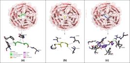
Citral isomer and Keap1 protein complexes (3D diagrams). Legend: (A): Geranial-Keap1 complex 3D schematic; (B): Neral-Keap1 complex 3D schematic; (C): co-crystallized ligand-Keap1.
DISCUSSION
The role of ROS produced by dysfunctional mitochondria during renal I/R is recognized as an important pathophysiological mechanism [26]. ROS increase during reperfusion intensifies the inflammatory response, causing damage to the cell membrane and DNA and triggering mitochondrial alterations and the apoptotic cell death pathway. Numerous mechanisms of tissue protection against oxidative damage, immune response and inflammation have been proposed [27-29]. Regarding that, natural substances with defense mechanisms against I/R injury are highlighted, such as monoterpenes, widely found in essential oils from several plants [30,31].
This study sought to investigate the cytoprotective and antioxidant effects of CCEO against I/R-induced kidney injury. The oil chemical composition analysis of CCEO showed that geranial and neral isomers, also known as citral, are the main components of essential oil, which are involved in its antioxidant effect. These findings corroborate previous studies with lemon grass essential oil terpenoids [32,33]. The LCC-MK2 proximal tubular epithelial cell line was used in this model in order to mimic the damage caused by the I/R process on the kidney. In this study, we have seen that CCEO, in a concentration-dependent manner, increased cell viability after ischemia, reduced membrane fragmentation, prevented cell death by apoptosis by reducing KIM-1, and decreased ultrastructural changes.
KIM-1 acts as a phagocytic receptor for apoptotic cells that must be replaced during cell repair [34]. It has been used as a sensitive early marker to assess the nephroprotective effect of bioactive substances, including those from medicinal plants [35]. In our study, we have seen that CCEO was able to reduce this marker. This effect can be related to a reduction in the apoptotic pathway. The SEM results reinforce these findings. There was a reduction in apoptotic bodies after treatment.
Recently, non-phenolic components have been reported as strong antioxidants. Citral, for example, in the presence of ROS, can be a co-oxidizing agent and prevent oxidative damage [36]. Several mechanisms have justified citral cytoprotective roles [15,37]. This direct antioxidant effect may justify the partial protective effect of CCEO. However, previous studies have shown that scavenger antioxidants are ineffective in achieving complete recovery from damage after I/R [38]. Therefore, it is suggested that citral would act as an indirect antioxidant and anti-inflammatory, acting on signaling pathways such as Nrf2 and MAPK, which might justify the mechanisms behind the effects of CCEO concentrations [16,17].
Nrf2 is a transcription factor responsible for regulating cellular redox balance by increasing the expression of antioxidant and detoxification enzymes. Its expression is regulated by a repressor protein called Keap1, which prevents Nrf2 translocation to the nucleus, promoting its degradation by ubiquitination [39]. So, substances that impair the Nrf2-Keap1 interaction promote cell protection against I/R-induced oxidative damage [40]. Additionally, recent evidence shows that Nrf2 upregulation might suppress Nf-κβ factor transcription, inhibiting Ras and MAPK [8].
To assess the potential citral/neral-Keap1 interactions, molecular docking simulations were conducted. This assay showed that the geranial isomer had a greater number of hydrophobic bonds than the neral isomer. Previously, geranial was described as a potential Nrf2 pathway activator [41]. Additionally, only the geranial was able to bind to TYR 572 and TYR 344 residues. These are key residues for Keap1 interaction, involved in potential Nrf2 activity, supporting our hypothesis that geranial would be active in this pathway [42]. Otherwise, neral interacted with the residues ALA366 and ILE559, commonly described as potential Keap1 inhibitors [27,43].
Further, we demonstrated that citral isomers have common amino acids with the Keap1 co-crystallized ligand. The ability of citral isomers to be in the same binding site as other Nrf2 activators suggests their Keap1-Nrf2 activator potential. Some characteristics, such as ∆G (>-6kcal/mol) and the presence of abundant oxygen, have been shown to be necessary for potential Nrf2 pathway activators through Keap1 inhibition [27]. However, it does not completely exclude the possibility of activating this pathway. Some natural electrophilic compounds activate Nrf2-regulated genes and increase the cytoprotective response [44]. This may be one of the main antioxidant mechanisms of citral.
MAPK pathway activation has been related to an intense response to tubular epithelial cell injury during ischemic injury with KIM-1 increase [34]. It is activated along with Nfκβ transcription factor in response to stress and inflammation and has become a target for medicines with anti-inflammatory potential [45].
Although the antioxidant mechanism is important in the effect of C. citratus essential oil, it was found that the oil has reparative effects independent of antioxidant action. The results of this study showed that the citral isomers have a greater affinity with the Keap1 protein. Finally, this study sought to clarify the possible C. citratus essential oil cytoprotective mechanisms of action. The search for new substances able to act in cell signaling pathways may help to better understand I/R injury pathophysiology and be important in developing new drugs against this condition. Although this was an in vitro study and further tests are needed to deepen the findings of the work, the results presented are positive, as they point to the translational potential of the plant and its components as medicinal agents.
CONCLUSION
CCEO showed a cytoprotective and antioxidant effect in I/R-induced acute kidney injury model. This might be related to the difference in affinity of the citral to some intracellular signaling pathways. This was a preliminary study of the possible interactions between this monoterpene isomers and Nrf2- Keap1 proteins. New works should be developed to understand the role of these pathways in citral cytoprotective action.
-
Funding:
This research received no external funding.
Data Availability Statement:
The data presented in this study are available on request from the corresponding author due to data privacy.
Acknowledgments:
The authors would like to thank CNPq (Conselho Nacional de Pesquisa e Desenvolvimento [Brazilian National Research Council] and CAPES (Coordenação de Aperfeiçoamento de Pessoal de Nível Superior [Coordination for the Improvement of Higher Education Personnel] for the support granted to perform this work. Laboratory of Clinical and toxicological analysis - Prof. PhD. Eurico Litton Pinheiro de Freitas-LACT for their support during the experiments. We also are grateful to Analytic Center - UFC/CT - Infra/Pro and Capes Equipment.
REFERENCES
-
1 Yousefi-Manesh H, Hemmati S, Shirooie S, Nabavi SM, Talebzadeh Bonakdar A, Fayaznia R, et al. Protective effects of hydroalcoholic extracts from an ancient apple variety “Mela Rosa dei Monti Sibillini” against renal ischemia/reperfusion injury in rats. Food Funct. 2019 Nov 1 ;10(11):7544-52. Available from: https://www.researchgate.net/publication/336687413_Title_Protective_effects_of_the_hydroalcoholic_extracts_from_the_ancient_apple_variety_’Mela_Rosa_dei_Monti_Sibillini’_against_renal_ischemiareperfusion_injury_in_rats
» https://www.researchgate.net/publication/336687413_Title_Protective_effects_of_the_hydroalcoholic_extracts_from_the_ancient_apple_variety_’Mela_Rosa_dei_Monti_Sibillini’_against_renal_ischemiareperfusion_injury_in_rats -
2 Güvenç M, Cellat M, Uyar A, Özkan H, Gokcek İ, İsler CT, et al. Nobiletin Protects from Renal Ischemia-Reperfusion Injury in Rats by Suppressing Inflammatory Cytokines and Regulating iNOS-eNOS Expressions. Inflammation. 2020 Feb 1 ;43(1):336-46. Available from: https://pubmed.ncbi.nlm.nih.gov/31705353/
» https://pubmed.ncbi.nlm.nih.gov/31705353/ -
3 Guo A, Wang W, Shi H, Wang J, Liu T. Identification of Hub Genes and Pathways in a Rat Model of Renal Ischemia-Reperfusion Injury Using Bioinformatics Analysis of the Gene Expression Omnibus (GEO) Dataset and Integration of Gene Expression Profiles. Med Sci Monit. 2019 Nov 8; 25:8403. Available from: https://pmc.ncbi.nlm.nih.gov/articles/PMC6863034/
» https://pmc.ncbi.nlm.nih.gov/articles/PMC6863034/ -
4 Goiato RG, Gandolfi JV, Cavallari V, Amendola C, Ajeje ET, Junior JMS, et al. Acute kidney injury during the perioperative: incidence and impact of extrarenal organ dys-function in the outcomes. Arch Health Sci. 2019 Nov 15 ;26(2):84-8. Available from: https://ahs.famerp.br/index.php/ahs/article/view/104
» https://ahs.famerp.br/index.php/ahs/article/view/104 -
5 Arfian N, Wahyudi DAP, Zulfatina IB, Citta AN, Anggorowati N, Multazam A, et al. Chlorogenic Acid Attenuates Kidney Ischemic/Reperfusion Injury via Reducing Inflammation, Tubular Injury, and Myofibroblast Formation. Biomed Res Int. 2019 Sep 2; 2019:5423703. Available from: https://pubmed.ncbi.nlm.nih.gov/31662982/
» https://pubmed.ncbi.nlm.nih.gov/31662982/ -
6 Cordeiro PM, Fernandes SM, da Fonseca CD, Watanabe M, Lopes SM, de Fatima Fernandes Vattimo M. Effects of Justicia acuminatissima, or Amazonian Sara Tudo, on ischemic acute kidney injury: an experimental study. Rev Esc Enferm USP. 2019 Aug 19; 53: 03487. Available from: https://www.revistas.usp.br/reeusp/article/view/182803
» https://www.revistas.usp.br/reeusp/article/view/182803 -
7 Singh S, Nagalakshmi D, Sharma KK, Ravichandiran V. Natural antioxidants for neuroinflammatory disorders and possible involvement of Nrf2 pathway: A review. Heliyon. 2021 Feb 1 ;7(2):06216. Available from: https://pmc.ncbi.nlm.nih.gov/articles/PMC7890213/
» https://pmc.ncbi.nlm.nih.gov/articles/PMC7890213/ -
8 Ahmed SMU, Luo L, Namani A, Wang XJ, Tang X. Nrf2 signaling pathway: Pivotal roles in inflammation. Biochim Biophys Acta Mol Basis Dis. 2017 Feb 1 ;1863(2):585-97. Available from: https://pubmed.ncbi.nlm.nih.gov/27825853/
» https://pubmed.ncbi.nlm.nih.gov/27825853/ -
9 Kaulmann A, Bohn T. Carotenoids, inflammation, and oxidative stress--implications of cellular signaling pathways and relation to chronic disease prevention. Nutr Res. 2014 ;34(11):907-29. Available from: https://pubmed.ncbi.nlm.nih.gov/25134454/
» https://pubmed.ncbi.nlm.nih.gov/25134454/ -
10 Aranda A, Sequedo L, Tolosa L, Quintas G, Burello E, Castell J V., et al. Dichloro-dihydro-fluorescein diacetate (DCFH-DA) assay: a quantitative method for oxidative stress assessment of nanoparticle-treated cells. Toxicol In Vitro. 2013 Mar ;27(2):954-63. Available from: https://pubmed.ncbi.nlm.nih.gov/23357416/
» https://pubmed.ncbi.nlm.nih.gov/23357416/ -
11 Shah G, Shri R, Panchal V, Sharma N, Singh B, Mann AS. Scientific basis for the therapeutic use of Cymbopogon citratus, stapf (Lemon grass). J Adv Pharm Technol Res. 2011 ;2(1):3. Available from: https://pmc.ncbi.nlm.nih.gov/articles/PMC3217679/
» https://pmc.ncbi.nlm.nih.gov/articles/PMC3217679/ -
12 Ekpenyong CE, Akpan E, Nyoh A. Ethnopharmacology, phytochemistry, and biological activities of Cymbopogon citratus (DC.) Stapf extracts. Chin J Nat Med. 2015 May 1 ;13(5):321-37. Available from: https://pubmed.ncbi.nlm.nih.gov/25986281/
» https://pubmed.ncbi.nlm.nih.gov/25986281/ -
13 Bayala B, Bassole IHN, Maqdasy S, Baron S, Simpore J, Lobaccaro JMA. Cymbopogon citratus and Cymbopogon giganteus essential oils have cytotoxic effects on tumor cell cultures. Identification of citral as a new putative anti-proliferative molecule. Biochimie. 2018 Oct 1; 153:162-70. Available from: https://pubmed.ncbi.nlm.nih.gov/29501481/
» https://pubmed.ncbi.nlm.nih.gov/29501481/ -
14 Boeira CP, Piovesan N, Flores DCB, Soquetta MB, Lucas BN, Heck RT, et al. Phytochemical characterization and antimicrobial activity of Cymbopogon citratus extract for application as natural antioxidant in fresh sausage. Food Chem. 2020 Jul 30; 319:126553. Available from: https://www.sciencedirect.com/science/article/pii/S0308814620304155
» https://www.sciencedirect.com/science/article/pii/S0308814620304155 -
15 Mota CMD, Rodrigues-Santos C, Carolino ROG, Anselmo-Franci JA, Branco LGS. Citral-induced analgesia is associated with increased spinal serotonin, reduced spinal nociceptive signaling, and reduced systemic oxidative stress in arthritis. J Ethnopharmacol. 2020 Mar 25 ;250. Available from: https://pubmed.ncbi.nlm.nih.gov/31846747/
» https://pubmed.ncbi.nlm.nih.gov/31846747/ -
16 Yang SM, Hua KF, Lin YC, Chen A, Chang JM, Kuoping Chao L, et al. Citral Is Renoprotective for Focal Segmental Glomerulosclerosis by Inhibiting Oxidative Stress and Apoptosis and Activating Nrf2 Pathway in Mice. PLoS One. 2013 Sep 16 ;8(9):74871. Available from: https://journals.plos.org/plosone/article?id=10.1371/journal.pone.0074871
» https://journals.plos.org/plosone/article?id=10.1371/journal.pone.0074871 -
17 Ka SM, Lin JC, Lin TJ, Liu FC, Chao LK, Ho CL, et al. Citral alleviates an accelerated and severe lupus nephritis model by inhibiting the activation signal of NLRP3 inflammasome and enhancing Nrf2 activation. Arthritis Res Ther. 2015 Nov 19 ;17(1). Available from: https://pubmed.ncbi.nlm.nih.gov/26584539/
» https://pubmed.ncbi.nlm.nih.gov/26584539/ -
18 Hull RN, Cherry WR, Tritch OJ. Growth characteristics of monkey kidney cell strains LLC-MK1, LLC-MK2, and LLC-MK2 (NCTC-3196) and their utility in virus research. Journal of Experimental Medicine. 1962 May 1 ;115(5):903-18. Available from: http://rupress.org/jem/article-pdf/115/5/903/1649443/903.pdf
» http://rupress.org/jem/article-pdf/115/5/903/1649443/903.pdf -
19 Kurian G, Pemaih B. Standardization of in vitro Cell-based Model for Renal Ischemia and Reperfusion Injury. Indian J Pharm Sci. 2014 Jul 1 ;76(4):348. Available from: https://pmc.ncbi.nlm.nih.gov/articles/PMC4171872/
» https://pmc.ncbi.nlm.nih.gov/articles/PMC4171872/ -
20 Mosmann T. Rapid colorimetric assay for cellular growth and survival: application to proliferation and cytotoxicity assays. J Immunol Methods. 1983 Dec 16 ;65(1-2):55-63. Available from: https://pubmed.ncbi.nlm.nih.gov/6606682/
» https://pubmed.ncbi.nlm.nih.gov/6606682/ -
21 Schmid I, Uittenbogaart CH, Keld B, Giorgi J V. A rapid method for measuring apoptosis and dual-color immunofluorescence by single laser flow cytometry. J Immunol Methods. 1994 Apr 15;170(2):145-57. Available from: https://pubmed.ncbi.nlm.nih.gov/8157993/
» https://pubmed.ncbi.nlm.nih.gov/8157993/ -
22 Khandrika L, Koul S, Meacham RB, Koul HK. Kidney Injury Molecule-1 Is Up-Regulated in Renal Epithelial Cells in Response to Oxalate In Vitro and in Renal Tissues in Response to Hyperoxaluria In Vivo. PLoS One. 2012 Sep 12 ;7(9):44174. Available from: https://journals.plos.org/plosone/article?id=10.1371/journal.pone.0044174
» https://journals.plos.org/plosone/article?id=10.1371/journal.pone.0044174 -
23 Chihanga T, Ma Q, Nicholson JD, Ruby HN, Edelmann RE, Devarajan P, et al. NMR spectroscopy and electron microscopy identification of metabolic and ultrastructural changes to the kidney following ischemia-reperfusion injury. Am J Physiol Renal Physiol. 2018 Feb 2 ;314(2):154. Available from: https://pmc.ncbi.nlm.nih.gov/articles/PMC5866452/
» https://pmc.ncbi.nlm.nih.gov/articles/PMC5866452/ -
24 Bass DA, Parce JW, Dechatelet LR, Szejda P, Seeds MC, Thomas M. Flow cytometric studies of oxidative product formation by neutrophils: a graded response to membrane stimulation. J Immunol. 1983 Apr;130(4):1910-7. Available from: https://pubmed.ncbi.nlm.nih.gov/6833755/
» https://pubmed.ncbi.nlm.nih.gov/6833755/ -
25 O’Connor JE, Vargas JL, Kimler BF, Hernandez-Yago J, Grisolia S. Use of Rhodamine 123 to investigate alterations in mitochondrial activity in isolated mouse liver mitochondria. Biochem Biophys Res Commun. 1988 Feb 29;151(1):568-73. Available from: https://pubmed.ncbi.nlm.nih.gov/3348796/
» https://pubmed.ncbi.nlm.nih.gov/3348796/ -
26 Rezk AM, Ibrahim IAAEH, Mahmoud MF, Mahmoud AAA. Quercetin and lithium chloride potentiate the protective effects of carvedilol against renal ischemia-reperfusion injury in high-fructose, high-fat diet-fed Swiss albino mice independent of renal lipid signaling. Chem Biol Interact. 2021 Jan 5 ;333. Available from: https://pubmed.ncbi.nlm.nih.gov/33159969/
» https://pubmed.ncbi.nlm.nih.gov/33159969/ -
27 Li Z, Zhu J, Wan Z, Li G, Chen L, Guo Y. Theaflavin ameliorates renal ischemia/reperfusion injury by activating the Nrf2 signalling pathway in vivo and in vitro. Biomedicine & Pharmacotherapy. 2021 Feb 1; 134:111097. Available from: https://www.sciencedirect.com/science/article/pii/S0753332220312907
» https://www.sciencedirect.com/science/article/pii/S0753332220312907 -
28 Hu X, Ding C, Ding X, Fan P, Zheng J, Xiang H, et al. Inhibition of myeloid differentiation protein 2 attenuates renal ischemia/reperfusion-induced oxidative stress and inflammation via suppressing TLR4/TRAF6/NF-kB pathway. Life Sci. 2020 Sep 1; 256:117864. Available from: https://pubmed.ncbi.nlm.nih.gov/32474021/
» https://pubmed.ncbi.nlm.nih.gov/32474021/ -
29 Liang S, Xu Z, Ruan Y, Niu T, Guo W, Jiang W, et al. Isoquercitrin Attenuates Renal Ischemia/Reperfusion Injury Through Antioxidation, Anti-inflammation, and Antiapoptosis in Mice. Transplant Proc. 2020 Apr 1 ;52(3):1014-9. Available from: https://pubmed.ncbi.nlm.nih.gov/32115238/
» https://pubmed.ncbi.nlm.nih.gov/32115238/ -
30 Britto RM de, Silva-Neto JA da, Mesquita TRR, Vasconcelos CML de, de Almeida GKM, Jesus ICG de, et al. Myrtenol protects against myocardial ischemia-reperfusion injury through antioxidant and anti-apoptotic dependent mechanisms. Food Chem Toxicol. 2018 Jan 1; 111:557-66. Available from: https://pubmed.ncbi.nlm.nih.gov/29208507/
» https://pubmed.ncbi.nlm.nih.gov/29208507/ -
31 Dai M, Wu L, Yu K, Xu R, Wei Y, Chinnathambi A, et al. D-Carvone inhibit cerebral ischemia/reperfusion induced inflammatory response TLR4/NLRP3 signaling pathway. Biomed Pharmacother. 2020 Dec 1; 132:110870. Available from: https://pubmed.ncbi.nlm.nih.gov/33080468/
» https://pubmed.ncbi.nlm.nih.gov/33080468/ -
32 Gonzales APPF, Yoshioka ETO, Mathews PD, Mertins O, Chaves FCM, Videira MN, et al. Anthelminthic efficacy of Cymbopogon citratus essential oil (Poaceae) against monogenean parasites of Colossoma macropomum (Serrasalmidae), and blood and histopathological effects. Aquac. 2020 Nov 15; 528:735500. Available from: https://doi.org/10.1016/j.aquaculture.2020.735500
» https://doi.org/10.1016/j.aquaculture.2020.735500 -
33 Almeida KB, Araujo JL, Cavalcanti JF, Romanos MT V., Mourão SC, Amaral ACF, et al. In vitro release and anti-herpetic activity of Cymbopogon citratus volatile oil-loaded nanogel. Rev Bras Farmacogn. 2018 Jul 1 ;28(4):495-502. Available from: https://www.scielo.br/j/rbfar/a/3CK3shKrrH6wKtywVZxN6MF/?lang=en
» https://www.scielo.br/j/rbfar/a/3CK3shKrrH6wKtywVZxN6MF/?lang=en -
34 Zhang Z, Cai CX. Kidney injury molecule-1 (KIM-1) mediates renal epithelial cell repair via ERK MAPK signaling pathway. Mol Cell Biochem. 2016 May 1 ;416(1-2):109-16. Available from: https://pubmed.ncbi.nlm.nih.gov/27084535/
» https://pubmed.ncbi.nlm.nih.gov/27084535/ -
35 Sampaio TL, Menezes RRPPB de, da Costa MFB, Meneses GC, Arrieta MCV, Chaves Filho AJM, et al. Nephroprotective effects of (-)-α-bisabolol against ischemic-reperfusion acute kidney injury. Phytomedicine. 2016 Dec 15 ;23(14):1843-52. Available from: https://pubmed.ncbi.nlm.nih.gov/27912887/
» https://pubmed.ncbi.nlm.nih.gov/27912887/ -
36 Baschieri A, Ajvazi MD, Tonfack JLF, Valgimigli L, Amorati R. Explaining the antioxidant activity of some common non-phenolic components of essential oils. Food Chem. 2017 Oct 1; 232:656-63. Available from: https://pubmed.ncbi.nlm.nih.gov/28490124/
» https://pubmed.ncbi.nlm.nih.gov/28490124/ -
37 Bouzenna H, Hfaiedh N, Giroux-Metges MA, Elfeki A, Talarmin H. Biological properties of citral and its potential protective effects against cytotoxicity caused by aspirin in the IEC-6 cells. Biomed Pharmacother. 2017 Mar 1; 87:653-60. Available from: https://pubmed.ncbi.nlm.nih.gov/28088731/
» https://pubmed.ncbi.nlm.nih.gov/28088731/ -
38 Weng XF, Li ST, Song Q, Zhu Q, Song DD, Qin ZH, et al. Protective Effect of Nicotinamide Adenine Dinucleotide Phosphate on Renal Ischemia-Reperfusion Injury. Kidney Blood Press Res. 2018 Jun 1 ;43(3):651-63. Available from: https://pubmed.ncbi.nlm.nih.gov/29734167/
» https://pubmed.ncbi.nlm.nih.gov/29734167/ -
39 Dodson M, De La Vega MR, Cholanians AB, Schmidlin CJ, Chapman E, Zhang DD. Modulating NRF2 in disease: Timing is everything. Annu Rev Pharmacol Toxicol. 2019 Jan 1; 59:555. Available from: https://pubmed.ncbi.nlm.nih.gov/30256716/
» https://pubmed.ncbi.nlm.nih.gov/30256716/ -
40 Zhao Y, Liu X, Fu X, Mo Z, Jiang Y, Yan Y. Protective effects of epigallocatechin gallate against ischemia reperfusion injury in rat skeletal muscle via activating Nrf2/HO-1 signaling pathway. Life Sci. 2019 Dec 15 ;239. Available from: https://pubmed.ncbi.nlm.nih.gov/31678278/
» https://pubmed.ncbi.nlm.nih.gov/31678278/ -
41 Nakamura Y, Miyamoto M, Murakami A, Ohigashi H, Osawa T, Uchida K. A phase II detoxification enzyme inducer from lemongrass: Identification of citral and involvement of electrophilic reaction in the enzyme induction. Biochem Biophys Res Commun. 2003 Mar 14 ;302(3):593-600. Available from: https://pubmed.ncbi.nlm.nih.gov/12615076/
» https://pubmed.ncbi.nlm.nih.gov/12615076/ -
42 Londhe AMH, Gadhe CG, Lim SM, Pae AN. Investigation of Molecular Details of Keap1-Nrf2 Inhibitors Using Molecular Dynamics and Umbrella Sampling Techniques. Mol. 2019 Nov 12 ;24(22):4085. Available from: https://www.mdpi.com/1420-3049/24/22/4085/htm
» https://www.mdpi.com/1420-3049/24/22/4085/htm -
43 Adelusi TI, Abdul-Hammed M, Idris MO, Oyedele QK, Adedotun IO. Molecular dynamics, quantum mechanics and docking studies of some Keap1 inhibitors - An insight into the atomistic mechanisms of their antioxidant potential. Heliyon. 2021 Jun 1 ;7(6). Available from: https://pubmed.ncbi.nlm.nih.gov/34195424/
» https://pubmed.ncbi.nlm.nih.gov/34195424/ -
44 Mou Y, Wen S, Li YX, Gao XX, Zhang X, Jiang ZY. Recent progress in Keap1-Nrf2 protein-protein interaction inhibitors. Eur J Med Chem. 2020 Sep 15; 202:112532. Available from: https://pubmed.ncbi.nlm.nih.gov/32668381/
» https://pubmed.ncbi.nlm.nih.gov/32668381/ -
45 Kyriakis JM, Avruch J. Mammalian MAPK signal transduction pathways activated by stress and inflammation: a 10-year update. Physiol Rev. 2012 Apr 1 ;92(2):689-737. Available from: https://pubmed.ncbi.nlm.nih.gov/22535895/
» https://pubmed.ncbi.nlm.nih.gov/22535895/
-
Editor-in-Chief:
Paulo Vitor Farago
-
Associate Editor:
Jane Manfron