 Supplementation with inorganic iron has no impact on the morphology of the hypopharyngeal glands in honey bees (Apis mellifera L.)
ABSTRACT.
Nutrition plays a major role in honey bees health, development and performance. Industrial agriculture, deforestation and climate change, however, are known to have negative impacts in natural habitats, compromising access to abundant and diversified sources of pollen and nectar, essential for honey bees. Beekeepers commonly use alternative supplementation feed, particularly during the fall and winter when resources are scarce to avoid or reduce colony loss as a consequence of starvation. The goal of the present study was to evaluate the effects of diets containing an inorganic iron source on the morphology of hypopharyngeal glands (HG) in 6-day-old honey bees during the off-season. Twelve colonies were distributed into four groups and supplemented, or not, with inorganic iron at concentrations of 0, 25, 50, and 100 ppm, diluted in sugar syrup. Six-day-old bees were then collected from each treatment, and their heads were prepared for the evaluation of the area and acini number of the HG. No significant differences were observed in the analyzed parameters of HG, regardless of the iron concentrations used (p < 0.05). In conclusion, providing an inorganic iron source during the off-season for colony nutrition does not modulate the development of the hypopharyngeal glands in 6-day-old nurse bees.
Keywords:
beekeeping; hypopharyngeal glands; morphology; nutrition
Introduction
Bees are indispensable insects to the planet through their products (honey, propolis, pollen, among others) or through the pollination services they provide to natural forests or commercially valuable crops (Klein et al., 2007). For a bee colony to carry out its activities properly, a balanced diet acquired through the collection of natural resources is fundamental, being nectar and pollen the most important elements foraged by the bees. An adequate pollen consumption, for example, is associated with lower contamination by gut parasite Nosema ceranae; it improves bees’ resistance to xenobiotics, increases bees’ tolerance to transportation stress, parasitic pressure and it contributes to general colony immunity and development (Mattila & Otis, 2006). On the other hand, negative effects of poor nutrition can include higher levels of Nosema spp. infection, lower brood and adult population, lower egg-laying rates and greater damage caused by the parasite Varroa spp. (Branchiccela et al., 2019; Mattila & Otis, 2006). Nectar, on its turn, is mostly responsible for providing bee’s sufficient energy to fly, maintain the nest’s internal temperature and subsidize beeswax production, essential for colony reproduction and food storage.
For managed hives, during periods of scarcity, it is important for beekeepers to provide the necessary nutrients for the colony's survival since supplementary diets can contribute to brood development, enhance biological activity and favor immunity and natural defenses (Ullah et al., 2021). Without it, bee colonies are more susceptible to disturbance and consequently to impair the economic viability of commercial beekeeping.
While nutrition studies focused on the important aspects of carbohydrates and protein from nectar and pollen respectively are well documented (Di Pasquale et al., 2013; Branchiccela et al., 2019; Brodschneider & Crailsheim, 2010), there is still much to be discovered regarding the importance of fats, vitamins and minerals for honey bees. The challenge to properly include such nutrients in defined diets has been pointed out as an important step on what comes to further understanding the importance of subtle elements in bee’s performance and health (Herbert Jr & Shimanuki, 1978).
Minerals are fundamental for the normal functioning of living organisms, as they act as structural components and participate in important metabolic processes (Soetan et al., 2010; Araújo et al., 2019). Iron for instance is a trace mineral that plays a crucial role in various metabolic processes, including oxygen transport, maintaining the immune system, electron transfer, and DNA and RNA synthesis (Locke & Nichol, 1992; Soetan et al., 2010). It is also important for the formation of iron granules in the abdomen of bees, which may be involved in their ability to perceive the Earth's magnetic field, particularly during foraging activities (Liang et al., 2016; Wright et al., 2018).
The lack of a proper nutrition can compromise iron’s absorption, especially if pollen is scarce as it accounts for the main source of minerals in the bee’s diet. Pollen deprivation could then negatively impact bee physiology and, consequently, the entire colony. The presence of diseases can also play a role in damaging iron absorption. Recent studies have found that Nosema spp., for example, can hijack iron from its host, causing an important deficiency potentially damaging for colonies (Rodríguez-García et al., 2021).
It is known that the nutritional content of available pollen significantly varies depending on the species and seasons of the year (Somerville & Nicol, 2002; Di Pasquale et al., 2013; Keller et al., 2005). Furthermore, unpublished data from our research group showed lower iron concentrations in bee-collected pollen during the winter compared to other months, and this deficiency could influence the development of honey bee glands, which are essential for colony maintenance (Seeley, 1995).
Among the most important glands in hymenoptera, particularly for the honey bee, is the hypopharyngeal gland (HG). In this insect, the hypopharyngeal glands are twin structures found on either side of the head, located anterior to the brain and between the compound eyes. These glands are made up of thousands of secretory units, each linking to a secretory duct in worker bees. Its main function is related to the production and secretion of Royal Jelly proteins (Ahmad et al., 2021).
Royal jelly is a substance produced not only by the hypopharyngeal but also by the mandibular glands located in the heads of nurse honey bees. It is a complex substance composed of water, sugars, proteins, lipids, vitamins, and minerals, used to feed all larvae younger than 3 days old and the queen throughout her life (Deseyn & Billen, 2005). Additionally, it is responsible for caste differentiation, stimulating the ovaries, and accelerating the development of the queen's body (Kamakura, 2011).
Understanding the variables that could affect the development of hypopharyngeal glands is crucial for gaining a better understanding of colony biology and behavior. While many studies emphasize the importance of proteins for honey bees and their effects on hypopharyngeal glands and colony maintenance (Standifer, 1960; Hrassnigg & Crailsheim, 1998; Pernal & Currie, 2000; DeGrandi-Hoffman et al., 2010), little is known about the response to different levels of minerals in the diet, especially under field conditions, which better reflect beekeeping practices.
The secretory activity of hypopharyngeal glands is correlated with the size of acini. Thus, more developed glands have a greater capacity for producing royal jelly (Deseyn & Billen, 2005). Hypopharyngeal glands reach their maximum size and activity at the sixth day of a bee's life, following the consumption of large quantities of pollen, which shows a positive correlation between proper nutrition and HG development (DeGrandi-Hoffman et al., 2010).
The objective of this research was to evaluate the supplementation of inorganic iron during the off-season and its effects on the number and area of acini in the hypopharyngeal glands of six-day-old honey bees. The main differentiating factor of this research was the experimental protocol conducted under field conditions, and it was found that providing an inorganic iron source during the off-season for colony nutrition does not modulate the development of the hypopharyngeal glands in six-day-old nurse bees.
Material and methods
Treatment groups
Twelve colonies of A. mellifera with a standardized number of brood and food frames (three brood frames and two frames of bee bread and honey) were used. The colonies were divided into four groups: T0Fe: which received sugar syrup without iron supplementation; T25Fe: sugar syrup with supplementation of 25 ppm; T50Fe: with 50 ppm and T100Fe: with 100 ppm of inorganic iron. These concentrations were selected based on previous studies that related 50 ppm as the honeybee requirements (Hebert Jr & Shimanuki, 1978).
The source used was ferrous sulfate heptahydrate (containing 31.10% iron - FeSO4·7H2O), which was diluted in sugar syrup (1:1), homogenized by Vortex Mixer equipment and provided with a Boardman feeder (500 mL per week). The syrup was consumed within 24 hours by the experimental colonies, ensuring the stability of ferrous sulfate (Swamy et al., 1979). The research was conducted during the off-season (May, June, and July) at the experimental apiary.
The levels of Fe were determined using Atomic Absorption Spectrometry (FAAS) and were found to be 25.695, 47.796, and 104.175 ppm in the Fe25, Fe50, and Fe100 groups, respectively.
Collection of nurse bees and morphological analysis
Monthly, frames were removed from each treatment, and were individually wrapped in tissue. The frames were placed in an incubator at a temperature of approximately 32°C and a relative humidity of about 60%, until the emergence of individuals. The worker bees that emerged were marked on the pronotum region with a nontoxic pen (Posca Paint Pens, Mitsubishi, Japan), totaling approximately 50 bees marked per colony.
The marked bees were reintroduced into their original colonies, and these colonies continued to receive supplementation with different iron concentrations until the collection of the marked bees, which occurred at six days of age. Then, 10 six-day-old nurse bees were collected each month using the methodology described by Bovi et al. (2017), totaling 120 bees in the study (40 per month). The heads of the six-day-old nurse bees were dehydrated in a series of ethanol and embedded in methacrylate resin (HistoResin©; Leica Microsystems) following the methodology described by Zaluski et al. (2017). Using a rotating microtome, semi-serial sections of 3µm thick were made with a 30µm gap in between to guarantee that the acini were measured only once. Slides were stained with hematoxylin and eosin and the images captured using a Leica DMC 2900 camera connected to a Leica DM 2500 microscope and analyzed with Leica QWin V3 software.
All the acini of hypopharyngeal glands were counted and measured. The values (µm²) then were divided in categories as described by Zaluski et al. (2017), which were < 29,999 / 30,000 - 59,999 and 60,000 - 89,999 µm² for statistical analysis.
Statistical analysis
The data were compared using non-parametric Kruskal-Wallis ANOVA followed by Dunn’s test using GraphPad Prism 6. Means were considered statistically different when p < 0.05.
Results
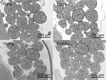
Figure 1 displays the secretory units of the hypopharyngeal glands of 6-day-old nurse bees supplemented with different levels of inorganic iron.

Histological section of the hypopharyngeal glands of 6-day-old nurse bees (Apis mellifera L.) during the off-season. Ducts are marked with asterisks (*) and acini are circled to emphasize. T0Fe: without supplementation; T25Fe: 25 ppm; T50Fe: 50 ppm; T100Fe: 100 ppm of inorganic iron supplementation. Stained with Hematoxylin and Eosin.
Figure 2 shows the number and mean area (µm²) of acini in the hypopharyngeal glands of 6-day-old nurse bees. No significant differences were observed among treatments in the number and area of acini of hypopharyngeal glands in 6-day-old honey bees supplemented with 0, 25, 50, and 100 ppm of inorganic iron.

Number (x102) and mean area (x103) of the acini of hypopharyngeal glands of 6-day-old nurse bees during the off-season. T0Fe: without supplementation; T25Fe: 25 ppm; T50Fe: 50 ppm; T100Fe: 100 ppm of inorganic iron supplementation.
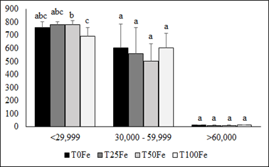
The numbers of acini of hypopharyngeal glands within the categories < 29,999 µm², 30,000 - 59,999 µm², and > 60,000 µm² are represented in Figure 3. While group T100Fe showed significantly fewer numbers of acini within the category < 29,999 µm² compared to T50Fe, no significant differences were observed when the treatments were compared to T0Fe.

Number (x102) and mean area (x103) of the acini of hypopharyngeal glands of 6-day-old nurse bees during the off-season. T0Fe: without supplementation; T25Fe: 25 ppm; T50Fe: 50 ppm; T100Fe: 100 ppm of inorganic iron supplementation.
Discussion
Overall, the supplementation of inorganic iron, under the experimental conditions, did not show evident effects on the modulation of the structure of the hypopharyngeal glands (HG), even with similar concentrations suggested by Hebert and Shimanuki (1978) and adopted in this research.
There are many factors that might modulate the development of the HG, especially in experiments conducted in field conditions, where honey bees have free access to food resources. Studies have shown that stressful managements (Bovi et al., 2017), pesticides exposition (Hatjina, et al., 2013; Zaluski et al., 2017), quantity and quality of food (Di Pasquale et al., 2013; Omar et al., 2017) and mite infestations (Pinto et al., 2011; Power et al., 2021.; Morfin et al., 2023) are capable of impairing the HG structure. Additionally, although iron is an essential micronutrient in the organism of insects such as honey bees (Wright et al., 2018, high concentrations of free iron can be toxic to the organism, causing lipid peroxidation and damage to cell wall structure, leading to cell death (Jumarie et al., 2017). However, the iron concentrations studied in this experiment did not have adverse effects in the HG morphology. On the other hand, Sousa et al. (2022) verified that high iron concentrations in the diet promotes different feeding habits, as a means to mitigate the risk of mineral intoxication.
At the end of the experiment, the frequency within categories between T0Fe and the other treatments did not differ significantly. However, it was observed that T100Fe showed a significantly lower number of acini within the category < 29,999 µm² compared to T50Fe. The acini size in nurse bees is correlated with their activity (Deseyn & Billen, 2005; Škerl & Gregorc, 2015), and the groups that presents greater number of acini within the categories 30,000-59,999 µm² and > 60,000 µm² and lower number of acini within the category < 29,999 µm² can be considered more active and represent a more appropriate method for measuring acini that display an irregular form (Zaluski et al., 2017). Despite this, the mean areas (µm²) of the acini of the groups were not affected by the different distribution among categories.
A possible explanation for the absence of differences could also be the low availability of inorganic iron used in this experiment. The source of inorganic iron used in this study was ferrous sulfate diluted in sugar syrup. It is possible that the ferrous state of iron (Fe + 2) in the syrup was oxidized to ferric state (Fe + 3), an insoluble form of free iron, making its absorption more difficult (López & Martos, 2004). Otherwise, it appears that iron might be less essential for hypopharyngeal gland (HG) development, which could explain the unaffected morphology observed in this research.
Another possible explanation could be the transport of absorbed iron to the abdomen of the bees. A study conducted by our research group investigated the effects of the same levels of inorganic iron on nurse bees and observed an accumulation of iron in the abdomen of bees (Araújo et al., 2019). An increase in iron content in the abdomen of the bees was noticed, possibly due to the formation of iron granules in the abdomen, which are thought to be responsible for magnetoreception-the sensitivity to Earth’s magnetic field, crucial during foraging activities (Liang et al., 2016).
The HG of nurse bees secretes the major protein fraction of the components of royal jelly (Wright, 2018), and their size indicates their activity, reflecting the amount of protein secreted. Additionally, an improvement in the quality of royal jelly produced by colonies supplemented with 100 mg L-1 of inorganic iron under field conditions was observed. This was evidenced by an increase in protein content and the number of protein spots in two-dimensional electrophoresis (2D-PAGE) analysis. These results suggest that the supplementation of 100 mg L-1 of inorganic iron could be beneficial during pollen shortage periods to enhance the quality of royal jelly and the colony’s nutrition without impairing the morphology of HG in 6-day-old worker bees (Araújo et al., 2019).
Conclusion
In conclusion, the supplementation of inorganic iron during the off-season did not significantly alter the development of hypopharyngeal glands in 6-day-old nurse bees.
Data availability
Does not apply
References
-
Ahmad, S., Khan, S. A., Khan, K. A. & Li, J.. (2021). Novel Insight into the Development and Function of Hypopharyngeal Glands in Honey Bees. Frontiers in Physiology, 11, 615830. https://doi.org/10.3389/fphys.2020.615830
» https://doi.org/https://doi.org/10.3389/fphys.2020.615830 -
Araújo, W. L. P., Negrão, A. F., Vieira, J. C. S., Bittarello, A. C., Padilha, P. M. & Orsi, R. O.. (2019). Supplementation with an inorganic iron source modulates the metalloproteomic profile of the royal jelly produced by Apis mellifera L. Biological Trace Element Research, 195(2), 1-10. https://doi.org/10.1007/s12011-019-01863-8
» https://doi.org/https://doi.org/10.1007/s12011-019-01863-8 -
Bovi, T. S., Onari, P., Santos, S. A. A., Justulin, L. A. & Orsi, R. O.. (2017). Apitoxin harvest impairs hypopharyngeal gland structure in Apis mellifera honey bees. Apidologie, 48(6), 755-760. https://doi.org/10.1007/s13592-017-0520-8
» https://doi.org/https://doi.org/10.1007/s13592-017-0520-8 -
Branchiccela, B., Castelli, L. & Corona, M.. (2019). Impact of nutritional stress on the honeybee colony health. Scientific Reports, 9, 10156. https://doi.org/10.1038/s41598-019-46453-9
» https://doi.org/https://doi.org/10.1038/s41598-019-46453-9 -
Brodschneider, R. & Crailsheim, K.. (2010). Nutrition and health in honey bees. Apidologie, 41, 278-294. https://doi.org/10.1051/apido/2010012
» https://doi.org/https://doi.org/10.1051/apido/2010012 -
DeGrandi-Hoffman, G., Chen, Y., Huang, E. & Huang, M. H.. (2010). The effect of diet on protein concentration, hypopharyngeal gland development and virus load in worker honey bees (Apis mellifera L.). Journal of Insect Physiology, 56(9), 1184-1191. https://doi.org/10.1016/j.jinsphys.2010.03.017
» https://doi.org/https://doi.org/10.1016/j.jinsphys.2010.03.017 -
Deseyn, J. & Billen, J.. (2005). Age-dependent morphology and ultrastructure of the hypopharyngeal gland of Apis mellifera workers (Hymenoptera, Apidae). Apidologie, 36(1), 49-57. https://doi.org/10.1051/apido:2004068
» https://doi.org/https://doi.org/10.1051/apido:2004068 -
Di Pasquale, G., Salignon, M., Le Conte, Y., Belzunces, L. P., Decourtye, A., Kretzschmar, A., Suchail, S., Brunet, J. L. & Alaux, C. (2013). Influence of pollen nutrition on honey bee health: do pollen quality and diversity matter? Plos One, 8(8), e72016. https://doi.org/10.1371/journal.pone.0072016
» https://doi.org/https://doi.org/10.1371/journal.pone.0072016 -
Hatjina, F., Papaefthimiou, C., Charistos, L., Dogaroglu, T., Bouga, M., Emmanouil, C. & Arnold, G.. (2013). Sublethal doses of imidacloprid decreased size of hypopharyngeal glands and respiratory rhythm of honeybees in vivo. Apidologie, 44(4), 467-480. https://doi.org/10.1007/s13592-013-0199-4
» https://doi.org/https://doi.org/10.1007/s13592-013-0199-4 -
Herbert Jr, E. W. & Shimanuki, H.. (1978). Mineral requirements for brood-rearing by honeybees fed a synthetic diet. Journal of Apicultural Research, 17(3), 118-122. https://doi.org/10.1080/00218839.1978.11099916
» https://doi.org/https://doi.org/10.1080/00218839.1978.11099916 -
Hrassnigg, N. & Crailsheim, K.. (1998). The influence of brood on the pollen consumption of worker bees (Apis mellifera L. ). Journal of Insect Physiology, 44(5-6), 393-404. https://doi.org/10.1016/S0022-1910(98)00022-5
» https://doi.org/https://doi.org/10.1016/S0022-1910(98)00022-5 -
Jumarie, C., Aras, P. & Boily, M.. (2017). Mixtures of herbicides and metals affect the redox system of honey bees. Chemosphere, 168, 163-170. https://doi.org/10.1016/j.chemosphere.2016.10.056
» https://doi.org/https://doi.org/10.1016/j.chemosphere.2016.10.056 -
Kamakura, M.. (2011). Royalactin induces queen differentiation in honeybees. Nature, 473, 478-483. https://doi.org/10.1038/nature10093
» https://doi.org/https://doi.org/10.1038/nature10093 -
Keller, I., Fluri, P. & Imdorf, A.. (2005). Pollen nutrition and colony development in honey bees: part 1. Bee World, 86(1), 3-10. https://doi.org/10.1080/0005772X.2005.11099641
» https://doi.org/https://doi.org/10.1080/0005772X.2005.11099641 -
Klein, A. M., Vaissière, B. E., Cane, J. H., Steffan-Dewenter, I., Cunningham, S. A., Kremen, C. & Tscharntke, T.. (2007). Importance of pollinators in changing landscapes for world crops. Proceedings. Biological Sciences, 274(1608), 303-313. https://doi.org/10.1098/rspb.2006.3721
» https://doi.org/https://doi.org/10.1098/rspb.2006.3721 -
Liang, C. H., Chuang, C. L., Jiang, J. A. & Yang, E. C.. (2016). Magnetic sensing through the abdomen of the honey bee. Scientific Reports, 6, 23657. https://doi.org/10.1038/srep23657
» https://doi.org/https://doi.org/10.1038/srep23657 -
Locke, M. & Nichol, H.. (1992). Iron economy in insects: transport, metabolism, and storage. Annual Review of Entomology, 37(1), 195-215. https://doi.org/10.1146/annurev.en.37.010192.001211
» https://doi.org/https://doi.org/10.1146/annurev.en.37.010192.001211 -
López, M. A. & Martos, F. C.. (2004). Iron availability: An updated review. International Journal of Food. Science Nutrition, 55(8), 597-606. https://doi.org/10.1080/09637480500085820
» https://doi.org/https://doi.org/10.1080/09637480500085820 -
Mattila, H. R. & Otis, G. W.. (2006). Influence of Pollen Diet in Spring on Development of Honey Bee (Hymenoptera: Apidae) Colonies. Journal of Economic Entomology, 99(3). https://doi.org/10.1093/jee/99.3.604
» https://doi.org/https://doi.org/10.1093/jee/99.3.604 -
Morfin, N., Goodwin, P. H. & Guzman-Novoa, E. (2023). Varroa destructor and its impacts on honey bee biology. Frontiers in Bee Science. https://doi.org/10.3389/frbee.2023.1272937
» https://doi.org/https://doi.org/10.3389/frbee.2023.1272937 -
Omar, E., Abd-Ella, A. A., Khodairy, M. M., Moosbeckhofer, R., Crailsheim, K. & Brodschneider, R.. (2017). Influence of different pollen diets on the development of hypopharyngeal glands and size of acid gland sacs in caged honey bees (Apis mellifera). Apidologie, 48(4), 425-436. https://doi.org/10.1007/s13592-016-0487-x
» https://doi.org/https://doi.org/10.1007/s13592-016-0487-x -
Pernal, S. F. & Currie, R. W.. (2000). Pollen quality of fresh and 1-year-old single pollen diets for worker honey bees (Apis mellifera L. ). Apidologie, 31(3), 387-409. https://doi.org/10.1051/apido:2000130
» https://doi.org/https://doi.org/10.1051/apido:2000130 - Pinto, F., Souza, G., Sanches, M. & Serrão, J.. (2011). Parasitic Effects of Varroa destructor (Acari: Varroidae) on hypopharyngeal glands of Africanized Apis mellifera (Hymenoptera: Apidae). Sociobiology, 58, 769-778.
-
Power, K., Martano, M., Altamura, G., Piscopo, N. & Maiolino, P.. (2021). Histopathological features of symptomatic and asymptomatic honeybees naturally infected by Deformed wing virus. Pathogens, 10(7), 874. https://doi.org/10.3390/pathogens10070874
» https://doi.org/https://doi.org/10.3390/pathogens10070874 -
Rodríguez-García, C., Heerman, M. C., Cook, S. C., Evans, J. D. & DeGrandi-Hoffman, G.. (2021). Transferrin-mediated iron sequestration suggests a novel therapeutic strategy for controlling Nosema disease in the honey bee, Apis mellifera. PLOS Pathogens, 17(2). https://doi.org/10.1371/journal.ppat.1009270
» https://doi.org/https://doi.org/10.1371/journal.ppat.1009270 - Seeley, T. D. (1995). The wisdom of the hive: the social physiology of honey bee colonies. Harvard University Press.
- Gregorc, A.. (2015). Characteristics of hypopharyngeal glands in honeybees (Apis mellifera carnica) from a nurse colony. Slovenski Veterinarski Zbornik, 52(2), 67-74.
- Soetan, K. O., Olaiya, C. O. & Oyewole, O. E.. (2010). The importance of mineral elements for humans, domestic animals and plants: A review. African Journal of Food Science, 4(5), 200-222.
-
Somerville, D. C. & Nicol, H. I.. (2002). Mineral content of honeybee-collected pollen from southern New South Wales. Australian Journal of Experimental Agriculture, 42(8), 1131-1136. https://doi.org/10.1071/EA01086
» https://doi.org/https://doi.org/10.1071/EA01086 -
Standifer, L. N., McCaughey, W. F., Todd, F. E. & Kemmerer, A. R.. (1960). Relative availability of various proteins to the honey bee. Annals of the Entomological Society of America, 53(5), 618-625. https://doi.org/10.1093/aesa/53.5.618
» https://doi.org/https://doi.org/10.1093/aesa/53.5.618 -
Sousa, R. T., Darnell, R. & Wright, G. A.. (2022). Behavioural regulation of mineral salt intake in honeybees: a self-selection approach. Philosophical Transactions of the Royal Society B: Biological Sciences, 377(1853). https://doi.org/10.1098/rstb.2021.0169
» https://doi.org/https://doi.org/10.1098/rstb.2021.0169 -
Swamy, M. S. R., Prasad, T. P. & Sant, B. R.. (1979). Thermal analysis of ferrous sulphate heptahydrate in air. Journal of Thermal Analysis, 16, 471-478. https://doi.org/10.1007/BF01910710
» https://doi.org/https://doi.org/10.1007/BF01910710 -
Ullah, A., Shahzad, M. F., Iqbal, J. & Baloch, M. S.. (2021). Nutritional effects of supplementary diets on brood development, biological activities and honey production of Apis mellifera L. Saudi Journal of Biological Sciences, 28(12), 6861-6868. https://doi.org/10.1016/j.sjbs.2021.07.067
» https://doi.org/https://doi.org/10.1016/j.sjbs.2021.07.067 -
Wright, G. A., Nicolson, S. W. & Shafir, S.. (2018). Nutritional Physiology and Ecology of Honey Bees. Annual Review of Entomology, 63(1), 327-344. https://doi.org/10.1146/annurev-ento-020117-043423
» https://doi.org/https://doi.org/10.1146/annurev-ento-020117-043423 -
Zaluski, R., Justulin, L. A. & Orsi, R. O.. (2017). Field-relevant doses of the systemic insecticide fipronil and fungicide pyraclostrobin impair mandibular and hypopharyngeal glands in nurse honeybees (Apis mellifera). Scientific Reports, 7(1), 15217. https://doi.org/10.1038/s41598-017-15581-x
» https://doi.org/https://doi.org/10.1038/s41598-017-15581-x