 Advanced surgical intervention and comprehensive anesthetic management in the rehabilitation of a Crab-eating raccoon (Procyon cancrivorus) following severe polytrauma
ABSTRACT.
The Crab-eating raccoon (Procyon cancrivorus) is widely distributed in Brazil's diverse ecosystems. This case report describes the treatment and rehabilitation of a 6.8 kg male Procyon cancrivorus with severe polytrauma, including fractures in both pelvic limbs, following a ballistic injury. Physical examination revealed body score condition score of one out of nine, significant bruising, lacerations, hypocolored and dehydrated mucous membranes, and muscle atrophy. The animal presented anemia and leukocytosis. Radiographs revealed oblique and comminuted fractures in the proximal and middle diaphysis regions of the femur, and metallic foreign body in the right femur and cervical region at the level of C1. Surgical intervention was performed under general anesthesia, using a combination of propofol, midazolam, and sevoflurane, with epidural anesthesia for pain management. Osteosynthesis involved the application of a 2.7 mm bridge plate and locking screws to the right femur, and cerclage wires and a plate to the left femur. Postoperative care included antibiotics, analgesics, and anti-inflammatory medications. Suture dehiscence and infection occurred at 42 days post-surgery, requiring additional wound care and antibiotic treatment. Bacteriology identified multidrug-resistant Enterococcus sp. and Enterobacter cloacae. The anesthetic combination and epidural anesthesia, along with the osteosynthesis technique, confirmed efficacy in recovery and rehabilitation.
Keywords:
osteosynthesis; veterinary surgery; wildlife rehabilitation; ballistic injury
Introduction
The Crab-eating raccoon (Procyon cancrivorus) is a mammal of the order Carnivora, family Procyonidae, found extensively throughout South America (Leuchtenberger et al., 2023). The expansion of road networks exacerbates habitat disruption, forcing wildlife to adapt to new environments and increasing the risk of vehicle collisions, which often result in significant wildlife mortality (Litvaitis & Tash, 2008; Garcês et al., 2021). This species is notably common among roadkill incidents in the coastal plain of Rio Grande do Sul e Santa Catarina (Cherem et al., 2007; Coelho et al., 2008). Bone fractures are frequent injuries resulting from such collisions, often affecting various body parts, with the humerus and femur being particularly susceptible to fractures (Navas-Suárez et al., 2022).
Illegal hunting driven by cultural factors, and the need to protect domestic animals from attacks, is prevalent in Brazil. Hunters frequently employ compressed air guns and lead ammunition (Fernandes-Ferreira et al., 2012; Freitas et al., 2013; Lau, 2016). Ballistic projectiles can cause fractures, bruises, perforations and tissue contusion (Silva et al., 2020). Understanding the biomechanics forces involved in the pathological process is essential for selecting the most suitable implant and fixation technique for each individual case. This study aims to report the clinical and surgical treatment of a Procyon cancrivorus that suffered injuries from a ballistic trauma.
Material and methods
A 6.8 kg male Procyon cancrivorus was referred to the Veterinary Hospital at the Universidade Federal de Minas Gerais presenting with polytrauma (fractures in both left and right pelvic limbs). The animal had a history of lethargy, severe bruising and ataxia, and had received emergency care prior to the referral. Upon initial examination, the body condition score (BCS) was assessed as one out of nine. Bruising was observed on the medial aspect of the pelvic limbs (10 x 10 cm), along with lacerations on the foot and metatarsus (2 x 2 cm) (Figure 1). Sedation was hand-administered using a combination of dexmedetomidine (17 µg kg-1 IM), ketamine (6 mg kg-1 IM) after physical restraint. While vital parameters were within normal ranges, the mucous membranes were hypocolored and dehydrated. The animal exhibited prominent vertebrae and atrophied pelvic limb muscles (Figure 1).
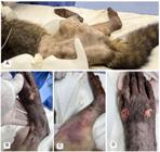
. Procyon cancrivorus at the Veterinary Hospital. (A): Low body score condition observed in Procyon cancrivorus. (B): Skin laceration on the right pelvic limb. (C): Areas of suffusion in the medial area of the pelvic limb. (D): Skin lacerations with bone exposure on the pelvic limb.
Samples were collected for blood count and biochemistry evaluation, revealing abnormalities including low hematocrits (22%), reduced red blood cells (4.15x10⁶ uL-1), high number of white blood cells (27.7x10³ uL-1), increased neutrophils (26.038x10³ uL-1), and elevated number of lymphocytes (13.85x10³ uL-1). Phosphorus (7.81mg dL-1) and blood urea nitrogen (65.35mg dL-1) were also high.
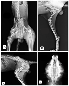
Radiographic examinations revealed highly comminuted fractures in the proximal and middle diaphysis regions of the right pelvic limb, together with metallic fragments suggestive of ballistic projectile in adjacent soft tissues (Figure 2A and 2C). Similarly, comminuted fractures were observed in the mid-diaphysis region of the contralateral limb, with a ballistic projectile detected in adjacent soft tissues (Figure 2B and 2C). No significant alterations were observed in Focused Assessment with Sonography in Trauma (A-FAST), thoracic and skull radiographic projections, except for the presence of ballistic projectiles in the soft tissues of the cervical region at the level of C1 (Figure 2D) and in the subcutaneous tissue of the left thoracic region at the level of T8.
Surgical therapy was deemed necessary in this case. Under sedative effect, general anesthesia was induced using a combination of propofol (5mg kg-1 IV) and midazolam (0.3 mg kg-1 IV). Orotracheal intubation was performed with tube number 5 after instilling 2% lidocaine (0.2 mL) in the periglottic region. After intubation, general anesthesia was maintained with sevoflurane (2-2,6%) diluted in oxygen 40-50%, in the circular valve circuit, and under mechanical ventilation in volume-controlled mode (Vt 10 mL kg-1, PEEP 5 cm H2O, RR to maintain EtCO2 35-45 mmHg). Anesthesia depth monitoring was done by clinical parameters (Guedel anesthesia stages and plans) and ECG, NIBP, SpO2, EtCO2, temperature and glucose were also monitored. Due to the evident clinical dehydration fluid therapy was maintained with LR in 10 mL kg-1 h-1 rate. Epidural anesthesia was administered at L7/S1 with ropivacaine 0.75% (0.2 mL kg-1) and morphine (0.1 mg kg-1), neuraxial block was performed with a 20 G Tuohy needle. Fluid bolus (LR 10 mL kg-1 IV in 10 minutes) were done 2 times and ephedrine bolus (0.1 mg kg-1 IV) for 4 times during the entire 5 hours of anesthesia procedure to maintain mean arterial blood pressure above 60 mmHg.
Antisepsis was performed using 2% chlorhexidine for degerming followed by 0.5% alcohol chlorhexidine. The distal part of the limb was isolated with sterile tubular mesh and covered with a sterile drape, exposing only the limb to be operated on. A craniolateral incision extending from the greater trochanter to the lateral condyle of the right femur was made (Figure 3A). Subcutaneous tissue was dissected, and an incision of the fascia lata was performed. The vastus lateralis was retracted cranially and biceps femoris caudally until the fracture site was visualized (Figure 3B and 3C). After removing all fibrous tissue from the region, distraction and alignment of the bone fragments were performed, followed by the positioning of a 2.7 mm bridge plate (Figure 3D and 3E) over the fracture site. Drilling was performed over the plate holes using a 2.0 mm pneumatic drill, followed by the placement of six locking screws (Figure 3F). For the synthesis, myorrhaphy was performed using poliglecaprone 25 2-0 in a Sultan pattern, followed by closure of the fascia with poliglecaprone 2-0 in a Reverdin pattern. The subcutaneous was reduced using a simple continuous pattern, and dermorrhaphy was performed with nylon 3-0 in a simple interrupted pattern.

Radiographic projections of Procyon cancrivorus. (A): Dorsoventral projection revealing bilateral Oblique fracture with two bone fragments in femur. (B): Right mediolateral projection showing highly comminuted fractures in the proximal and middle diaphysis regions and a fragmented metallic foreign body in the right femur. (C): Oblique fracture with two bone fragments in the mid-diaphysis region and presence of a metallic foreign body in the left femur. (D): Dorsoventral projection showing a metallic structure suggestive of a ballistic projectile located in the soft tissues of the cervical region at the level of C1.

Intraoperative osteosynthesis of the right femur in Procyon cancrivorus: Incision from the greater trochanter to the lateral condyle of the right femur (a). Exposure of the fracture site (b) and (c). Bridge plate (d). Positioning and implantation of the plate over the fractured bone (e) and (f).
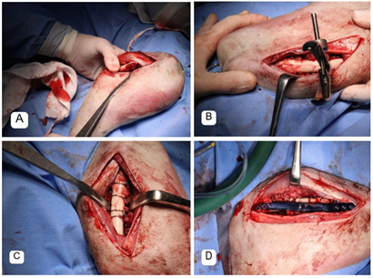
After completing the procedure on the right limb, the animal was positioned in right lateral recumbency for the procedure on the left pelvic limb. The same antisepsis technique described previously was used. A lateral incision extending from the greater trochanter to the lateral condyle of the left femur was made. Subcutaneous tissue was dissected, and an incision of the fascia lata was performed. The vastus lateralis retracted cranially and the biceps femoris caudally until the fracture site was visualized (Figure 4A). Blood clots were observed around the fracture site. After removing all fibrous tissue from the region, distraction and alignment of the bone fragments were performed (Figure 4B), placing four 0.8 mm cerclage wires around the femur, involving the bone fragments (Figure 4C). A 2.7 mm plate was positioned over the fracture site (Figure 4D). Drilling was performed over the plate holes using a 2.0 mm pneumatic drill, followed by the placement of six locking screws. Synthesis was performed using the same technique as in the contralateral limb.

Intraoperative osteosynthesis of the left femur in Procyon cancrivorus: Exposure of the fracture site (a). Alignment of the bone fragments (b). Implantation of cerclage wires (c). Implantation of the plate on the fractured bone (d).
During the surgical intervention, it was decided not to remove the ballistic projectiles from the affected areas as they were outside the surgical field obtained in the fracture correction, thus avoiding additional complications such as damage to adjacent structures and increased tissue trauma. Postoperatively, radiographic projections were obtained to confirm the proper positioning of the implants (Figure 5).

Post-surgical radiographs in Procyon cancrivorus. (A) 2.7 mm bridge plate and 6 locking screws in the right femur in the right mediolateral projection. (B) 0.8 mm cerclage wires around the femur and 2.7 mm neutralization plate with 6 locking screws in the left femur in left mediolateral projection.
The final intraoperative medications administered were Meloxicam (0.2 mg kg-1 SC), Dipyrone (25 mg kg-1 IV), Morphine (0.1 mg kg-1 IM) and Cefovecin (8 mg kg-1 SC). Postoperatively, the animal was treated with Gabapentin (70 mg kg-1 BID PO for 30 days), Dipyrone (25 mg kg-1 TID PO for 4 days), Tramadol (8 mg kg-1 TID PO for 4 days) and Meloxicam (0.75 mg SID PO for 3 days). The animal recovered from anesthesia after 1 hour.
A 16 days post-surgery consultation showed good recovery and muscle gain in the pelvic limbs allowing better locomotion. The radiographic evaluation upon return, compared to the day of the procedure, revealed a reduction in dead space between bone fragments. The hematocrit increased by 40%. The leukocytes decreased to 19.2 x 10³ uL-1, lymphocytes to 2.8 x 10³ uL-1, and neutrophils to 15.168 x 10³ uL-1. A 42 days post-surgery revealed suture dehiscence in both limbs with the presence of exudative secretion. The affected areas were cleaned with pressurized saline solution 0.9%, iodopovidone for degerment debridement, and closure of dead space with a simple continuous pattern suture using Caprofyl 3-0. Dermorrhaphy was performed using a simple interrupted pattern with 3-0 nylon. A swab sample was obtained from both limbs and cultured in Stuart medium for bacteriology and antibiogram.
Bacteriological culture identified Enterococcus sp., Enterobacter cloacae, and Staphylococcus sp. Antibiotic therapy was initiated with amoxilin and clavulanate (12.5 mg kg-1 BID-1 PO-1) due to sensitivity of the agents. The animal presented multiresistant strains of Enterococcus sp. and Enterobacter cloacae. The treatment was successful and the animal was then directed to physiotherapy and rehabilitation to return to the wild.
Results and discussion
Conflicts with humans in anthropized landscapes are an increasing problem for wildlife. The Crab-eating raccoon (Procyon cancrivorus), although adaptable to various environments, is particularly vulnerable to car collisions and retaliatory hunting, which often result in severe injuries such as fractures (Reid et al., 2016). Surgical interventions are frequently required to treat these injuries and rehabilitate the wildlife, thereby contributing to the conservation of the species.
In this case, a free-ranging P. cancrivorus was rescued after being found on the road and underwent surgical intervention. The Crab-eating raccoon is widely distributed across South America and is classified as Least Concern (LC) by the IUCN Red List (Leuchtenberger et al., 2023). However, its population is declining due to habitat fragmentation, retaliatory hunting, vehicle collisions, and conflicts with domestic animals (Garcês et al., 2021).
The initial physical examination revealed a cachectic body condition of one out of nine and dehydration. Blood samples collected for hematology and biochemistry evaluation showed pronounced anemia, leukocytosis, neutrophilia and lymphocytosis, which may indicate chronic infection and tissue necrosis (Siegel & Walton, 2020). Follow-up tests showed significant improvement, with hematocrit levels increasing by 40%, and reductions in leukocytes (19.2 x 10³ uL-1), lymphocytes (2.8 x 10³ uL-1), and neutrophils (15.168 uL-1), indicating the efficacy of the antibiotic treatment and therapeutic measures employed despite suture dehiscence due to wound infection.
Surgical treatment is crucial for femur fractures and may involve interlocking nails, plates with screws, cerclage rings, or a combination of methods (Lovrić et al., 2020; Günay et al., 2022). Successful rehabilitation examples included the release of five panthers back into the wild after surgery for long-bone fractures (Au Yong et al., 2018). For instance, one panther with an open, comminuted femoral fracture was stabilized with an 8-mm intramedullary interlocking nail, bolts, and cerclage wires, while another received double plating (Au Yong et al., 2018). Proper fracture healing requires strict confinement, which can be challenging in wildlife management (Au Yong et al., 2018). Using screws for fracture stabilization is effective against bending, rotation, and compression forces and is a cost-effective alternative to intramedullary wires combined with plates, offering similar biomechanical stability (Lovrić et al., 2020).
The surgical plan involved positioning of a 2.7 mm bridge plate and association of 1.5 mm Steinmann pin over the fracture site of the right femur and a 2.7 mm bridge plate associated with cerclage wires and 1.5 mm Steinmann pin on the left femur. The use of bridging plate techniques to treat comminuted femoral fractures resulted in a markedly shorter surgery time, also shorter anesthetics episode for patient (Johnson et al., 1998). The findings of this report substantiates the efficacy of this technique for this species and establishes shorter time for recovery and better chances.
In this case, the anesthesia protocol has also yielded a positive outcome. Epidural anesthesia was administered at the L7/S1 level using a combination of ropivacaine and morphine. This method has been shown to provide effective analgesia for both hind and front legs in dogs and has also been utilized for limb fractures in foxes (Valverde et al., 1989; Anagnostou et al., 2015). The neuroaxial block was successful, as the animal showed no signs of pain and was able to stand on its feet a few days after surgery.
Results
The increasing conflicts between wildlife and human activities in anthropized landscapes pose significant threats to species like the Crab-eating raccoon (Procyon cancrivorus). This case report highlights the critical role of surgical intervention in treating severe injuries such as fractures resulting from retaliatory hunting. The successful rehabilitation and recovery of the injured P. cancrivorous underscore the importance of advanced veterinary care and proper postoperative management in wildlife conservation efforts. Furthermore, the study illustrates the need for effective strategies to mitigate human-wildlife conflicts, ensuring the survival and well-being of vulnerable species.
Conclusion
This case demonstrates the effectiveness of bilateral femoral fracture stabilization and anesthetic management in the treatment of severe ballistic trauma in a free-ranging Procyon cancrivorus. Despite the complexity of the injuries and postoperative wound infection, appropriate surgical management, anesthesia, antimicrobial therapy, and rehabilitation resulted in satisfactory clinical recovery. This report highlights the importance of multidisciplinary veterinary care in improving functional outcomes and supporting the rehabilitation and potential release of severely injured wildlife.
Data availability
The data supporting the findings of this case report are included within the article. No additional datasets were generated or analyzed during the current study.
References
-
Anagnostou, T., Flouraki, E., Kostakis, C., Komnenou, A. & Prassinos, N.. (2015). Anesthetic management of a 4-month-old Red Fox (Vulpes vulpes) for orthopedic surgery. Journal of Zoo Wildlife Medicine, 46(1), 155-157. https://doi.org/10.1638/2014-75R1.1
» https://doi.org/https://doi.org/10.1638/2014-75R1.1 -
Reid, F., Helgen, K. & González-Maya, J. F. (2016). Procyon cancrivorus. The IUCN red list of threatened species 2016: e.T41685A45216426. Available in https://www.iucnredlist.org/species/41685/45216426
» https://www.iucnredlist.org/species/41685/45216426 -
Au Yong, J. A., Lewis, D. D., Citino, S. B., Cunningham, M. W., Cross, A. R., Farese, J. P. & Pablo, L. S.. (2018). Surgical management of appendicular long-bone fractures in free-ranging Florida panthers (Puma concolor coryi) six cases (2000-2014). Journal of Zoo Wildlife Medicine, 49(1), 162-171. https://doi.org/10.1638/2017-0058R1
» https://doi.org/https://doi.org/10.1638/2017-0058R1 -
Günay, E. & Altundağ, Y.. (2022). Humerus fracture treatment in a raccoon after dog attack. Acta Veterinaria Eurasia, 48(2), 156-160. https://doi.org/10.5152/actavet.2022.21039
» https://doi.org/https://doi.org/10.5152/actavet.2022.21039 - Siegel, A. & Walton, R. M. (2020). Hematology and biochemistry of small mammals. In K. E. Quesenberry & J. W. Carpenter (Eds.), Ferrets, Rabbits, and Rodents. (pp. 569-582). Saunders.
- Cherem, J. J., Kammers, M., Ghizoni-Jr, I. R. & Martins, A.. (2007). Mamíferos de médio e grande porte atropelados em rodovias do Estado de Santa Catarina, sul do Brasil. Biotemas, 20(3), 81-96.
-
Coelho, I. P., Kindel, A. & Coelho, A. V. P.. (2008). Roadkills of vertebrate species on two highways through the Atlantic Forest Biosphere Reserve, southern Brazil. European Journal of Wildlife Research, 54(4), 689-699. https://doi.org/10.1007/s10344-008-0197-4
» https://doi.org/https://doi.org/10.1007/s10344-008-0197-4 -
Fernandes-Ferreira, H., Mendonça, S. V., Albano, C. & Ferreira, F. S.. (2012). Hunting, use and conservation of birds in Northeast Brazil. Biological Conservation, 21(1), 221-244. https://doi.org/10.1007/s10531-011-0179-9
» https://doi.org/https://doi.org/10.1007/s10531-011-0179-9 - Freitas, S. H., Dória, R. G. S., Minto, B. W., Nardi, A. B., Melo, M. M., Camargo, L. M., Santos, M. D., Shimano, A. C., & Ambrósio, C. E. (2013). Haste intramedular modificada no tratamento de fratura diafisária de fêmur em cão - relato de caso. Revista Brasileira de Medicina Veterinária, 35(4), 323-328.
- Garcês, A., Prada, J., Silva, A., Silva, F., Santos, C. & Pires, I.. (2021). Pattern and distribution of fatal injuries in wildlife vehicle collision (2010-2019). Revista Electronica de Veterinaria, 22(1), 22-26.
-
Johnson, A. L., Smith, C. W. & Schaeffer, D. J.. (1998). Fragment reconstruction and bone plate fixation versus bridging plate fixation for treating highly comminuted femoral fractures in dogs: 35 cases (1987-1997). Journal of the American Veterinary Medical Association, 213(8), 1157-1161. https://doi.org/10.2460/javma.1998.213.08.1157
» https://doi.org/https://doi.org/10.2460/javma.1998.213.08.1157 -
Lau, R. A.. (2016). Caça esportiva e os efeitos do chumbo nas aves do sul do Brasil. Oecologia Australis, 20(4), 417-424. https://doi.org/10.4257/oeco.2016.2004.02
» https://doi.org/https://doi.org/10.4257/oeco.2016.2004.02 -
Litvaitis, J. & Tash, J.. (2008). An approach toward understanding wildlife-vehicle collisions. Journal of Environmental Management, 42(4), 688-697. https://doi.org/10.1007/s00267-008-9108-4.
» https://doi.org/https://doi.org/10.1007/s00267-008-9108-4. -
Lovrić, L., Kreszinger, M. & Pećin, M.. (2020). Surgical treatment of canine femoral fractures - a review. World’s Veterinary Journal, 10(2), 137-145. https://doi.org/10.36380/scil.2020.wvj18
» https://doi.org/https://doi.org/10.36380/scil.2020.wvj18 -
Navas-Suárez, P. E., Diaz-Delgado, J., Caiaffa, M. G., Silva, M. C., Yogui, D. R., Alves, M. H., Cereda, J. F., Silva, M. P., Cremer, M. J., Ascensão, F. J. P. M., Lorigados, C. A. B., Medici, E. P., Desbiez, A. L. J. & Catão-Dias, J. L.. (2022). Characterization of traumatic injuries due to motor vehicle collisions in neotropical wild mammals. Journal of Comparative Pathology, 197(1), 1-18. https://doi.org/10.1016/j.jcpa.2022.06.003
» https://doi.org/https://doi.org/10.1016/j.jcpa.2022.06.003 -
Leuchtenberger, C., Cheida, C. C., Dias, D. M., Abra, F. D. & Oliveira, T. G. (2023). Procyon cancrivorus. Sistema de avaliação do risco de extinção da biodiversidade - SALVE. Instituto Chico Mendes de Conservação da Biodiversidade. https://salve.icmbio.gov.br/salve/api/fichaHash/307749472f615945727464564f756c3154506a545a413d3d
» https://salve.icmbio.gov.br/salve/api/fichaHash/307749472f615945727464564f756c3154506a545a413d3d -
Silva, L. T. R., Santos, N. C. T., Valença, Y. M., Barros e Silva, T. C., Silva, N. L. G., Maschka, F. G. & Oliveira, A. A. F.. (2020). Análise das lesões promovidas por armas de ar comprimido em aves silvestres no nordeste do Brasil. Revista Brasileira de Ciências Criminais, 9(1), 36-42. https://doi.org/10.15260/rbc.v9i1.438
» https://doi.org/https://doi.org/10.15260/rbc.v9i1.438 -
Valverde, A., Dyson, D. H. & McDonell, W. N.. (1989). Epidural morphine reduces halothane MAC in the dog. Canadian Journal of Anaesthesia, 36(1), 629-632. https://doi.org/10.1007/BF03005412
» https://doi.org/https://doi.org/10.1007/BF03005412
Edited by
-
Associate Editor in charge:
Leandro Dalcin Castilha

 Source: Itbelongs to the collection of Universidade Federal de Minas Gerais.
 Source: It belongs to the collection of Universidade Federal de Minas Gerais.
 Source: It belongs to the collection of Universidade Federal de Minas Gerais.
 Source: It belongs to the collection of Universidade Federal de Minas Gerais.
 Source: It belongs to the collection of Universidade Federal de Minas Gerais.