 Anticancer activity and histological changes in albino male mice injected with gold nanoparticles
ABSTRACT.
Cancer is a global health problem and the second-leading cause of death worldwide. Gold nanoparticles (GNPs) are used in a wide range of medical fields, particularly in cancer diagnosis and treatment, therapy, and anti-inflammation. However, the question that always arises is whether GNPs affect normal, healthy cells when used as treatment. Therefore, GNPs were used in this research to treat muscle and brain cancer and to demonstrate its effect on tissues in living organs in healthy mice. The objectives were to determine the efficiency of GNPs in killing cancer cells through cytotoxicity assay, examine the effect of GNPs on different healthy organ tissues, and show the influence of different GNP concentrations, cell types, and incubation times through in vitro experiments. Two cell lines, AMGM (brain cancer cell line) and RD (muscle cancer cell line), at different concentrations (4.5×10¹² - 6×10¹⁰ particles mL-1) with two incubation periods (24 and 72h) were used in this study. Ten adult male albino mice were randomly distributed into two groups. The experiment was extended for 10 days to study the effect of a single dose of 250 µl of GNPs (4.5×10¹² particles mL-1) administered through intraperitoneal injection by examining the histological changes of the liver, kidney, and spleen of the mice. In the cytotoxicity experiment, high GNP concentrations had the greatest effect at 24h, and the RD cell line was more sensitive than the AMGM cell line at 72h. Histological tests of the mouse organs revealed different changes such as severe hepatic granulomas, renal apoptosis, amyloidosis of spleen tissues, and inflammation with fibrosis of intestinal tissues. GNPs are an influential factor in cancer therapy, and their effects vary with cell line type, exposure time, and dose. They also cause histological harm (fibrosis, inflammation, and apoptosis) on vital organs.
Keyword:
gold nanoparticles; cytotoxicity; histological changes; albino mice
Introduction
According to the Global Cancer Statistics in 2024 (Bray et al., 2024), approximately 9.7 million people die from cancer worldwide in 2022. By 2050, the number of patients with cancer will increase to 35 million worldwide (Siegel et al., 2024). The signs and symptoms of cancer vary depending on the individual’s immunity and what part of the body is affected. The traditional treatments for cancer include chemotherapy, radiation, and surgery, which have many side effects, each strategy has ongoing issues, because the tumor's heterogeneity, surgery has difficulty removing it completely, chemotherapy deals with drug resistance and negative effects and, radiation therapy has precise challenges and restricted access in some places (Zafar et al., 2025). Hence, cancer detection and treatment methods, especially at an early stage, have become the main focus of the researchers.
The shape, physical, chemical, and mechanism of noble metal nanoparticles have been the basis for the development and synthesis of numerous nanomedicines for cancer (Zhao et al., 2022). Nobel metals such as gold, silver, and platinum have attracted the attention of researchers due to their unique properties and potential applications in different fields.
Gold nanoparticles (GNPs) emerge as critical agents in the development of treatments for many diseases, particularly cancer, and in a variety of technical fields, demonstrating their versatility in drug transport, photothermal therapy, and diagnostics. Their diverse applications demonstrate their promise to transform healthcare, environmental monitoring, catalysis, and other fields by providing novel solutions to complex problems (Hossain et al., 2024).
Studies have focused on controlling the size and shape of nanoparticles to facilitate their entry into normal and cancer cells. GNPs function well against a range of infections and have gained popularity because of their anti-inflammatory, antibacterial, anticancer, and antioxidant activities (Ali et al., 2020; Abdallaha, 2021; Farman, 2024; Chick, 2025). In addition to their physiochemical properties, nanoparticles with a neutral or negative surface charge have a long circulation period, and positively charged nanoparticles have a strong attraction for cells and good cellular absorption; this trade-off represents a substantially problem in optimizing surface charge for theranostic applications (Ow et al., 2025). Furthermore, due to their nanosize and stability, nanoparticles can pass through cell membranes, interact with intracellular components, and finally trigger cell death during the multiplication phase (Dheyab et al., 2023).
This study aimed to assess the efficiency of GNPs as anticancer by conducting an MTT assay of two cell lines. The histological changes occurring in vital organs after GNP injection were also examined using male mice. Finally, the toxicity of nanomaterials, even if taken in small concentrations, was assessed by in vitro and in vivo studies.
Materials and methods
Gold nanoparticles (GNPs) was purchased from Sigma Aldrich Company with 10 nm, spherical shape and at concentration 6× 10¹² particles mL-1.
Cell culture
Two cancer cell lines, human cerebral glioblastoma- Multiforme primary culture (AMGM) and Rhabdomyosarcoma cell lines (RD) were kindly provided by the Iraqi Center for Cancer and Medical Genetic Research (ICCMGR, Baghdad, Iraq) and used throughout this study. RD cell line was cultivated on a minimal essential medium (MEM, Sigma), the AMGM cell line was propagated and maintained on Rosswell Park Memorial Institute medium (RPMI-1640, US biological) (Freshney, 2002). These media were incubated at 37ºC in a humidified 5% CO2 incubator (Heracell 150, Thermo Electron Corp.) with 10% fetal bovine serum (FBS) (Cellgro, USA) and 1% penicillin/streptomycin (Cellgro, USA) added. Once the cells reached 80-90% confluence, as seen under an inverted microscope (Nicon Eclipse TS100), they were subcultured. Using the trypan blue (Pharma, Sweden) exclusion test, cell viability was evaluated and determined to be more than 99% (Phelan, 1999).
Cytotoxicity measurement of gold nanoparticles (GNPs)
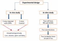
The inhibition of proliferation rate (IR%) was used to analyze the cytotoxicity assay for cancer cell lines. Plant 2 x 10⁴ cells per well in 200 µL of standard growth medium without antibiotics that has been enhanced with FBS in a 96-well tissue culture plate. The cells should be kept in a CO2 incubator at 37°C until they are 60-80% confluent. Usually, this will take 18 to 24 hours. Next, the cells were exposed to varying concentrations of GNPs (4.5×10¹²-6×10¹⁰ particles mL-1) in 100 µL, and it was incubated for 24 and 72 hours at 37ºC.
The untreated cells were used as a control. Following incubation, 20 µL of MTT [3-(4, 5- dimethylthiazol-2-yl-2-2.5- diphenyl tetrazolium bromide)] was added, and the cells were incubated for an additional three hours at 37ºC. At 492 nm, the absorbance of both treated and untreated cells was determined. The formula used to determine the inhibitory rate of cell growth was IR%= A-B/A×100, where A is the absorbance of untreated cells and B is the absorbance of treated cells. The GNP concentration was represented on the X-axis, while the IR% values were plotted on the Y-axis. Graph pad Prism software was used to do non-linear regression analysis and identify the concentrations that resulted in a 50% decrease in cell growth (IC 50) (Gao et al., 2003).
Histopathological experiment
Ten albino mice 5-7 weeks in age, weighing 25-30 g each, animals will be divided into two groups, five mice as control and the other five mice injected with IP/daily with GNPs at concentration of 4.5 ×10¹² P mL-1 for 10 days. All mice received sterilized food and water for 10 days.
After 10 days, every mouse was murdered by periodically dislocating its neck and scarifying to take the liver, kidney, intestine and spleen. The organs will be dissected, washed in phosphate buffer saline, fixed in formalin (10%) to prevent any changes in tissue structure and for histological assessment. Tissues will be subject to histopathological examination that will be carried out according to (Bancroft & Gamble, 2007). Slides were then examined by taking different fields randomly (10, 20 and 40x), so, the macroscopic abnormal changes (necrosis, congestion, degeneration, regeneration, swelling, lesions and others) of the organ were recorded.
The procedure of the histopathological experiment was approved by our college committee for animal protection as per the guidelines of the ethical approval registered under the number 980 in 2024.
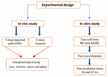
Experimental design (Figure 1)
Statistical analysis
The Statistical Packages of Social Sciences-SPSS-2019 (George & Mallery, 2024) program was used to detect the effect of difference factors (Cell lines, concentrations with three replication and times) in study parameters. LSD-Least significant difference was used to significant compare between means (ANOVA two ways) in this study.
Results
Cytotoxicity results
The findings of the present study demonstrated that the GNPs considerably reduced AMGM cell growth when compared to untreated control cells, and also found that the growth inhibition seemed to be concentration and exposure duration dependent.
Table 1 shows high inhibition rates for the AGMG cell line at (52.3 and 56.7%) for the concentrations (4.5×10¹² and 1.8×10¹¹ particles mL-1) at 24 and 72h of exposure time. However, significant difference (p<0.05) was observed between concentrations and exposure times at the concentrations (5.4×10¹¹ and 6×10¹⁰ particles mL-1). Non-significant variations were found after 24 and 72h of exposure time.
Similar to these results, the highest inhibition rate for the RD cell line was 65.7% after exposure to the concentration (4.5×10¹² particles mL-1) at 24h and 85.2% after exposure to the concentration (1.8×10¹¹ particles mL-1) at 72h. Significant difference (p<0.05) was observed between concentrations and exposure times (Table 2).
Table 3 illustrates the compression between the two cell lines exposed to different GNP concentrations for 24h. At the concentrations (4.5×10¹², 1.8×10¹¹and 6×10¹⁰ particles mL-1) of GNPs, the inhibition rates for the RD cell line were higher than those for the AMGM cell line. Meanwhile, GNPs at the concentrations (1.5×10¹² and 5.4×10¹¹ particles mL-1) had a great effect on the AMGM cell line. Therefore, significant difference was observed for all the concentrations expect the fourth concentration (p<0.05).
The inhibition rate for the RD cell line was higher than that for the AMGM cell line after 27h of exposure. Significant variation in inhibition rate (p<0.05) was observed under all the concentrations during this exposure time (Table 4).
Histopathological study results
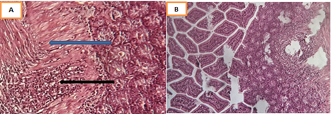
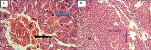
A histological study was conducted on the organ tissues of BALB male mice injected with GNPs. The intestinal tissues showed severe inflammatory reactions, with the lymphocytes in lamina propria and granular regions indicating the inflammation of the lymphoid tissues of intestinal tract (Figure 2). Meanwhile, the histological changes of liver appeared in the form of granuloma reaction, hepatocyte vacuolation, cytoplasmic degeneration, necrotic foci, Kupffer cell activation, hemorrhage, and infiltration of inflammatory cells. In addition, severe granuloma reaction was found near the congested blood vessels, and some inflammatory cells, mainly monocytes, appeared inside the central vein (Figure 3).
The changes in kidney tissues included apoptosis in epithelial lining renal tubules after sloughing into the lumen. Other cells showed swelling with occlusion of their lumen. However, some cells were not affected and showed no clear changes (Figure 4).
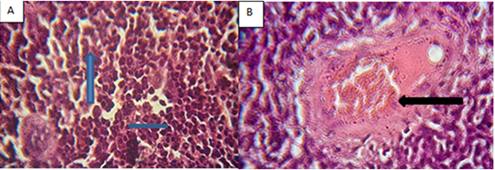
Figure 5 shows the changes in spleen tissues. Immunity reaction appeared as megakaryocyte activation with emargination of lymphoma follicles. Dark pink materials suspected as amyloidosis appeared in other fields.

A. Showed histological changes of intestine tissues of injected mice with AuNPs, intestine tissue showed severe inflammatory reaction (black arrow), mainly inflation of lymphocytes in lamina propria and glandular regions indicating inflammation of lymphoid tissue of intestinal tract with fibrosis (blue arrow), (20x, H & E). B. Clear lose in glandular region due to sever inflammatory response in lamina properia layer (20x, H & E).
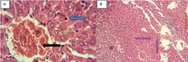
A. Showed histological changes of liver tissue in injected mice with GNPs, necrosis with inflammatory infiltration, mainly kupffer cells (blue arrow) and outside congested (black arrow) central vein, hepatitis vacuolation (green arrow) (40x, H & E), B. Showed sever granuloma,s reaction (purple arrow) and inflammatory cells (brown arrow) (20x, H & E).
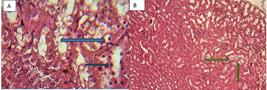
A. Showed histological changes in kidney tissue of injected mice with GNPs, Clear apoptosis in epithelial lining renal tubules (blue arrow) after slouphing in to lumen others showed swelling with occlusion their lumen (40X, H & E), B: Degeneration of some renal appears empty contained desquamated epithelial cells (green arrow), (10X, H & E).

A. Showed histological changes of spleen tissues of injected mice with GNPs, immunity reaction appears as megakaryocytes (blue arrow) activation (20x, H & E), B: Showed emarigenation of lymphomas follicles with dark pink materials (black arrow) suspected amyloidosis as well as appears in others fields clearly (40x, H & E).
Discussion
GNPs have the potential to be used in cancer treatment due to their radiation sensitivity, biostability, and simplicity of surface modification.
In this study, we show that GNPs can inhibit the activation effect of RD and AMGM cell lines. This inhibition may occur through several pathways, such as changes in cytoskeletons. This phenomenon is linked to the breakdown of actin filaments caused by GNPs or the suppression of the migration and proliferation of stimulated endothelial cells by downregulating vascular endothelium growth factors and altering cell shape (Pan et al., 2014).
This study observed the time dependence of the inhibition rate. The value increased after 72h of exposure compare with that after 24h. This phenomenon may be due to the effect of GNPs on the can cer cells by suppressing DNA synthesis in the S phase and consequently decreasing the number of cells that enter the G2/M phase. Hence, apoptosis is induced, especially with prolonged incubation time. In addition, GNPs were detected inside the cytostomes, nucleus, and endoplasmic reticulum and widely distributed in the cytoplasm of necrotic cells at 3, 12, and 24h. The GNPs were also internalized in the cells’ cytoplasm and gathered in the cytoplasmic vacuoles at 24h (Tsai et al., 2011; Huang et al., 2014). As long as the GNPs enter the cells and kill them within 24 h, they can kill a great quantity of cells within 72h.
Mustafa et al. (2011) investigated how the concentration of nanoparticles affects the uptake of MC3T3-E1 osteoblastic cells. Even after the incubation period, the particles appeared to be concentrated around the nucleus and did not penetrate the nuclear area. This phenomenon could be attributed to the two-layered structure of the nuclear membrane.
According to the transmission electron microscopy (TEM) study, GNPs can independently disperse in the plasma membrane at low concentrations. However, at high concentrations, the cells ingest GNPs through endocytosis, consuming the large clusters on the apical surface of cell plasma membranes.
Sawalha et al. (2020) pointed that because their density rises, nanoparticle agglomerates create an internalized cluster around the cell membrane, instead of single particles diffused at high concentrations and possibly ingested through endocytosis. This finding explains why the low concentrations in the present study caused greater inhibition compared with the high concentrations.
This study found that GNPs were more effective on the RD cell line than on the AMGM cell line. Other works suggested that GNPs can stop the growth of various cancer cell lines; hence, the inhibition varies depending on the type of cell line and GNPs have less impact on healthy cells (Hussein, 2016, Aldabbagh et al., 2024). By conducting an immunohistochemistry assay, (Hussein et al., 2023) proved that FOXP1 can be used as a biomarker for the prognosis outcome of breast and other solid tumors. Treatment with GNPs plus laser is more effective against the tumors compared with GNPs alone. The physicochemical interactions of gold atoms with the nitrogen bases and phosphate groups of DNAs have been linked to the cytotoxic effect of GNPs (Siddique & Chow, 2020; Mostafavi et al., 2022). Small GNPs exhibit improved renal clearance, tumor tissue permeability, cell uptake, and effective entry into nuclei, so surface modifications and functionalization are critical in improving the compatibility of ultrasmall GNPs in physiological environments, maximizing their effectiveness for cancer cell killing, and reducing damage to healthy tissues (Panwar et al., 2024). The difference in response to GNPs between the RD and AMGM cell lines may be due to the difference in their capacities to respond to varying ROS levels. In addition, the RD cell line might be relatively sensitive to the DNA-damaging agents.
Many histological changes occurred in the organ tissues due to GNP injection. These changes were evident when we examined the liver cells, which showed signs that the hepatocytes and Kupffer cells were harmed by GNP toxicity and were unable to handle the residues that accumulated as a result of the structural and metabolic disruptions caused by GNPs (Abdelhalim & Jarrar, 2012; Kassab et al., 2021). In the kidney, the histological section showed many alterations such as apoptosis, swelling, and sloughing, which may be due to oxidative stress and cellular interaction induced by the GNPs and led to tubular degeneration and a necrotic state (Fadia et al., 2022). By contrast, (Peres et al., 2023) pointed that GNP treatment prevents tubule-interstitial injury induced by subclinical acute kidney injures. Therefore, these particles are not nephrotoxic to healthy mice. Low concentrations of GNPs are more effective than high concentrations, whether in cells or in tissues, perhaps because GNPs at low concentrations easily enter cells and tissues and cause cellular damage. Meanwhile, GNPs at high concentrations may cause aggregation, which hinders their entry into cells.
The pathological alterations in the spleen manifested as immunity reaction due to distorted lymphoid architecture, minimized lymphoid follicles, and appearance of megakaryocyte and amyloidosis (Ibrahim et al., 2018). Some researchers showed that GNPs specifically target the spleen, resulting in notable pathological alterations to its architecture that starts on day 2 and continues until day 8 for medium- and large-sized (Ibrahim et al., 2018).
GNPs may exhibit toxicity through oxidative mechanisms, which include membrane damage, molecular damage, inflammation, DNA damage, and eventually cell death, or nonoxidative mechanisms, which include morphology disruption, mitochondrial damage, DNA damage, and cell death pathways. However, a previous study suggested the contrary and reported that glutathione and malondialdehyde, two indicators of oxidative stress, were not substantially affected by any of the GNP dosages in any of the organs (Niżnik et al., 2024). We believe that nanoparticles negatively affect the histological structure of body organs in humans and animals and particularly the cellular structure. They also exhibit positive effects on cancer cells, but only at specific concentrations. The emerging controversy over the toxicity of nanoparticles, especially GNPs, limits their widespread use at high concentrations in animals, especially albino mice, which are sensitive and intolerant to any external symptoms.
Conclusion
GNPs have a killer effect on different types of cancer cells and a pathological effect on vital organs even at low concentrations. The detrimental effects of GNPs on various organs of mice depend on their size and dose. This study also highlights the importance of in vivo and long-term research to fully comprehend the effects of prolonged exposure to GNPs.Various investigations are required to help and understand the importance and achievements of GNPs as potent tools for next-generation cancer treatment and their effectiveness in different organ tissues. Experiments employing different animals, concentrations, doses, and other factors are warranted to establish the effect of GNPs in vitro and in vivo. The toxicity of GNPs may result from the chemicals used as a reducing agent in gold synthesis. Therefore, the current study suggests the use of biologically synthesized GNPs, which may have a less toxic effect.
Data availability
Data is available with the corresponding author and it will be made available on request
References
-
Abdallaha, B. F., Younus, M. A. & Ibraheem, I. J. (2021). Preparation, characterization, antimicrobial and antitumor activity of chitosan schiff base/PVA/PVP Au, Ag nanocomposite in treatment of breast cancer cell line. Nanomedicine Research Journal, 6(4), 369-384. https://doi.org/ https://doi.org/10.22034/nmrj.2021.04.007
» https://doi.org/https://doi.org/ https://doi.org/10.22034/nmrj.2021.04.007 -
Abdelhalim, M. A. K. & Jarrar, B. M.. (2012). Histological alterations in the liver of rats induced by different gold nanoparticle sizes, doses and exposure duration. Journal of Nanobiotechnology, 10(1), 1-9. https://doi.org/10.1186/1477-3155-10-5.
» https://doi.org/https://doi.org/10.1186/1477-3155-10-5. -
Ali, M. M., Ali, M. M. & Mohammed, A. M.. (2020). Synthesis of gold nanoparticles by using batch method and study its antibacterial activity. Baghdad Science Journal, 17(2), 663-641. https://doi.org/10.21123/bsj.2020.17.2(SI).0633
» https://doi.org/https://doi.org/10.21123/bsj.2020.17.2(SI).0633 -
Aldabbagh, M., Abdallaha, B. F., Abdulla, N. Y. & Ibraheem, I. J.. (2024). Synthesis, characterization and anticancer activity of chitosan schiff base/PEG blend doped with gold and silver nanoparticles in treatment of breast cancer cell line MCF-7. Iraqi Journal of Pharmaceutical Sciences, 33(2), 101-111. https://doi.org/10.31351/vol33iss2pp101-111
» https://doi.org/https://doi.org/10.31351/vol33iss2pp101-111 - Bancroft, J. D. & Gamble, M. (2007). Theory and practice of histological techniques. Churchill Livingstone.
- Chick, C. N., Takano, M., Meva, F. E. A. & Usuki, T. J. M. A.. (2025). Synthesis of gold nanoparticles using Eutrema japonicum (Wasabi): antioxidant and anti-inflammatory studies. Materials Advances, 6(1), 2365-2370.
-
Bray, F., Laversanne, M., Sung, H., Ferlay, J., Siegel, R. L., Soerjomataram, I. & Jemal, A.. (2024). Global cancer statistics 2022: GLOBOCAN estimates of incidence and mortality worldwide for 36 cancers in 185 countries. CA: A Cancer Journal for Clinicians, 74(3), 229-263. https://doi.org/10.3322/caac.21834
» https://doi.org/https://doi.org/10.3322/caac.21834 -
Dheyab, M. A., Aziz, A. A., Oladzadabbasabadi, N., Alsaedi, A., Braim, F. S., Jameel, M. S., Ramizy, A., Alrosan, M. & Almajwal, A. M. J. M. (2023). Comparative analysis of stable gold nanoparticles synthesized using sonochemical and reduction methods for antibacterial activity. Molecules, 28(9), 3931. https://doi.org/10.3390/molecules28093931
» https://doi.org/https://doi.org/10.3390/molecules28093931 - Fadia, B. S., Mokhtari-Soulimane, N., Meriem, B., Wacila, N., Zouleykha, B., Karima, R., Soulimane, T., Tofail, S. A., Townley, H. & Thorat, N. D.. (2022). Histological injury to rat brain, liver, and kidneys by gold nanoparticles is dose-dependent. ACS Omega, 7(24), 20656-20665.
-
Farman, M. A. & Abood, W. N.. (2024). Evaluation of anti-bacterial activity of gold nanoparticles in-vitro. Diyala Journal for Veterinary Sciences, 2(4), 53-60. https://doi.org/10.71375/djvs.2024.02404
» https://doi.org/https://doi.org/10.71375/djvs.2024.02404 -
Freshney, R. I.. (2002). Cell line provenance. Cytotechnology, 39(2), 55-67. https://doi.org/10.1023/A:1022949730029
» https://doi.org/https://doi.org/10.1023/A:1022949730029 -
Gao, S., Yu, B., Li, Y., Dong, W. & Luo, H.. (2003). Antiproliferative effect of octreotide on gastric cancer cells mediated by inhibition of Akt/PKB and telomerase. World Journal of Gastroenterology, 9(10), 2362-2365. https://doi.org/10.3748/wjg.v9.i10.2362
» https://doi.org/https://doi.org/10.3748/wjg.v9.i10.2362 - George, D. & Mallery, P. (2024). IBM SPSS statistics 29 step by step: a simple guide and reference. Routledge.
-
Hossain, A., Rayhan, M. T., Mobarak, M. H., Rimon, M. I. H., Hossain, N., Islam, S. & Al Kafi, S. A. J. R. I. C.. (2024). Advances and significances of gold nanoparticles in cancer treatment: a comprehensive review. Chemistry, 8(1), 101559. https://doi.org/10.1016/j.rechem.2024.101559
» https://doi.org/https://doi.org/10.1016/j.rechem.2024.101559 -
Huang, Y., Yang, Y., Yang, K., Shieh, H., Wang, T., Hwu, Y. & Chen, Y.. (2014). Pegylated gold nanoparticles induce apoptosis in human chronic myeloid leukemia cells. BioMed Research International, 2014, 182353. https://doi.org/10.1155/2014/182353
» https://doi.org/https://doi.org/10.1155/2014/182353 -
Hussein, S. I.. (2016). Study the effect of gold nanoparticles on cancer and normal cells (in vitro study). Iraqi Journal of Cancer and Medical Genetics, 9(2), 144-149. https://doi.org/10.29409/ijcmg.v9i2.186
» https://doi.org/https://doi.org/10.29409/ijcmg.v9i2.186 -
Hussein, S. I., Al-Yasiri, A. Y., Hassan, H. F., Kashman, B. M. & Azeez, R. A.. (2023). Immunohistochemistry technique for effect of gold nanoparticles, laser, and photodynamic therapy on FoxP1 level in infected mice with mammary adenocarcinoma. Lasers in Medical Science, 38(1), 106. https://doi.org/10.1007/s10103-023-03765-7
» https://doi.org/https://doi.org/10.1007/s10103-023-03765-7 -
Ibrahim, K. E., Al-Mutary, M. G., Bakhiet, A. O. & Khan, H. A.. (2018). Histopathology of the liver, kidney, and spleen of mice exposed to gold nanoparticles. Molecules, 23(8), 1848. https://doi.org/10.3390/molecules23081848
» https://doi.org/https://doi.org/10.3390/molecules23081848 -
Kassab, A. A., Moustafa, K. A. A., Ragab, M. H. & Ragab, A. M. H.. (2021). The biological effect of different doses of gold nanoparticles on the liver of female rats: a histological and immunohistochemical study. Egyptian Journal of Histology, 44(2), 489-502. https://doi.org/10.21608/ejh.2020.37340.1335
» https://doi.org/https://doi.org/10.21608/ejh.2020.37340.1335 -
Mostafavi, E., Zarepour, A., Barabadi, H., Zarrabi, A., Truong, L. B. & Medina-Cruz, D.. (2022). Antineoplastic activity of biogenic silver and gold nanoparticles to combat leukemia: beginning a new era in cancer theragnostic. Biotechnology Reports, 34(1), e00714. https://doi.org/10.1016/j.btre.2022.e00714
» https://doi.org/https://doi.org/10.1016/j.btre.2022.e00714 -
Mustafa, T., Watanabe, F., Monroe, W., Mahmood, M., Xu, Y., Karmakar, A., Casciano, D., Ali, S. & Biris, A. S.. (2011). Impact of gold nanoparticles concentration on their cellular uptake by MC3T3-E1 mouse osteoblastic cells as analyzed by transmission electron microscopy. Journal of Nanomedic & Nanotechnol, 2(6), 1-7. https://doi.org/10.4172/2157-7439.1000118
» https://doi.org/https://doi.org/10.4172/2157-7439.1000118 -
Niżnik, Ł. Noga, M., Kobylarz, D., Frydrych, A., Krośniak, A., Kapka-Skrzypczak, L. & Jurowski, K.. (2024). Gold nanoparticles (AuNPs) - toxicity, safety and green synthesis: a critical review. International Journal of Molecular Sciences, 25(7), 4057. https://doi.org/10.3390/ijms25074057
» https://doi.org/https://doi.org/10.3390/ijms25074057 -
Ow, V., Lin, Q., Wong, J. H. M., Sim, B., Tan, Y. L., Leow, Y., Goh, R. & Loh, X. J. J. N.. (2025). Understanding the interplay between pH and charges for theranostic nanomaterials. Nanoscale, 17(1), 6960-6980. https://doi.org/10.1039/d4nr03706e
» https://doi.org/https://doi.org/10.1039/d4nr03706e -
Pan, Y., Wu, Q., Liu, R., Shao, M., Pi, J., Zhao, X. & Qin, L.. (2014). Inhibition effects of gold nanoparticles on proliferation and migration in hepatic carcinoma-conditioned HUVECs. Bioorganic & Medicinal Chemistry Letters, 24(2), 679-684. https://doi.org/10.1016/j.bmcl.2013.11.045
» https://doi.org/https://doi.org/10.1016/j.bmcl.2013.11.045 -
Panwar, C., Phillips, A., Sangal, A. & Gupta, D.. (2024). Synthesis of gold nanoparticles and their applications in cancer therapy. Biointerface Research in Applied Chemistry, 14(1), 1-27. https://doi.org/10.33263/BRIAC141.012
» https://doi.org/https://doi.org/10.33263/BRIAC141.012 -
Peres, R. A., Silva-Aguiar, R. P., Teixeira, D. E., Peruchetti, D. B., Alves, S. A., Leal, A. B. C., Castro, G. F., Ribeiro, N. B., Guimarães, F. V., Pinheiro, A. A. S., Martins, M. A. & Caruso-Neves, C.. (2023). Gold nanoparticles reduce tubule-interstitial injury and proteinuria in a murine model of subclinical acute kidney injury. Biochimica et Biophysica Acta-General Subjects, 1867(4), 130314. https://doi.org/10.1016/j.bbagen.2023.130314
» https://doi.org/https://doi.org/10.1016/j.bbagen.2023.130314 - Phelan, M. C. (1999). Techniques for mammalian cell tissue culture. Current Protocols in Toxicology, 2(suppl. 38), A.3B.1-A.3B. 19.
-
Sawalha, S. M., Ghadhban, E. & Al-Dahan, Z. T.. (2020). Impact of Gold nanoparticles sizes and concentrations on the Rhabdomyosarcoma cells. IOP Conference Series: Materials Science and Engineering, 870(1), 012022. https://doi.org/10.1088/1757-899X/870/1/012022
» https://doi.org/https://doi.org/10.1088/1757-899X/870/1/012022 -
Siddique, S. & Chow, J. C.. (2020). Gold nanoparticles for drug delivery and cancer therapy. Applied Sciences, 10(11), 3824. https://doi.org/10.3390/app10113824
» https://doi.org/https://doi.org/10.3390/app10113824 -
Siegel, R. L., Giaquinto, A. N. & Jemal, A.. (2024). Cancer statistics, 2024. CA: A Cancer Journal for Clinicians, 74(1), 12-49. https://doi.org/10.3322/caac.21820
» https://doi.org/https://doi.org/10.3322/caac.21820 -
Tsai, Y., Huang, Y., Chao, Y., Hu, K., Chin, L., Chou, S., Hour, A., Yao, Y., Tu, C., Liang, Y., Tsai, C., Wu, H., Tan, S. & Liang, Y.. (2011). Identification of the nanogold particle-induced endoplasmic reticulum stress by omic techniques and systems biology analysis. ACS Nano, 5(12), 9354-9369. https://doi.org/10.1021/nn2027775
» https://doi.org/https://doi.org/10.1021/nn2027775 -
Zafar, A., Khatoon, S., Khan, M. J., Abu, J. & Naeem, A. J. D. O.. (2025). Advancements and limitations in traditional anti-cancer therapies: a comprehensive review of surgery, chemotherapy, radiation therapy, and hormonal therapy. Discover Oncology, 16(1), 607. https://doi.org/10.1007/s12672-025-02198-8
» https://doi.org/https://doi.org/10.1007/s12672-025-02198-8 -
Zhao, R., Xiang, J., Wang, B., Chen, L. & Tan, S.. (2022). Recent advances in the development of noble metal nps for cancer therapy. Bioinorganic Chemistry and Applications, 2022, 2444516. https://doi.org/10.1155/2022/2444516
» https://doi.org/https://doi.org/10.1155/2022/2444516