Spermatogonial stem cell markers and distribution in different regions of the testis of adult Nile tilapia (Oreochromis niloticus)
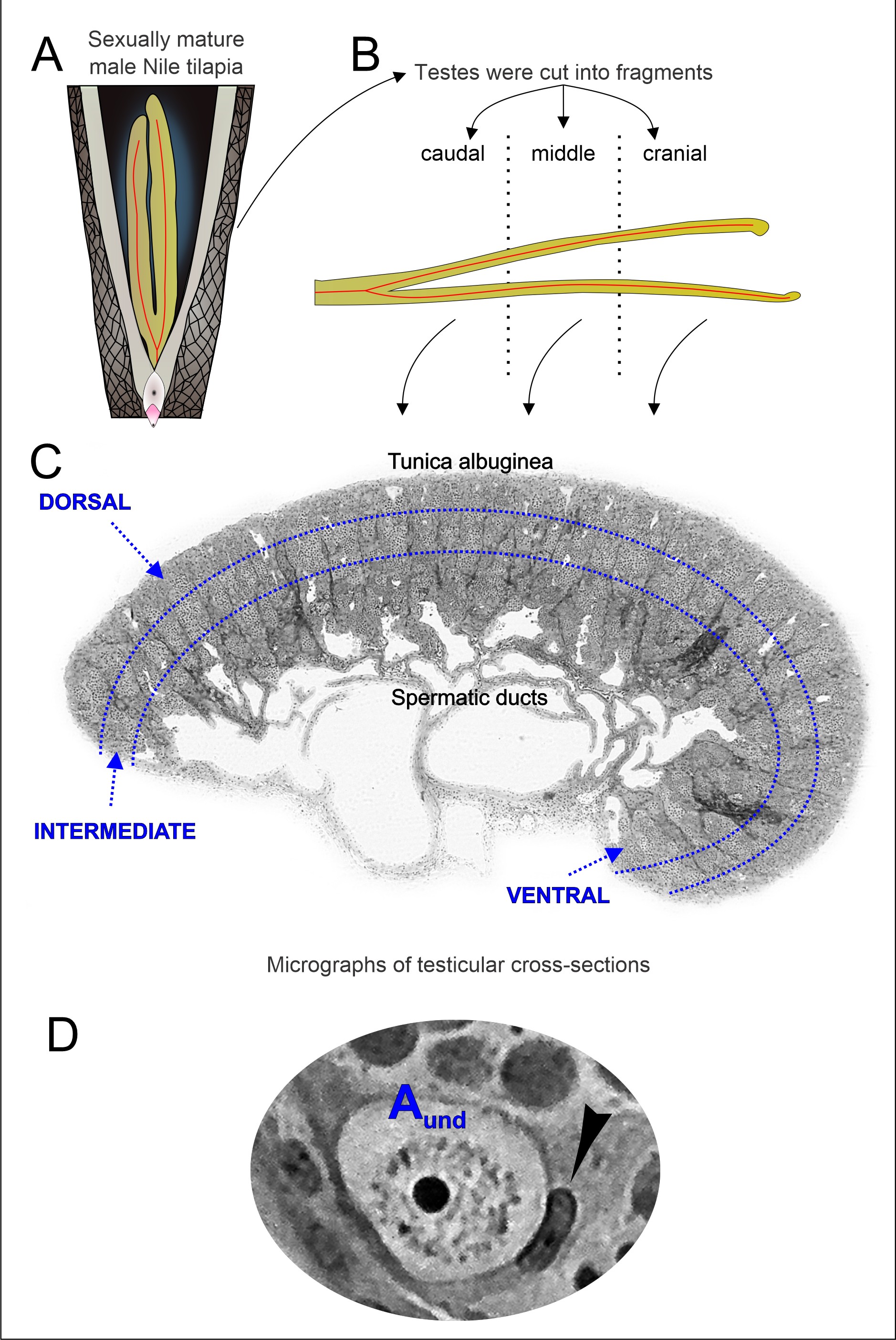
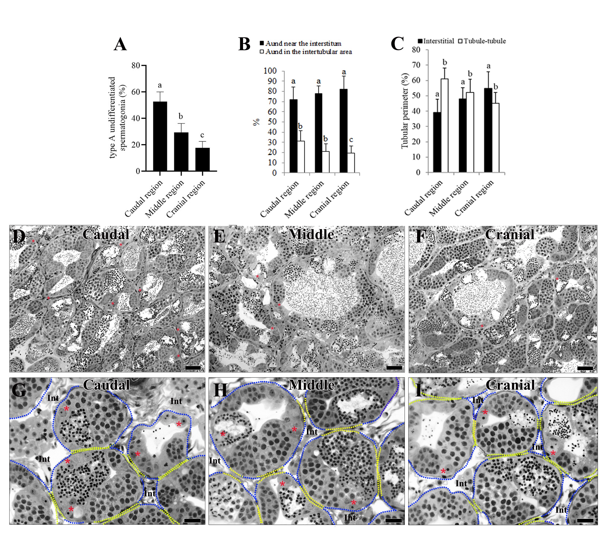
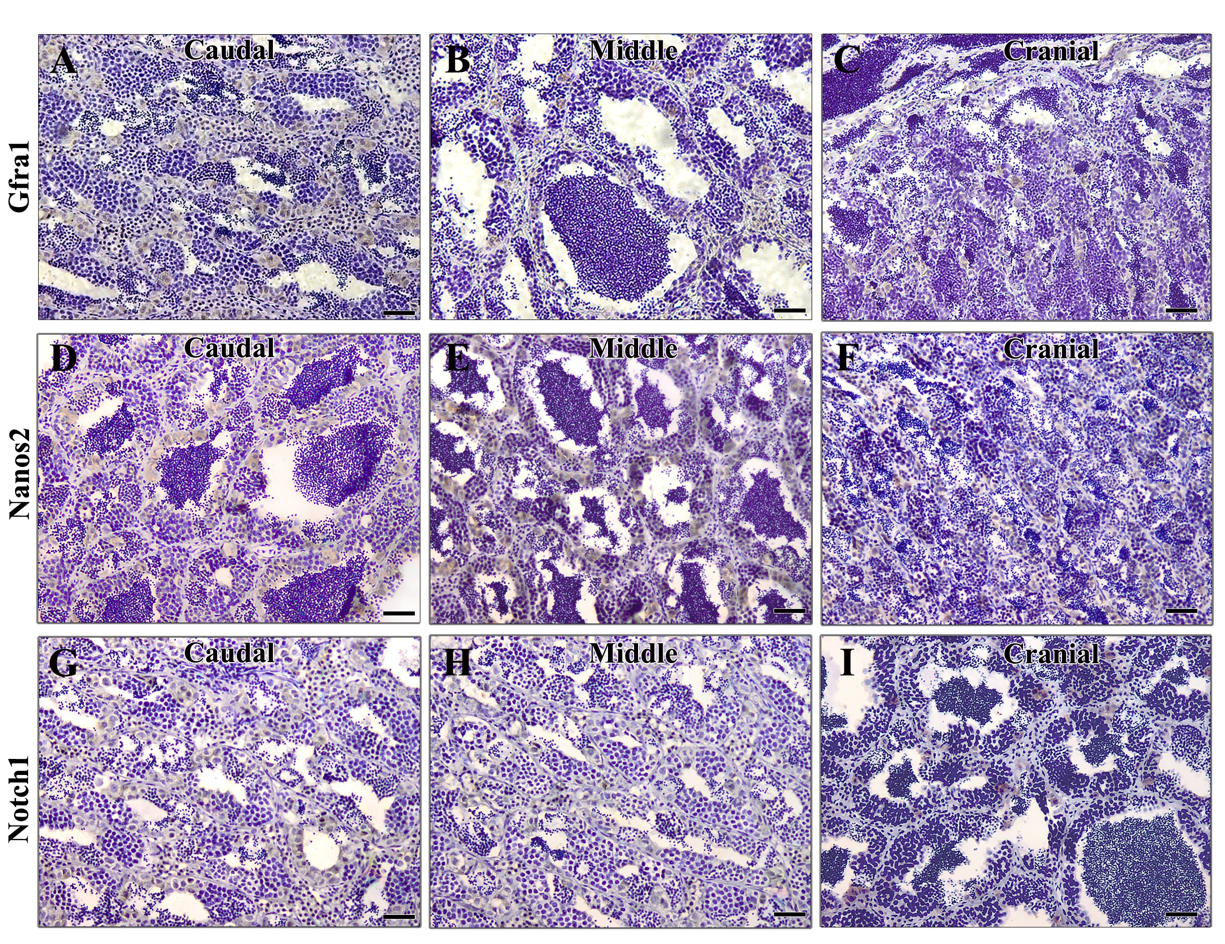
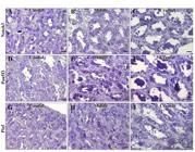
Highly conserved vertebrate molecular markers, such as Gfra1, Pou5f3, Notch1, Plzf, and Nanos2, represent important candidates for evaluating spermatogonial stem cells (SSCs) physiology and preferential location in the fish testis. In tilapia, germ cell cysts are distributed within seminiferous tubules oriented along the dorso–ventral axis, while the adult testis continues to grow along both the longitudinal (cranio–caudal) and dorso–ventral axes. Herein we investigated the spatial distribution of undifferentiated spermatogonia (Aund) expressing established SSC-associated markers in the testes of sexually mature Nile tilapia. Topographical analysis along the longitudinal axis showed that Aund were predominantly located in regions of the seminiferous tubules adjacent to the interstitial compartment, with approximately 53% of Aund in the caudal region and 18% in the cranial region of the testis. Accordingly, most Aund positive for Gfra1, Plzf, and Nanos2 (>70%; p<0.05) were preferentially detected in the caudal region. Analysis along the dorso–ventral axis showed that Aund positive for Plzf, Gfrα1, Nanos2, Nanos1, Notch1, and Notch3 were more frequently observed in areas closer to the tunica albuginea compared with regions adjacent to the efferent ducts. Together, these findings demonstrate pronounced regional differences in the distribution of Aund along both the cranio–caudal and dorso–ventral axes of the Nile tilapia testis, highlighting a spatially organized germinal architecture that may be relevant for the maintenance of the germinal epithelium and post-pubertal testicular growth. To our knowledge, this is the first study to systematically describe the spatial distribution of Aund-associated markers in a teleost species.
Keywords:
Tilapia (O. niloticus); spermatogonial stem cells (SSCs); SSCs niche; SSCs markers; testicular development
 Thumbnail
 Thumbnail
 Thumbnail
 Thumbnail
 Thumbnail
 Thumbnail
 Thumbnail