Standardization of techniques and conditions for in vitro ovulation induction in Astyanax altiparanae
Abstract
This study aimed to establish in vitro culture conditions for inducing ovulation in lambari (Astyanax altiparanae). All fish received a priming dose of either 0.6 mg/kg or 100 IU hCG/kg (human chorionic gonadotropin) in vivo. In experiment 1, 0.6 mg and 5.4 mg of carp pituitary extract (CPE)/ kg of ovarian fragment were tested as resolving doses. In experiment 2, prostaglandin F2α (PGF2α) analogue at 100 ng/mL was added to 5.4 mg CPE/kg as the resolving dose. Since ovulation did not occur, in experiment 3 and 4, we compared diverse forms of obtaining follicles, comparing manual follicle dissociation and collagenase at 100, 200, and 400 CDU/mL. Additionally, in experiment 4, the resolving dose of CPE and hCG was replaced by 1 µg/mL 17α,20β-dihydroxy-4-pregnen-3-one (DHP). Ovulation was successful only in Experiment 4, using mechanically dissociated follicles with a priming dose of either CPE or hCG and 1 µg/mL DHP as the resolving dose. Key findings include that 5% CO2 is unnecessary, mechanical dissociation of ovarian fragments is optimal, a priming dose of CPE or hCG is required, and DHP at 1 µg/mL is effective. These results establish a standardized protocol for in vitro ovulation induction in A. altiparanae, offering a valuable tool to study ovarian function and spawning failure in tropical species.
Keywords:
fish ovulation; spawning induction; DHP; PGF2alpha; pituitary extracts
Introduction
Spawning failures affect many species worldwide, leading to significant losses in aquaculture (Mylonas et al., 2010). One key advantage of in vitro studies is the ability to efficiently test a wide range of hormone types and doses in a short period. These systems enable researchers to gather data on a smaller, controlled scale (i.e., in vitro), reducing the need for live fish and minimizing hormone use. The direct application of hormonal agents in vitro cultures, which influence final oocyte maturation (FOM) and ovulation, has been widely used and has contributed to the development of effective protocols for various species produced globally (Jalabert and Szöllösi, 1975; Goetz and Theofan, 1979; Hagiwara et al., 2014; Tang et al., 2016, 2017). However, such approaches remain largely unexplored for native Latin American (LA) species. For lambari (Astyanax altiparanae), a small species important in aquaculture, especially as bait, and one for which the spawning rate requires enhancement (Roza de Abreu et al., 2021, 2022; Ariki et al., 2023), in vitro studies offer a valuable opportunity to test various hormones while reducing the need for euthanizing large numbers of individuals.
In recent years, we have demonstrated that a common issue in hypophysation processes is the successful resumption of meiosis, evidenced by germinal vesicle breakdown (GVBD) and oocyte maturation, but with no subsequent ovulation. GVBD oocytes often remain in the ovaries, a problematic phenomenon observed in species such as pacu (Piaractus mesopotamicus) (Criscuolo-Urbinati et al., 2012; Kuradomi and Batlouni, 2018; Sato et al., 2021 and 2023), matrinxã (Brycon amazonicus) (Hainfellner et al., 2012), piau três pintas (Leporinus fridrici) (Souza et al., 2020), piauçu (Leporinus macrocephalus) (Pereira et al., 2018), and lambari (Astyanax altiparanae) (Roza de Abreu et al., 2021, 2022; Ariki et al., 2023).
Ovulation, defined as the rupture of the ovarian follicle and release of a mature oocyte into the ovarian ducts (Jalabert, 2005), is mediated by luteinizing hormone (LH), which regulates the expression of nuclear progesterone receptors (nPR) and prostaglandin receptors (ptger4b) (Tang et al., 2016, 2017). Prostaglandins play a critical role in ovulation in fish (Hagiwara et al., 2020), and their synthesis occurs during both natural and induced ovulation in non-mammalian vertebrates (Takahashi et al., 2013). Much of the understanding of their hormonal function has been derived from studies about hormonal function comes from studies on species like carp (Jalabert et al., 1977; Levavi-Zermonsky and Yaron, 1986), trout (Bobe et al., 2006), and yellow perch (El Mohajer et al., 2021).
Based on this knowledge, we proposed modifying the conventional hypophysation protocol by incorporating prostaglandin F2α (PGF2α), a potent inducer of follicle rupture (review in Batlouni et al., 2024). This revised protocol proved effective in pacu (Criscuolo-Urbinati et al., 2012) and holds potential for application for use in other species (ongoing studies). However, subsequent studies showed that PGF2α levels alone did not fully account for spawning failures in LA migratory species (Kuradomi and Batlouni, 2018; Sato et al., 2023), suggesting the need for further exploration of the mechanisms of actions and effective doses of gonadotropins, steroids, prostaglandins and other substances such as melatonin and cortisol in vitro.
The mechanisms underlying FOM and ovulation in native LA fish, such as lambari, remain largely unknown. Classical in vitro studies, conducted primarily on species from the Northern Hemisphere (Joy and Singh, 2013; Kagawa et al., 2013) and experimental model species (Lister and Van Der Kraak, 2009; Knight and Van Der Kraak, 2015), have shown that substances involved in FOM and ovulation exhibit varying efficacy mechanism of action across species. This body research has been critical for refining hormonal induction protocols for those species. Since the 1970s, studies have been developed in diverse fish species European (Jalabert et al., 1977; Jalabert and Szöllösi, 1975; Fujimori et al., 2011; Hagiwara et al., 2014; Tang et al., 2016, 2017) have used in vitro systems to investigate ovulation failures and the associated mechanisms. Through these investigations, we seek to provide new insights into the hormonal mechanisms regulating ovulation in this species.
Methods
Preparation for In vitro experimentation and culture conditions
Experimental design
Experiment 1
The objective of Experiment 1 was to establish baseline conditions for culturing A. altiparanae ovarian follicles and to evaluate the induction of FOM and ovulation using protocols adapted from other species. To this end, an initial priming dose was administered in vivo to ensure the fish acquired oocyte maturational competence (Patino and Thomas, 1990), and subsequent resolving doses were applied in vitro. Previous in vivo studies on A. altiparanae have shown that while priming doses do not induce germinal vesicle breakdown (GVBD) in vivo, they are essential for oocytes to acquire maturational competence (Roza de Abreu et al., 2021, 2022; Ariki et al., 2023).
In vivo part of the experiment
Three A. altiparanae females were selected based on external characteristics such as rounded bellies and reddish, prominent urogenital papillae (Lira et al., 2018). All females (n=3) were treated with in vivo priming injection of carp pituitary extract (CPE) at doses of 0.6 mg/kg body weight.
In vitro part of the experiment
Five hours after the priming dose, the females were euthanized, and their ovaries were placed in Hank's balanced salt solution (HBSS) (Vitrocell Embriolife H0345), approximately 25 mL per dish/ovary. The ovaries were rinsed multiple times to remove blood and debris using sterile disposable pipettes. Nine ovarian fragments (approximately 0.1 g each) were collected from each female, yielding a total of 27 fragments, which were individually placed in a sterile, three-compartment Petri dish. Each fragment was placed in an experimental unit (plate compartment) containing 5 mL of medium with the corresponding diluted treatment.
Inside a laminar flow cabinet, the ovarian fragments underwent mild mechanical disintegration by repeatedly aspirating them with a sterile pipette. The HBSS was then replaced with Leibovitz L-15 culture medium (Vitrocell Embriolife L0131) supplemented with gentamicin (0.1 g/L, Sigma BP 918-1). The ovarian fragments were then randomly distributed into three in vitro treatments, as described below and in Figure 1A:

Experimental design of experiments 1 – 4. A) Experiment 1: 1st in vivo dose with 0.6mg/kg CPE and after 12 hours, in vitro incubation in CO2. C: control; T1: treatment 1; T2: treatment 2; B) Experiment 2: 1st in vivo dose with 0.6mg/kg CPE and after 12 hours, in vitro incubation in CO2; C: control; T1: Treatment 1; T2: Treatment 2; C) Experiment 3: Test to identify the best way to dissociate the ovarian fragments. C: Control (intact ovarian fragments); MD: Mechanical follicle isolation; C100: 100 CDU/mL Collagenase 100 CDU/mL in L-15 medium; C200: 200 CDU/mL Collagenase 100 CDU/mL in L-15 medium; C400: 400 CDU/mL Collagenase 100 CDU/mL in L-15 medium; D) Experiment 4: At the in vivo dose, CPE and hCG were tested and all were incubated in vitro with DHP. FD: fragment with in vitro incubation with DHP; FED: Fragment with 1st in vivo dose with EBHC and in vitro incubation with DHP; FHD: Fragment with 1st in vivo dose with hCG and in vitro incubation with DHP; DMD: Mechanical digestion of follicles with in vitro incubation with DHP; DMED: Mechanical digestion of follicles with 1st in vivo dose with EBHC and in vitro incubation with DHP; DMHD: Mechanical digestion of follicles with 1st in vivo dose with hCG and in vitro incubation with DHP; DCD: Collagenase digestion of follicles with in vitro incubation with DHP; DCED: Collagenase digestion of follicles with 1st in vivo dose with EBHC and in vitro incubation with DHP; DCHD: Collagenase digestion of follicles with 1st dose in vivo with hCG and in vitro incubation with DHP. Legend: CPE: Carp pituitary extract.
-
Control (C): After receiving an in vivo dose of 0.6 mg/kg CPE, the ovarian fragments were cultured in L-15 medium only.
-
Treatment 1 (T1): Following the in vivo priming dose (0.6 mg/kg CPE), fragments received an additional 0.6 mg/kg CPE in vitro (the mass of the fragment was considered for the calculation).
-
Treatment 2 (T2): After the in vivo priming dose (0.6 mg/kg CPE), fragments received a higher in vitro dose of 5.4 mg/kg CPE.
After overnight incubation (12 hours) in the microprocessor CO2 incubator (5% CO2) at a temperature of 26–28ºC, the fragments were separated for the following morning for evaluation.
Follicles of all replicates were removed from the culture medium and subjected to analysis with Serra's Liquid, which allows the presence and position of the nucleus to be visualized. The presence of oocytes without a nucleus, with an eccentric nucleus and with a central nucleus was observed. Part of oocytes were separated for the for histological analysis to compare with stereoscopic evaluation
Experiment 2
This experiment followed the same protocol as Experiment 1, but with the addition of cloprostenol (a synthetic analogue of PGF2α) in vitro, a well-known potent inducer of ovulation in fish (Jalabert and Szöllösi, 1975). Based on in vivo studies (Criscuolo-Urbinati et al., 2012), 100 ng/mL of PGF2α was added to Treatment 2. The experimental groups were as follows, as described below and in Figure 1B:
-
Control (C):In vivo priming with 0.6 mg/kg CPE and in vitro culture with L-15 medium only.
-
Treatment 1 (T1):In vivo priming with 0.6 mg/kg CPE and in vitro treatment with 5.4 mg/kg CPE.
-
Treatment 2 (T2):In vivo priming with 0.6 mg/kg CPE and in vitro treatment with 5.4 mg/kg CPE plus 100 ng/ml PGF2α.
Experiment 3
Due to the lack of clear responses in Experiments 1 and 2, structural modifications were implemented in Experiment 3. CO2 was removed from the incubator based on studies suggesting that higher pH levels in the culture medium are beneficial (Welch et al., 2017). Additionally, collagenase (Type II, Sigma-Aldrich) was introduced to aid in follicle isolation (Lacerda et al., 2018), and Trypan Blue staining was used to assess oocyte viability. The experimental design consisted of five groups, each with three replicates, as described below and in Figure 1C:
-
Control (C): No manipulation (intact 0.1 g ovarian fragments).
-
Treatment 1 (MD): Mechanical follicle isolation.
-
Treatment 2 (C100): 100 CDU/mL Collagenase 100 CDU/mL in L-15 medium.
-
Treatment 3 (C200): 200 CDU/mL Collagenase 100 CDU/mL in L-15 medium.
-
Treatment 4 (C400): 400 CDU/mL Collagenase 100 CDU/mL in L-15 medium.
After 30 minutes of mechanical or enzymatic treatment, follicles were evaluated for viability (blue for dead, bright for live) and nuclear status.
Experiment 4
Considering results obtained in Experiment 3, the experiment 4 tested new ovarian fragment preparation methods, including mechanical and enzymatic incubation without CO2. Additionally, human chorionic gonadotropin (hCG) – Sigma-Aldrich, at 500 IU/kg, was introduced as priming dose. For resolving doses, 1 µg/mL 17α,20β-dihydroxy-4-pregnen-3-one (DHP- Sigma-Aldrich) was used for inducing FOM and ovulation induction, replacing pituitaries extracts.
Ovarian fragments from three mature females (3 females and 9 treatments; n=27 fragments) were randomly assigned to different treatments, as outlined in Figure 1D. Twelve hours after the in vivo priming doses, ovarian fragments were subjected to in vitro culture conditions, including enzymatic digestion with 200 CDU of collagenase/mL for 30 minutes and incubation in L-15 medium, as described in Figure 1D.
All treatments began at 9 p.m., when the in vivo priming dose (CPE or hCG) was administered. The following day, 12 hours after the start of the experiment, the in vitro phase was initiated. For this, the euthanized females and, after the abdominal incision, 0.2 g ovarian fragments were obtuse and submerged in plastic petri dishes. Each plate initially contains 3 mL of culture medium L-15 (L-15 100%) (L0131, Vitrocell).
Nine experimental treatments were established by combining in vivo hormonal protocols and in vitro culture strategies:
-
DMD: (Mechanical dissociation) No in vivo hormonal induction. In vitro incubation with DHP.
-
DMED: (Mechanical dissociation) In vivo priming dose (0.6 mg/kg CPE). In vitro incubation with DHP.
-
DMHD: (Mechanical dissociation) In vivo priming dose (500 IU/kg hCG). In vitro incubation with DHP.
-
FD: (Fragment) No in vivo hormonal induction. In vitro incubation with DHP.
-
FED: (Fragment) In vivo priming dose (0.6 mg/kg CPE). In vitro incubation with DHP.
-
FHD: (Fragment) In vivo priming dose (500 IU/kg hCG). In vitro incubation with DHP.
-
DCD: (Collagenase digestion) No in vivo hormonal induction. In vitro incubation with DHP.
-
DCED: (Collagenase digestion) In vivo priming dose (0.6 mg/kg CPE). In vitro incubation with DHP.
-
DCHD: (Collagenase digestion) In vivo priming dose (500 IU/kg hCG). In vitro incubation with DHP.
For mechanical dissociation treatments (DMD, DMED, DMHD), ovarian fragments were manually dissociated in L-15 medium. In whole fragment treatments (FD, FED, FHD), intact ovarian fragment (0.1 g) was maintained in the medium without dissociation. For enzymatic digestion treatments (DCD, DCED, DCHD), ovarian fragments were incubated in L-15 containing 200 UCI/mL type II collagenase on a shaker (27 rpm) for 30 minutes, followed by addition of a stop solution (10% BSA in L-15) to halt enzymatic activity. In all treatments, the DHP (1 µg/mL) was added to the plates. The plates were then placed back on the shaker, maintaining a constant stirring at 27 rpm for 7 hours. After this period, all treatments were evaluated for the success of ovulation induction by counting different types of oocytes: without nucleus, with eccentric nucleus and GVBD. The oocytes without nucleus were considered ovulated, following widely used literature for these analyses (Jalabert and Szöllösi, 1975; Jalabert et al., 1977).
Ethics
All procedures were conducted at the Fish Reproduction Laboratory (CAUNESP), located in Jaboticabal, SP, Brazil, and were approved by the UNESP Animal Use Ethics Committee under protocol no. 1578/21.
Animals
The specimens used in this study were adult A. altiparanae obtained from captive breeding at the Reproduction Laboratory, CAUNESP, Jaboticabal (21º 15' 17" S, 48º 19' 20" W). The fish were housed in 200m2 earthen ponds with a stocking density of 50 fish/m3 and a water flow rate of 15-20 L/min. They were fed a commercial diet containing 36% crude protein twice daily until apparent satiety.
Results
Experiment 1
In the general analysis, all culture plates showed intense acidification of the medium, despite being contamination-free (Figure 2A). Mechanical digestion using pipettes did not dissociate the fragments adequately, preventing clear visualization of individual oocytes necessary for nuclear position counting (Figure 2B). However, all fragments contained the three oocyte types: central nucleus (CN), eccentric nucleus (EN), and absent nucleus (AN) (Figure 2B). The images presented are illustrative, as similar patterns were observed across all groups, with minor differences between treatments, but these differences could not be quantitatively analyzed due to the technical challenge of follicle separation. Histological evaluation confirmed the presence of these oocyte types (Figure 3).

Experiment 1. A) Tripartite plate with ovary fragments partially dissociated at the end of in vitro step of experiment 1; the yellowish culture medium indicates high acidification; B) Cluster of follicles with partial dissociation from treatment T1 (priming dose (0.6 mg/kg CPE) in vivo and 0.6 mg/kg carp pituitary extract in vitro. All replicas from all treatments showed the same aspect. The Serra liquid technique allowed the identification of the predominance of oocytes with a central nucleus (CN), as well as the presence of eccentric nucleus (EN) and with an absent nucleus (AN). Observe that the overlapping of oocytes does not allow the visualization of many of them. Scale bar: 2mm.
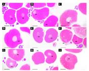
Illustrative images of different treatments of experiment 1. Histological evaluation of follicles at the end of experiment showing the similar distribution of central nucleus (cn), eccentric nucleus (en), and absent nucleus (an) oocytes, among control and treatments. A-C: Control; D-F: Treatment 1 (0.6 mg/kg CPE in vitro); G-I: Treatment 2 (in vitro dose of 6 mg/kg CPE). Arrow: pre-vitellogenic oocyte. Scale bar: 200μm.
Experiment 2
As in Experiment 1, no contamination was observed, but acidification of the culture medium persisted (data not shown). Dissociation of fragments remained inadequate (Figure 4A), and despite introducing prostaglandin in one treatment, no differences in oocyte types were observed across treatments (Figure 4AB). However, histological analysis revealed intact oocytes, including the theca and follicular layers, in all treatments (Figure 4C). Central, eccentric, and absent nuclei oocytes were observed randomLy, with no clear treatment effects. The lack of dissociation of the fragments prevented the counts from being evaluated so that the different types of oocytes could be quantified and compared between treatments, which can be observed in Figure 4A.

Illustrative images of different treatments of experiment 2. A) Cluster of oocytes in Serra's fluid, with inefficient cell separation. Observe that is possible to detect the presence of different type of oocytes; B) Through histological analysis, it is also possible to detect and confirm the random and similar presence of different types of oocytes in the different treatments and controls. Cn: central nuclei; ec: excentric nuclei; and an: absent nuclei; C) Intact follicular layers and theca in an oocyte with a central nucleus. Scale bar: A: 2mm; B-C: 200mm.
Experiment 3
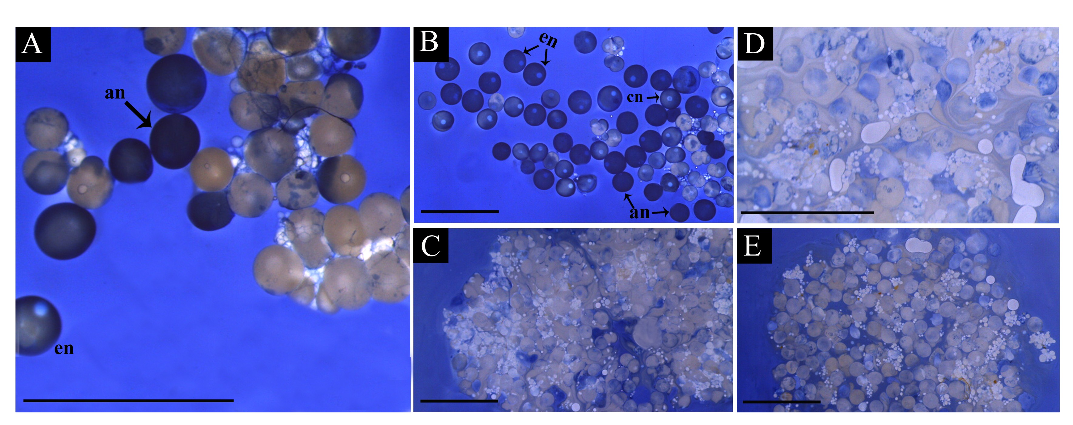
Three concentrations of collagenase (100, 200, and 400 IU/mL) were tested (Figure 5). Higher concentrations, especially 400 IU/mL, resulted in a friable appearance of the follicles (Figure 5E), suggesting collagenase digestion disrupted follicular structure. Although most oocytes were not stained with Trypan Blue, indicating viability, the friability of follicles treated with collagenase made these treatments unsuitable (Figure 5A). Follicles disintegrated easily with light handling (Figure 5CE). As discussed later, Trypan Blue may stain only the outer follicle layers without detecting oocyte viability. Cell survival rates are shown in Supplementary Figure 1.

Experiment 3. Evaluation of different types of ovarian follicle isolation. The blue color of some oocytes is due to the method of cell surveillance evaluation based on the use of Trypan blue. A) Control, no manipulation. Observe the presence of different oocyte types concerning the blue or bright color and that some oocytes are partially colored. In the same figure is possible to see that germinal vesicle can be central (cn), eccentric (en) or absent (an) and that due to coloration it can be bright (bn) or blue (bn); B) Mechanical dissociation of ovarian follicles. Observe that despite the regular aspect of cells, most cells have blue nuclei; C-E) Treatments with collagenase. Observe the irregular shape of cells, but despite that, most cells are bright. Treatment with 100 CDU/mL of collagenase type II; D) Treatment with 200 CDU/mL of collagenase type II; E) Treatment with 400 CDU/mL of collagenase type II. Scale bar: A: 2mm; B – E: 5mm.
Experiment 4
In all groups treated with enzymatic digestion, including controls, post-digestion analysis yielded minimal cellular material, with most samples exhibiting extensive dissolution (data not shown). In the limited instances where follicles were successfully obtained, marked cellular disorganization and loss of cell viability were observed, similar to experiment 3. Whole ovarian fragments were used in some treatments, but this approach proved unsuccessful, as ovulation-inducing substances apparently could not penetrate the follicles to trigger meiosis and ovulation, as will be discussed (Figure 6AC). In contrast, treatments using mechanical digestion achieved adequate follicle separation (Figure 6DF). Notably, the use of DHP, as the resolving dose, specifically in mechanically isolated follicles, effectively induced ovulation, resulting in the release of homogeneous oocytes without germinal vesicles (Figure 6EF).

Experiment 4. Obtaining in vitro ovulation with 1 μg/mL of 17α,20β-dihydroxy-4-pregnen-3-one (DHP). General appearance of the follicles after the end of experiment 4 of this study. A) FD treatment - Entire fragment, without hormonal induction in vivo, and incubation in vitro with 1 μg/mL DHP ; B) FED - Entire fragment with 1st dose in vivo with CPE and incubation with 1μg/mL DHP; C) FHD - Entire fragment with 1st dose in vivo with 500 UI/Kg hCG and incubation with 1μg/mL DHP; D) DMD - Follicles from a fish in which mechanical digestion, without hormonal induction in vivo and incubation in vitro with 1μg/mL DHP; E) DMED -Mechanical dissection, with 1st dose in vivo with CPE and incubation with 1μg/mL DHP; F) DMHD - Mechanical digestion, with 1st dose in vivo with 500 UI/Kg hCG and incubation with 1μg/mL DHP. Cn: Central nucleus; ec: excentric nucleus; an: absent nucleus. Scale bar: A, B) 3mm; C, E) 2mm; D) 2,5mm; F) 1mm.
Discussion
In this study, we standardized the conditions for maintaining and inducing A. altiparanae ovarian follicles to ovulate in vitro across four consecutive experiments. To achieve this, we first applied a priming dose in vivo, as done in spawning induction protocols for this species (Roza de Abreu et al., 2021, 2022; Ariki et al., 2023). Previous research showed that A. altiparanae spawns more effectively when hypophysation doses are fractionated: the priming dose initiates oocyte maturational competence, while the resolving dose induces FOM and ovulation, but only if the priming dose has been administered first (Roza de Abreu et al., 2021, 2022; Ariki et al., 2023). With this in mind, we applied the priming dose in vivo following the methodology of Patiño and Thomas (1990) and subsequently administered the resolving dose in vitro. This procedure proved effective, as none of the control oocytes (which received only the priming dose in vivo and were maintained solely in culture medium in vitro) showed signs of GVBD or ovulation. Instead, control oocytes maintained a central nucleus without morphological changes throughout all experiments, confirming the protocol's viability.
Leibovitz’s L-15 medium was selected for follicle culture based on its widespread use in teleosts, including Danio rerio (Welch et al., 2017; Mello et al., 2023), Clarias gariepinus (Kitanović et al., 2024), Sebastes schlegelii (Guo et al., 2024), and Chaeno gobius annularis (Kim et al., 2023). Considering the variability in optimal culture conditions among species and methodologies (as reviewed by Hartshorne, 1997), we tested several key factors for A. altiparanae, including 5% CO2 incubation, ovarian tissue dissociation method (mechanical vs. enzymatic), and the use of intact ovarian fragments versus dissociated follicles. Cell incubators with 5% CO2 are commonly used in cell cultures to mimic physiological CO2 pressure and concentration levels in tissues, reducing the harmful effects of reactive oxygen species (reviewed in Keeley and Mann, 2019). However, in our initial experiments, CO2 incubator led to significant medium acidification. Discontinuing CO2 resolved this issue, and in the fourth experiment, successful in vitro ovulation was achieved using 1 µg/mL DHP without CO2, consistent with findings in zebrafish (Welch et al., 2017). Although it remains unclear whether acidification directly inhibited ovulation, reduced pH is known to impact in vitro ovulation processes in fish (Goetz and Nagahama, 1985), warranting further research into the optimal pH environment for A. altiparanae follicle incubation. While ovulation was achieved using L-15 medium at standard pH in our study, future studies should also define optimal CO2 and O2 levels,(reviewed in Samokhin et al., 2022), temperature, osmolarity, and pH conditions for the ovaries of this species to better simulate in vivo conditions (reviews in Hartshorne, 1997; Keeley and Mann, 2019).
In the second experiment, we introduced a treatment combining 100 ng/mL PGF2α with the CPE dose to simulate a well-established in vivo reproduction protocol (Criscuolo-Urbinati et al., 2012; Sato et al., 2021). However, this approach also did not result in FOM or ovulation. Similar to the first experiment, we observed high acidification of the medium, and the results mirrored those of the control, with no ovulation achieved. These findings suggest that the extract dosage used in the first two experiments may warrant re-evaluation. Although the first and second experiments yielded similar (i.e., non-ovulatory) outcomes, the lack of successful ovulation does not imply that pituitary extracts are ineffective for in vitro ovulation in this species; rather, further optimization may be required.
Regarding pituitary extract dosages, when converting mg of CPE per kg of fragment to mg of CPE per mL of medium, the highest dose used in this study was 0.18 µg/mL CPE. In other species, higher doses of pituitary extracts have been effective for inducing in vitro hypophyseal activity. For instance, in Dicentrarchus labrax, researchers using CPE defined a "pituitary equivalent" (PE) as approximately 3 mg/mL of extract (1 PE/mL) (Sorbera et al., 1999). In their dose-response studies, a single dose ranging from 0.001 to 1 PE was effective in inducing ovulation, with 1 PE achieving nearly 90% oocyte maturation, while 0.001 PE was ineffective in triggering follicle maturation. Given that 1 PE corresponds to 3 mg/mL, this concentration is substantially higher than the 0.18 µg/mL dose used to mimic the resolving dose in lambari in our study. Similarly, in Siberian sturgeon follicles, the minimum effective dose of CPE to achieve 50% maturation was 7.4 µg/mL (Goncharov et al., 2001), which is ten times higher than the maximum dose tested in our study. These comparisons highlight the significant variability in the potency of pituitary extracts across species, suggesting that dose optimization is species-specific for effective in vitro maturation.
When comparing different substances, both dosage and latency period must be considered. In this study, we applied the same experimental duration for treatments with both pituitary extracts and DHP, despite their distinct latency periods in vitro. Typically, gonadotropins require longer to induce ovulation than DHP. For example, in Discentraxus labrax, the latency period for exogenous gonadotropins and DHP was approximately 45 and 15 hours, respectively (Sorbera et al., 1999). In Siberian sturgeon, a 50% ovulation rate with pituitary extracts was observed after about 36-38 hours of incubation (Goncharov et al., 2001). In contrast, the present study evaluated the response to CPE only 7 hours post-dose, aligning with the species’ in vivo ovulation timeline (Roza de Abreu et al., 2025). Therefore, future studies should extend the incubation period to fully assess the ovulation-inducing effects of pituitary extracts in lambari, adjusting both dose and incubation time.
This perspective is also relevant to the absence of ovulation observed with 100 ng/mL PGF2α in the third experiment. The concentration of PGF2α, derived from in vivo studies considering total body mass (Criscuolo-Urbinati et al., 2012), is relatively low for in vitro cultures. In fact, Jalabert and Szöllösi (1975) demonstrated that in vitro ovulation in trout could be achieved with PGF2α concentrations ranging from 1,000 to 5,000 ng/mL, which are 10 to 50 times higher than the dose used in our study. These findings strongly suggest that the dose of PGF2α employed in our experiment may have been insufficient to trigger ovulation, underscoring the need for dose optimization in in vitro protocols.
In the present study, Trypan Blue staining proved more effective for visualizing the outer follicular layers than for reliably assessing follicle viability, as staining was inconsistent and not clearly associated with oocyte death. Similar limitations were reported by Santos et al. (2017) in evaluate tambaqui follicle viability. By the fourth experiment, mechanical digestion was the most effective method for isolating lambari follicles. Notably, in this experiment, we observed promising signs of in vitro ovulation exclusively in treatments combining mechanical digestion with DHP. These results, helped confirm that the blue coloration of oocytes is not indicative of dead follicles. Although collagenase digestion produced morphologically brighter oocytes, it failed to yield viable follicles.
Given that DHP and other maturation-inducing steroids (MIS) typically induce FOM and ovulation more rapidly and effectively in vitro than gonadotropins or pituitary extracts (Jalabert and Szöllösi, 1975; Patiño and Thomas, 1990; Sorbera et al., 1999; Goncharov et al., 2001; Welch et al., 2017), we opted to replace CPE with 1 µg/mL DHP as the resolving dose in the fourth experiment. At this stage, improved experimental conditions were also established, opting to use DHP as an in vitro steroid inducer, similar to the application of prostaglandins in Anguilla japonica (Kagawa et al., 2003) or DHP in Perca fluviatilis (El Mohajer et al., 2021). Our study corroborates previous findings by Jesus et al. (2017), confirming DHP as a MIS for lambari, though the involvement of other MIS cannot be ruled out. DHP is widely recognized as a key MIS for many fish species (Takahashi et al., 2019), but additional investigations are needed to fully elucidate the specific MIS involved in lambari oocyte maturation. Based on our findings, mechanical digestion of ovarian fragments combined with an in vivo priming dose and in vitro DHP application as a resolving dose effectively induces in vitro ovulation. These findings provide a foundation for optimizing in vitro ovulation protocols in lambari and offer a potential framework for similar advancements in other fish species.
Conclusion
This study established a viable protocol to induce ovulation in vitro in A. altiparanae, highlighting the role of DHP and the efficiency of mechanical digestion. Future studies should focus on optimizing hormone doses and incubation parameters to further enhance the method's efficacy and applicability.
Supplementary Material
Supplementary material accompanies this paper.
Supplementary Figure 1


Acknowledgements
We extend our gratitude to the Fundação de Amparo à Pesquisa do Estado de São Paulo (FAPESP) for supporting this project in partnership with Fundação de Amparo à Pesquisa do Estado do Amazonas (FAPEAM) under grant nº 2020/08952-4, and FAPESP grant nº 2023/16571-9. Additionally, we thank FAPESP for funding the scholarship through grant nº 2022/13344-9 (Cristiane Fernanda Benevente). We also thank the PIBIC (CNPq) scientific initiation fellowship awarded to Queila Carla Ameno Alves and the research productivity fellowship awarded to Sergio Ricardo Batlouni by National Council for Scientific and Technological Development (CNPq), grant nº 311459/2022-6).
Data availability statement
Research data is only available upon request.
-
Financial support:
CFB received a scholarship from Sao Paulo Research Foundation – FAPESP (grant number #2022/13344-9). MRA received a fellowship from FAPESP (grant number #2022/16230-4). QCAA received a scholarship from National Council for Scientific and Technological Development – CNPq. SRB received a research productivity fellowship from CNPq (grant number #311459/2022-6). We extended our gratitude to the FAPESP for supporting this project in partnership with Amazonas Research Foundation (FAPEAM) (grant number #2020/08952-4) and FAPESP (grant number #2023/16571-9).
-
How to cite:
Benevente CF, Abreu MR, Alves QCA, Silva LMJ, Barbosa RP, Lacerda SMSN, Batlouni SR. Standardization of techniques and conditions for in vitro ovulation induction in astyanax altiparanae. Anim Reprod. 2026;23(1):e20250003. https://doi.org/10.1590/1984-3143-AR2025-0003
References
-
Ariki DGF, de Abreu MR, de Jesus Silva LM, Sato RT, Batlouni SR. Attempts for increasing Astyanax altiparanae spawning rates and percentage of responsive oocytes. Anim Reprod Sci. 2023;254:107262. https://doi.org/10.1016/j.anireprosci.2023.107262 PMid:37295049.
» https://doi.org/10.1016/j.anireprosci.2023.107262 -
Batlouni SR, Corrêa Filho RAC, Kuradomi RY, Spica LN, Abreu MR, Brasileiro LO, et al. Maintenance and management of breeders of Neotropical South American species in laboratories. In: Streit DP Jr, Zhang T, Paredes E, editors. Cryobiology for South American Neotropical Fish Species. Boca Raton: CRC Press; 2024. p. 142-70. https://doi.org/10.1201/9781003269441-7
» https://doi.org/10.1201/9781003269441-7 -
Bobe J, Montfort J, Nguyen T, Fostier A. Identification of new participants in the rainbow trout (Oncorhynchus mykiss) oocyte maturation and ovulation processes using cDNA microarrays. Reprod Biol Endocrinol. 2006;4:39. https://doi.org/10.1186/1477-7827-4-39 PMid:16872517.
» https://doi.org/10.1186/1477-7827-4-39 -
Criscuolo-Urbinati E, Kuradomi RY, Urbinati EC, Batlouni SR. The administration of exogenous prostaglandin may improve ovulation in pacu (Piaractus mesopotamicus). Theriogenology. 2012;78(9):2087-94. https://doi.org/10.1016/j.theriogenology.2012.08.001 PMid:23043951.
» https://doi.org/10.1016/j.theriogenology.2012.08.001 -
El Mohajer L, Bulteau R, Chevalier C, Selmi S, Fontaine P, Milla S. In vitro follicle culture shows that progestagens are the maturation-inducing hormones (MIH) and possible regulators of the ovulation-mediating hormone PGE2 in female Eurasian perch Perca fluviatilis. Fish Physiol Biochem. 2021;47(4):881-94. https://doi.org/10.1007/s10695-021-00946-5 PMid:33774768.
» https://doi.org/10.1007/s10695-021-00946-5 -
Fujimori C, Ogiwara K, Hagiwara A, Rajapakse S, Kimura A, Takahashi T. Expression of cyclooxygenase-2 and prostaglandin receptor EP4b mRNA in the ovary of the medaka fish, Oryzias latipes: possible involvement in ovulation. Mol Cell Endocrinol. 2011;332(1-2):67-77. https://doi.org/10.1016/j.mce.2010.09.015 PMid:20932877.
» https://doi.org/10.1016/j.mce.2010.09.015 -
Goetz FW, Nagahama Y. Effects of pH on in vitro ovulation of goldfish (Carassius auratus) oocytes. J Exp Zool. 1985;235(1):81-5. https://doi.org/10.1002/jez.1402350111 PMid:3863880.
» https://doi.org/10.1002/jez.1402350111 -
Goetz FW, Theofan G. In vitro stimulation of germinal vesicle breakdown and ovulation of Yellow Perch (Perca flavescens) oocytes. Effects of 17α-hydroxy-20β-digydroprogesterone and prostaglandins. Gen Comp Endocrinol. 1979;37(3):273-85. https://doi.org/10.1016/0016-6480(79)90001-7 PMid:456868.
» https://doi.org/10.1016/0016-6480(79)90001-7 -
Goncharov BF, Williot P, Le Menn F. Comparison of the effects of gonadotropic preparations of the carp and Stellate Sturgeon Pituitaries on in vivo and in vitro oocyte maturation in the Siberian Sturgeon Acipenser baeri Brandt. Ontogenez. 2001;32(5):384-92. https://doi.org/10.1023/A:1012364702687 PMid:11605413.
» https://doi.org/10.1023/A:1012364702687 -
Guo Q, Lyu L, Wen H, Li Y, Zuo C, Wang X, Jing X, Jiang T, Yang J, Qi X. Black rockfish (Sebastes schlegelii) nuclear progesterone receptor: Cloning, tissue distribution and potential role in ovoviviparity teleost pregnancy and parturition. Aquacult Rep. 2024;36:102068. https://doi.org/10.1016/j.aqrep.2024.102068
» https://doi.org/10.1016/j.aqrep.2024.102068 -
Hagiwara A, Ogiwara K, Katsu Y, Takahashi T. Luteinizing hormone-induced expression of Ptger4b, a prostaglandin E2 receptor indispensable for ovulation of the medaka Oryzias latipes, is regulated by a genomic mechanism involving nuclear progestin receptor. Biol Reprod. 2014;90(6):126-1. https://doi.org/10.1095/biolreprod.113.115485 PMid:24790162.
» https://doi.org/10.1095/biolreprod.113.115485 -
Hagiwara A, Ogiwara K, Sugama N, Yamashita M, Takahashi T. Inhibition of medaka ovulation by gap junction blockers due to its disrupting effect on the transcriptional process of LH-induced Mmp15 expression. Gen Comp Endocrinol. 2020;288:113373. https://doi.org/10.1016/j.ygcen.2019.113373 PMid:31874135.
» https://doi.org/10.1016/j.ygcen.2019.113373 -
Hainfellner P, Souza TG, Moreira RG, Nakaghi LSO, Batlouni SR. Gonadal steroids levels and vitellogenesis in the formation of oocytes in Prochilodus lineatus (Valenciennes) (Teleostei: characiformes). Neotrop Ichthyol. 2012;10(3):601-12. https://doi.org/10.1590/S1679-62252012005000021
» https://doi.org/10.1590/S1679-62252012005000021 -
Hartshorne GM. In vitro culture of ovarian follicle. Rev Reprod. 1997;2(2):94-104. https://doi.org/10.1530/ror.0.0020094 PMid:9414471.
» https://doi.org/10.1530/ror.0.0020094 -
Jalabert B, Szöllösi D. In vitro ovulation of trout oocytes: effect of prostaglandin on smooth muscle-like cells of the theca. Prostaglandins. 1975;9(5):765-78. https://doi.org/10.1016/0090-6980(75)90113-6 PMid:1162086.
» https://doi.org/10.1016/0090-6980(75)90113-6 -
Jalabert B, Breton B, Brzuska E, Fostier A, Wieniawski J. A new tool for induced spawning: the use of 17α-hydroxy-20β-dihydroprogesterone to spawn carp at low temperature. Aquaculture. 1977;10(4):353-64. https://doi.org/10.1016/0044-8486(77)90126-0
» https://doi.org/10.1016/0044-8486(77)90126-0 -
Jalabert B. Particularities of reproduction and oogenesis in teleost fish compared to mammals. Reprod Nutr Dev. 2005;45(3):261-79. https://doi.org/10.1051/rnd:2005019 PMid:15982453.
» https://doi.org/10.1051/rnd:2005019 -
Jesus LWO, Bogerd J, Vieceli FM, Branco GS, Camargo MP, Cassel M, et al. Gonadotropin subunits of the characiform Astyanax altiparanae: molecular characterization, spatiotemporal expression and their possible role on female reproductive dysfunction in captivity. Gen Comp Endocrinol. 2017;246:150-63. https://doi.org/10.1016/j.ygcen.2016.12.004 PMid:27940043.
» https://doi.org/10.1016/j.ygcen.2016.12.004 -
Joy KP, Singh V. Functional interactions between vasotocin and prostaglandins during final oocyte maturation and ovulation in the catfish Heteropneustes fossilis. Gen Comp Endocrinol. 2013;186:126-35. https://doi.org/10.1016/j.ygcen.2013.02.043 PMid:23510856.
» https://doi.org/10.1016/j.ygcen.2013.02.043 -
Kagawa H, Fujie N, Imaizumi H, Masuda Y, Oda K, Adachi J, Nishi A, Hashimoto H, Teruya K, Kaji S. Using osmotic pumps to deliver hormones to induce sexual maturation of female Japanese eels, Anguilla japonica. Aquaculture. 2013;388-91:30-4. https://doi.org/10.1016/j.aquaculture.2013.01.025
» https://doi.org/10.1016/j.aquaculture.2013.01.025 -
Kagawa H, Tanaka H, Unuma T, Ohta H, Gen K, Okuzawa K. Role of prostaglandin in the control of ovulation in the Japanese eel Anguilla japonica. Fish Sci. 2003;69(2):234-41. https://doi.org/10.1046/j.1444-2906.2003.00613.x
» https://doi.org/10.1046/j.1444-2906.2003.00613.x -
Keeley TP, Mann GE. Defining physiological normoxia for improved translation of cell physiology to animal models and humans. Physiol Rev. 2019;99(1):161-234. https://doi.org/10.1152/physrev.00041.2017 PMid:30354965.
» https://doi.org/10.1152/physrev.00041.2017 -
Kim DG, Hwang IJ, Baek HJ. Potent influence of exogenous melatonin on in vitro oocyte maturation in the Longchin Goby, Chaeno gobius annularis. Dev Reprod. 2023;27(3):127-35. https://doi.org/10.12717/DR.2023.27.3.127 PMid:38074459.
» https://doi.org/10.12717/DR.2023.27.3.127 -
Kitanović N, Marinović Z, Quyến NN, Kovács B, Müller T, Urbányi B, Horváth Á. In vitro maturation of African catfish (Clarias gariepinus, Burchell, 1822) oocytes results in viable larvae. Aquaculture. 2024;583:740617. https://doi.org/10.1016/j.aquaculture.2024.740617
» https://doi.org/10.1016/j.aquaculture.2024.740617 -
Knight OM, Van Der Kraak G. The role of eicosanoids in 17α,20β-dihydroxy-4-pregnen-3-one-induced ovulation and spawning in Danio rerio. Gen Comp Endocrinol. 2015;213:50-8. https://doi.org/10.1016/j.ygcen.2014.12.014 PMid:25573385.
» https://doi.org/10.1016/j.ygcen.2014.12.014 -
Kuradomi RY, Batlouni SR. PGF 2α and gonadal steroid plasma levels of successful and unsuccessful spawning Piaractus mesopotamicus (Teleostei, Characiformes) females. Aquacult Int. 2018;26(4):1083-94. https://doi.org/10.1007/s10499-018-0269-8
» https://doi.org/10.1007/s10499-018-0269-8 - Lacerda SMSN, Batlouni SR, Silva SBG, Homem CSP, França LR. Germ cells transplantation in fish: the Nile-tilapia model. Anim Reprod (AR). 2018;3(2):146-59.
-
Levavi-Zermonsky B, Yaron Z. Changes in gonadotropin and ovarian steroids associated with oocyte maturation during spawning induction in the carp. Gen Comp Endocrinol. 1986;62(1):89-98. https://doi.org/10.1016/0016-6480(86)90097-3 PMid:3781219.
» https://doi.org/10.1016/0016-6480(86)90097-3 -
Lira LVG, Kuradomi RY, Souza TG, Hainfellner P, Batlouni SR. Astyanax altiparanae ovarian maturation after spawning in water recycling systems. Bol Inst Pesca. 2018;44:1-10. https://doi.org/10.20950/1678-2305.2018.44.4.207
» https://doi.org/10.20950/1678-2305.2018.44.4.207 -
Lister AL, Van Der Kraak GJ. Regulation of prostaglandin synthesis in ovaries of sexually mature zebrafish (Danio rerio). Mol Reprod Dev. 2009;76(11):1064-75. https://doi.org/10.1002/mrd.21072 PMid:19551897.
» https://doi.org/10.1002/mrd.21072 -
Mello F, Alonso DJ, Faria NPVM, Marques VH, Oliveira EF, de Mello PH, et al. Alterations in gene expression and the fatty acid profile impact but do not compromise the in vitro maturation of zebrafish (Danio rerio) stage III ovarian follicles after cryopreservation. Animals (Basel). 2023;13(22):3563. https://doi.org/10.3390/ani13223563 PMid:38003179.
» https://doi.org/10.3390/ani13223563 -
Mylonas CC, Fostier A, Zanuy S. Broodstock management and hormonal manipulations of fish reproduction. Gen Comp Endocrinol. 2010;165(3):516-34. https://doi.org/10.1016/j.ygcen.2009.03.007 PMid:19318108.
» https://doi.org/10.1016/j.ygcen.2009.03.007 -
Patiño R, Thomas P. Induction of maturation of Atlantic croaker oocytes by 17α,20β,21-trihydroxy-4-pregnen-3-one in vitro: consideration of some biological and experimental variables. J Exp Zool. 1990;255(1):97-109. https://doi.org/10.1002/jez.1402550113 PMid:2391470.
» https://doi.org/10.1002/jez.1402550113 -
Pereira TSB, Boscolo CNP, Moreira RG, Batlouni SR. Leporinus elongatus induced spawning using carp pituitary extract or mammalian GnRH analogue combined with dopamine receptor antagonists. Anim Reprod. 2018;15(1):64-70. https://doi.org/10.21451/1984-3143-2017-AR983 PMid:33365097.
» https://doi.org/10.21451/1984-3143-2017-AR983 -
Roza de Abreu M, de Jesus Silva LM, Benevente CF, Barbosa RP, Mechaly AS, Batlouni SR. Hormonal regulation of oocyte maturation and ovulation in lambari (Astyanax altiparanae) induced by different pituitary extracts. Aquaculture. 2025;609:742843. https://doi.org/10.1016/j.aquaculture.2025.742843
» https://doi.org/10.1016/j.aquaculture.2025.742843 -
Roza de Abreu M, Silva LMJ, Figueiredo-Ariki DG, Sato RT, Kuradomi RY, Batlouni SR. Reproductive performance of lambari (Astyanax altiparanae) in a seminatural system using different protocols. Aquacult Res. 2021;52(2):471-83. https://doi.org/10.1111/are.14905
» https://doi.org/10.1111/are.14905 -
Roza de Abreu M, Silva LMJ, Figueiredo-Ariki DG, Sato RT, Kuradomi RY, Batlouni SR. The effect of LHRHa with and without dopamine antagonist on reproductive performance in lambari Astyanax altiparanae. Aquaculture. 2022;550:737883. https://doi.org/10.1016/j.aquaculture.2021.737883
» https://doi.org/10.1016/j.aquaculture.2021.737883 -
Samokhin P, Gardner GL, Moffatt C, Stuart JA. An inexpensive incubator for mammalian cell culture capable of regulating O2, CO2, and temperature. Oxygen (Basel). 2022;2(1):22-30. https://doi.org/10.3390/oxygen2010003
» https://doi.org/10.3390/oxygen2010003 -
Santos FBP, Affonso EG, Godoy LCD. Trypan blue staining is not efficient in determining oocyte viability in Colossoma macropomum and Brycon amazonicus. Bol Inst Pesca. 2017;44:121-9. https://doi.org/10.20950/1678-2305.2017.121.129
» https://doi.org/10.20950/1678-2305.2017.121.129 -
Sato RT, Abreu MR, Kuradomi RY, Silva LMJ, Batlouni SR. Effects of the time of administration and the concentration of exogenous prostaglandin F on ovulation in pacu (Piaractus mesopotamicus). Aquacult Int. 2023;1:1-10. https://doi.org/10.1007/s10499-023-01332-z
» https://doi.org/10.1007/s10499-023-01332-z -
Sato RT, Kuradomi RY, Calil MC, Silva LMJ, Roza de Abreu MR, Figueiredo-Ariki DG, Freitas GA, Batlouni SR. Resumption and progression of meiosis and circulating levels of steroids and prostaglandin F2α of Piaractus mesopotamicus induced by hypophysation with prostaglandin F2α Aquacult Res. 2021;52(3):1026-7. https://doi.org/10.1111/are.14957
» https://doi.org/10.1111/are.14957 -
Sorbera LA, Asturiano JF, Carrillo M, Cerda J, Kime DE, Zanuy S. In vitro oocyte maturation in the sea bass: effects of hCG, pituitary extract and steroids. J Fish Biol. 1999;55(1):9-25. https://doi.org/10.1111/j.1095-8649.1999.tb00652.x
» https://doi.org/10.1111/j.1095-8649.1999.tb00652.x -
Souza TG, Kuradomi RY, Rodrigues SM, Batlouni SR. Wild Leporinus friderici induced spawning with different doses of mGnRHa and metoclopramide or carp pituitary extract. Anim Reprod. 2020;17(1):e20190078. https://doi.org/10.21451/1984-3143-AR2019-0078 PMid:32399066.
» https://doi.org/10.21451/1984-3143-AR2019-0078 -
Takahashi T, Fujimori C, Hagiwara A, Ogiwara K. Recent advances in the understanding of teleost medaka ovulation: the roles of proteases and prostaglandins. Zool Sci. 2013;30(4):239-47. https://doi.org/10.2108/zsj.30.239 PMid:23537233.
» https://doi.org/10.2108/zsj.30.239 -
Takahashi T, Hagiwara A, Ogiwara K. Follicle rupture during ovulation with an emphasis on recent progress in fish models. Reproduction. 2019;157(1):R1-13. https://doi.org/10.1530/REP-18-0251 PMid:30394703.
» https://doi.org/10.1530/REP-18-0251 -
Tang H, Liu Y, Li J, Li G, Chen Y, Yin Y, Guo Y, Cheng CH, Liu X, Lin H. LH signaling induced ptgs2α expression is required for ovulation in zebrafish. Mol Cell Endocrinol. 2017;447:125-33. https://doi.org/10.1016/j.mce.2017.02.042 PMid:28254490.
» https://doi.org/10.1016/j.mce.2017.02.042 -
Tang H, Liu Y, Li J, Yin Y, Li G, Chen Y, Li S, Zhang Y, Lin H, Liu X, Cheng CH. Gene knockout of nuclear progesterone receptor provides insights into the regulation of ovulation by LH signaling in zebrafish. Sci Rep. 2016;6(1):28545. https://doi.org/10.1038/srep28545 PMid:27333837.
» https://doi.org/10.1038/srep28545 -
Welch EL, Eno CC, Nair S, Lindeman RE, Pelegri F. Functional manipulation of maternal gene products using in vitro oocyte maturation in zebrafish. J Vis Exp. 2017;(122):55213. https://doi.org/10.3791/55213 PMid:28518112.
» https://doi.org/10.3791/55213
Edited by
-
Editor-in-Chief:
Carlos Eduardo Ambrósio.