 NON-INVASIVE METHOD OF MONITORING INTRACRANIAL PRESSURE FOR THE EVALUATION OF HEPATIC ENCEPHALOPATHY
ABSTRACT
Background: Liver diseases often occur with hepatic encephalopathy (HE), whose pathophysiology may involve increased intracranial pressure (ICP). Tools for monitoring ICP and its pulse morphology can be useful for assessing HE. The use of a non-invasive and sensitive procedure would be extremely useful in managing these cases.
Objective: To evaluate the feasibility and performance of a new, non-invasive method of monitoring ICP, as an alternative to invasive methods, and to correlate the clinical diagnosis of HE with the morphological findings of ICP.
Methods: This is a cross-sectional analytical study, conducted in a tertiary hospital and pioneer in the application of Brain4Care® BWS equipment. The ICP pulse morphology is parallel to the arterial one, where there are three frequent peaks: percussion peak (P1), due to plasma extravasated by the choroid plexus; tidal wave (P2), due to the degree of intracranial compliance to the reflection of P1, and dicrotic notch (P3), due to the closure of the aortic valve. Normality indicates P1>P2>P3. These peaks determine intracranial compliance through their relationship with cerebral blood volume, where P2/P1 ratio >1 suggests a pathological morphology, with a sustained increase in ICP and decreased compliance. Another way to evaluate this would be by a change in the time-to-peak (TTP). These data were compared between patients with and without clinical signs indicative of HE. The study was approved by the Institution’s Research Ethics Committee (number 5.493.775).
Results: A total of 40 liver disease patients were evaluated, of which, at the time of collection, 20 did not have a clinical picture of HE (59.5±9.3 years; 70.0% male) and 20 had a clinical picture of HE (59.6±11.9 years; 65.0% male). The groups are demographically, clinically and laboratory similar; and statistically significant differences were identified in the morphological patterns of ICP between the groups evaluated, as well as trends in the parameters. The difference in the P2/P1 ratio was not significant (Mann Whitney: two-tailed P=0.2978); however, TTP proved to be a parameter with a statistically significant difference between the groups (Mann Whitney: two-tailed P=0.0282; median difference = 0.04). Analysis using the C statistic, using the ROC curve, suggested P2/P1=1.31 (AUROC: 0.5975) and TTP=0.22 (AUROC: 0.7013) as optimal cutoff points, where the presence of HE in liver disease patients would be associated with obtaining parameters below these thresholds.
Conclusion: The brain4care® BWS system proved to be feasible for use in liver disease patients with or without clinical signs of hepatic encephalopathy and was able to differentiate them. Pathophysiological explanations, however, still require better causality explanation and understanding of the intracerebral hydrodynamic picture in hepatic encephalopathy. Given the low sample power found, new studies need better clinical heterogeneity and longer-term follow-up for definitive conclusions.
Keywords:
Hepatic encephalopathy; intracranial pressure; liver diseases; brain4care
HIGHLIGHTS
•This study investigated the correlation of morphological findings from a non-invasive method of monitoring intracranial pressure with the clinical diagnosis of hepatic encephalopathy.
•40 liver disease patients without and with clinical signs suggestive of hepatic encephalopathy were compared demographically, clinically and laboratory wise - and statistically significant differences were found in the intracranial pressure pulse wave morphology parameters between them.
•ROC curve analysis suggested P2/P1=1.31 (AUROC: 0.5975) and TTP=0.22 (AUROC: 0.7013) as optimal cutoff points, where the presence of hepatic encephalopathy in liver disease patients would be associated with obtaining parameters below these thresholds.
•Even though the sensor was able to differentiate the groups, a larger case series, a longer-term follow-up, and a better understanding of the intracerebral hydrodynamic picture in hepatic encephalopathy are still required so that the method helps in defining management with the support of clinical parameters.
RESUMO
Contexto: Doenças hepáticas frequentemente cursam com encefalopatia hepática (EH), cuja fisiopatologia pode envolver aumento da pressão intracraniana (PIC). Ferramentas de monitorização da PIC e da sua morfologia de pulso podem ser úteis para avaliação da EH. A utilização de um procedimento não invasivo e sensível seria extremamente útil na condução desses casos.
Objetivo: Avaliar a factibilidade e o desempenho de um método inédito e não invasivo de monitorização da PIC, como alternativa aos métodos invasivos, e correlacionar o diagnóstico clínico de EH aos achados morfológicos da PIC.
Métodos: Trata-se de um estudo analítico transversal, conduzido em um hospital terciário e pioneiro na aplicação do equipamento BWS da brain4care®. A morfologia de pulso da PIC é paralela à arterial, onde existem três picos frequentes: pico de percussão (P1), pelo plasma extravasado pelo plexo coroide; onda de maré (P2), pelo grau de complacência intracraniana à reflexão de P1, e incisura dicrótica (P3), pelo fechamento da valva aórtica. A normalidade aponta P1>P2>P3. Esses picos determinam a complacência intracraniana através de sua relação com o volume sanguíneo cerebral, onde a relação de alturas P2/P1 >1 sugere uma morfologia patológica, com aumento sustentado da PIC e diminuição da complacência. Outra forma de avaliar isso seria por uma alteração no tempo até o pico (Time-to-Peak, TTP). Esses dados foram comparados entre pacientes sem e com quadro clínico indicativo de EH. O estudo foi aprovado pelo Comitê de Ética em Pesquisa da Instituição pelo parecer número 5.493.775.
Resultados: Foram avaliados 40 hepatopatas, dos quais, no momento da coleta, 20 não apresentavam quadro clínico de EH (59.5±9.3 anos; 70.0% masculinos) e 20 possuíam quadro clínico de EH (59.6±11.9 anos; 65.0% masculinos). Os grupos são demográfica, clínica e laboratorialmente semelhantes; e foram identificadas diferenças estatisticamente significantes nos padrões morfológicos da PIC entre os grupos avaliados, bem como tendências nos parâmetros. A diferença na relação P2/P1 não se mostrou significante (Mann Whitney: P bicaudal = 0.2978); porém TTP mostrou-se como um parâmetro com diferença estatisticamente significante entre os grupos (Mann Whitney: P bicaudal = 0.0282; diferença mediana = 0,04). A análise pela estatística C, por meio da curva ROC sugeriu como pontos de corte ótimos um P2/P1=1.31 (AUROC: 0.5975) e TTP = 0.22 (AUROC: 0.7013), onde a presença de EH em hepatopatas estaria associada à obtenção de parâmetros abaixo desses limiares.
Conclusão: A utilização do sistema BWS da brain4care® mostrou-se factível para utilização em pacientes hepatopatas sem ou com quadro clínico de EH e foi capaz de diferenciá-los. As explicações fisiopatológicas, porém, necessitam ainda de maior casuística, face ao baixo poder amostral constatado, melhor entendimento do quadro hidrodinâmico intracerebral na encefalopatia hepática, heterogeneidade clínica e necessidade de seguimento em mais longo prazo, para conclusões definitivas.
Palavras-chave:
Encefalopatia hepática; pressão intracraniana; hepatopatias; brain4care
INTRODUCTION
The liver disease
The liver is considered the largest gland in the body, with exocrine and endocrine functions, such that its dysfunction can cause considerable systemic damage. Bile secretion is the main digestive function of the liver. Furthermore, the liver is essential in regulating the metabolism of carbohydrates, proteins (including ammonia recycling) and lipids; in the storage of substances; and in the degradation and excretion of hormones. Other functions include the transformation and excretion of drugs; hemostasis (formation of clotting factors) and support for the immune response. Several etiologies affect this organ; however, it is possible to systematize them according to the period of involvement, didactically and non-exclusively, in chronic and acute diseases.
Chronic liver disease (CLD) can be mainly caused by viral infection such as hepatitis B and C; due to hepatitis induced by alcohol consumption; and due to the accumulation of fat in the liver (non-alcoholic hepatic steatosis). Chronic forms can evolve over time into cirrhosis (fibrosis), eventually requiring orthotopic liver transplantation. Alcohol is the most common underlying etiology associated with cirrhosis (45%) worldwide. Furthermore, from CLD, acute on chronic liver failure (ACLF) can occur, in which there is an acute decompensation of cirrhosis, associated with systemic involvement and encephalopathy. In this scenario, bacterial infections (35%), gastrointestinal hemorrhages (22%) and alcohol (19%) represent the most common etiologies globally responsible for the worsening of a chronic condition1.
In addition, acute liver failure (ALF) - also known as fulminant liver failure - can be defined as a clinical syndrome of encephalopathy and coagulopathy (with international normalized ratio [INR] >1.5) resulting from sudden massive loss of hepatocyte function. It represents a condition lasting up to 26 weeks, without previous CLD or ACLF2,3. The mortality rate varies between 30-50%4. Common causes of ALF include: acetaminophen poisoning (approximately 46% of cases; use is common in suicide attempts); prescription drug-induced injuries (approximately 11% of cases); liver ischemia; viral hepatitis (A, B and E, mainly) and autoimmune hepatitis; use of dietary and herbal supplements; hepatic venous thrombosis (Budd-Chiari Syndrome); acute hepatic steatosis of pregnancy (AFLP)/HELLP syndrome; Wilson’s disease; or even cardiac arrest2.
Hepatic encephalopathy: pathophysiology, clinic and classification
Still in the context of liver diseases, hepatic encephalopathy (HE) can be defined as a cognitive deficit or a change in the level of consciousness, possibly reversible, related to a functional disorder of the central nervous system (CNS) and normally associated with hepatocellular insufficiency. HE of any degree may mean the presence of cerebral edema, the probability of which increases as the condition worsens3,5. HE, which was previously seen as a terminal clinical sign, is now portrayed as a condition that can be treated.
Under the epidemiological scope, according to Stravitz et al., severe HE in ALF can occur in 54% of cases due to acetaminophen intoxication and 36% of cases due to drug-induced liver injury2. Also, mortality in ALF can reach 40 to 80% without a liver transplant, where cerebral edema and intracranial hypertension (ICH) secondary to hyperammonemia are responsible for 35% of deaths. These mortality rates are much higher than those of CLD5. In chronic disease, the prevalence of HE in cirrhosis diagnoses is 10-14% overall, 16-21% in patients with decompensated cirrhosis, and 10-15% in patients with transjugular intrahepatic portosystemic shunt (TIPS). HE eventually occurs in 30-40% of individuals with cirrhosis6.
Furthermore, to classify, as defined by the 11th World Congress of Gastroenterology, HE can occur in patients with ALF (type A); in patients with portosystemic shunt (type B); or in patients with CLD (type C). Type C HE, in turn, can be subclassified into: (I) episodic HE, when there is acute delirium or disturbance of consciousness in previously healthy patients from a neuropsychiatric point of view, and may be (Ia) precipitated (by triggering factors), (Ib) spontaneous (in the absence of these factors) or (Ic) recurrent (with more than two episodes per year); in (II) persistent HE, when there is a continuous and uninterrupted presence of neuropsychiatric signs and symptoms, generally extrapyramidal changes, dysarthria, personality, memory and sleep-wake cycle disorders, being graded into (IIa) mild, (IIb) severe and (IIc) treatment dependent; or even in (III) minimal HE, when there is a preclinical stage of HE in CLD, with deficits in neurological tests, but without evident changes in mental status7.
The pathophysiology of HE is complex, multifactorial and its mechanisms are not completely defined. However, some factors can be identified as predisposing to the progression and severity of neurological decline in CLD because they essentially form edema: inflammation, oxidative stress, excess bile acids, increased lactate and, mainly, hyperammonemia8.
Edema causes changes in chemical homeostasis and nerve impulse deficiencies. In this context, its formation can occur via (i) the vasogenic route, that is, when the blood-brain barrier (BBB) is broken and there is an influx of water into the cerebral interstitial compartment, which is hypertonic; or by (ii) cytotoxic route, when there is no disruption of the BBB. Along these lines, astrocytes, the main cells forming the BBB, play a crucial role in this dynamic - they protect neurons from toxic exogenous agents and control electrolyte homeostasis. Dysregulation of aquaporin four channels, accumulation of intracellular glutamine and increase in extracellular glutamate are possible mechanisms related to cerebral edema mediated by astrocytic inflammation8.
Furthermore, because of pathophysiological changes, the clinical presentation associated with HE has some common features. In relation exclusively to the neurological aspect of HE, apathy is an early sign, followed by hypersomnia and mental confusion. Patients may experience delirium, euphoria/mania, spatial disorientation, affected cognition, and short-term memory loss. There may be bradykinesia, but also muscular hypertonia with myoclonus and exaggerated tendon reflexes. Asterix (flapping) is more common in cases of CLD. Changes in pupillary reactions are rare. With an affected pontine or mesencephalic reflex, cerebral edema may also be suspected. Cushing’s Triad - systemic hypertension, bradycardia, and changes in respiratory rhythm - can also serve as an aid in detecting an increase in ICP. Thus, in general terms, as liver failure worsens, there is a proportional drop in the level of consciousness, which may progress to coma3.
Furthermore, since the clinical picture of HE has a very diverse presentation, the West-Haven criteria (WHC) for assessing mental status sought to compile this variety: those patients without abnormalities are classified as grade 0; grade 1 are patients who present changes in the sleep-wake cycle or sleep disorders, or brief periods of impaired consciousness; grade 2, patients with lethargy or apathy, with changes in behavior and slowed speech, and who may still present flapping; grade 3, patients with significantly reduced level of consciousness and stupor; and, finally, grade 4, individuals in a coma3,5,7.
A study analyzing 92 cases of ALF over 7 years detected the presence of cerebral edema in 31% of cases; did not identify cerebral edema in patients with grade 1 and 2 HE but reported the presence of edema in 88% of those with grade 3 or 49. Later, the same author reinforced the correlation, pointing out the presence of edema in more than 75% of patients with grade 4 HE3. Furthermore, another study indicated that cerebral edemas were identified in approximately 80% of patients with ALD, with 25-35% presenting edema when the HE was grade 3, and 65-75%, at Grade 410. Raschke et al. also pointed out that there is an increase in ICP in 85-95% cases of grade 3 or 4 HE11. Finally, Raghavan et al. described that cerebral edema and ICH occur in 75-80% of ALF patients with HE grade 3 or 412.
In this sense, the International Society for Hepatic Encephalopathy and Nitrogen Metabolism (ISHEN) also proposed a clinical grading - but reducing the variability intrinsic to the subjectivity of the analysis of WHC grades 1 and 2 and making it more objective for studies. It divides the HE syndrome into: (i) absent EH; in (ii) covert HE, whose patients present minimal HE and WHC grade 1; or in (iii) overt HE, whose patients present evident clinical abnormalities. This classification considers the patient’s mental state, the results of specialized tests and the presence or absence of asterixis (flapping)13.
Ochoa-Sanchez et al. pointed out that more than 80% of cirrhotic patients develop covert HE; while 30% of patients in ALF have apparent HE8. Lidofsky et al. reported overt HE in 12 to 67% of ALF patients14. Another study reports that minimal HE occurs in 20-80% of cirrhotic patients; in addition to pointing out that the chance of the first episode of HE is 5 to 25% within the first 5 years of diagnosis6.
Hepatic encephalopathy: diagnosis and management
Consequently, the diagnosis of HE is primarily clinical, focusing on the identification of specific clinical features. Concurrently, laboratory tests may be ordered to indicate an increase in transaminases, an increase in prothrombin time (or via INR), or even an increase in serum ammonia5. With regard to imaging tests, computed tomography (CT) should be performed as soon as possible to identify edema with mass effect, despite the low sensitivity for early edema. A patient with HE and a normal CT scan still presents a high risk of edema and a rapid increase in ICP, which must be monitored3. Non-invasive methods such as transcranial Doppler ultrasonography, optic nerve sheath diameter assessment and magnetic resonance imaging can also be used to estimate intracranial compliance (ICC), ICP and cerebral perfusion pressure (CPP)5.
With regard to the management and treatment of HE, it is recommended that (i) stabilization be achieved, (ii) modifiable precipitating factors be controlled, (iii) serum ammonia levels be lowered, (iv) ICP be managed, and (v) liver complications be addressed10.
Monitoring intracranial pressure and intracranial compliance
Several studies suggest that ICP monitoring may be clinically useful. It is indicated in all cases with grade 4 HE5. In ALF, whose definitive treatment is usually orthotopic liver transplantation, its preoperative and intraoperative function would be to control ICP to avoid brain stem herniation with consequent neurological damage; but it could also be used to classify receptors. However, despite the value of monitoring, complications with invasive monitoring have been reported in several studies, with intracranial hemorrhage being the most common of these14. Blei et al. reported that these complications may occur in 20% of cases, with fatal hemorrhage being reported in up to 4% of intraparenchymal catheter uses15; while Maloney et al. reported a hemorrhage rate of 3.8-22%4. The parallel coagulopathy combined with possible iatrogenesis is responsible for this considerable rate10. Also, the infection rate in intraventricular catheters still reaches 10%, depending on the duration16. Therefore, the use of invasive ICP monitoring is still controversial, considering the possibility of infection and hemorrhage intrinsic to the use of invasive methods4 - justifying the study of non-invasive ICP (ICPni) monitoring methods.
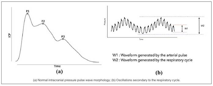
ICP, in turn, tends to be stabilized by an intrinsic characteristic of the brain, the ability to accommodate intracranial volume without significantly changing pressure, called intracranial compliance (ICC). Despite the attempt to stabilize ICP to aim for a stable CPP, ICP occurs in pulse waves (ICPW). These pulses occur as a reflection of the cardiac cycle - systole and diastole - in the cerebrospinal fluid. Analogous to the cardiac arterial pressure waveforms, the pressure pulse in the cerebrospinal fluid has components P1, P2 and P3: P1 is the percussion wave, due to the arterial pressure being transmitted from the choroid plexus to the intracranial ventricles; P2 would be the tidal wave and is inversely related to the ICC; and P3 would be the wave of closure of the dicrotic notch (closing of the aortic valve during diastole)17. In addition to isolated ICPW, the set of pulses oscillates secondary to the respiratory cycle: when we inhale deeply, intrathoracic pressure decreases and systemic venous return increases; however, this reduces the volume in the left heart chambers, which generates a specific decrease in ejection and a consequent drop in blood pressure (BP). This dynamic is reflected in a tendency for ICP to increase during expiration and vice versa (Figure 1). Furthermore, factors such as age, posture, time of day, and clinical condition affect average ICP; which varies in a range of 7-15 mmHg in healthy adults, 3-6 mmHg in children and 0.5-6 mmHg in infants18.

When the level of P2 exceeds P1, a considerable loss of ICC is expected. Also, several studies indicate that a pathological ICPW format is more suggestive than an increase in absolute ICP. For example, in idiopathic hydrocephalus, meningitis, hemorrhagic cerebrovascular accidents or traumatic brain injuries, an altered pulse wave is detected without necessarily an absolute increase in ICP18-20. Thus, the shape of the ICPW may serve as an early indicator of neurological decompensation - in addition to the absolute ICP itself (Figure 2).

Following these, Mascarenhas et al.21 innovated by questioning the Monro-Kellie Doctrine of 1820. This doctrine, developed by three anatomists, defined that the cranial cavity was filled mainly by three components - blood, fluid, and brain parenchyma - and that the increase in volume of one of these components would necessarily imply an increased ICP once the skull is considered an extremely rigid bone that cannot be expanded - at least after the fontanelles have closed. And to challenge the doctrine, the authors carried out a simple experiment: “in vitro”, they applied deformation sensors to the parietal region of human skulls and inflated a balloon inside the skull up to 100 mmHg, detecting a dilation directly and linearly proportional to the ICP. In the “in vivo” experiment, they performed postural maneuvers in Wistar rats to evaluate a variation in ICP and, with the same sensor, minimally invasive and placed externally to the skull, they were able to capture variations in cranial dilation21.
Subsequently, the same authors compared the gold standard of invasive ICP monitoring (ICPi) - the Codman intraparenchymal catheter - with the minimally invasive sensor attached to the skull of Wistar rats (ICPmi) and found a positive Pearson correlation coefficient (r) of 0.8±0.2 (0.31-0.99) after infusion of saline into the spinal canal22. Soon after, in another study, they tested a non-invasive device for measuring ICP (ICPni) against the gold standard ICPi method. With the device consisting of a band around the head, they found an r=0.8±0.2 (0.28-0.96)23. These studies led to the development of the brain4care® system, a non-invasive mechanical method of monitoring ICPni. Although this system does not measure absolute ICP, it is capable of monitoring volume and pressure variations over time based on the ICPW pulse morphology and its parameters, such as P2/P1 ratio, time to peak (TTP) and pulse amplitude, providing a high sensitivity for evaluating pulse wave formats and therefore avoiding the risks associated with the aforementioned invasive methods. This system has since been tested in various clinical contexts to evaluate its applicability in clinical diagnosis and prognosis.
METHODS
Study
This study aims to evaluate the feasibility and performance of a new non-invasive device for monitoring ICP and ICC as an alternative to invasive methods and to correlate the clinical diagnosis of HE with the morphologic findings of ICP.
This is an analytical cross-sectional and prospective study of diagnostic tests, conducted in a tertiary hospital that is a reference for liver transplantation, located in a city in the interior of the State of São Paulo (SP-Brazil). The study was carried out between July 2022 and October 2023. Eligible participants were those hospitalized, with ALF, CLD or ACLF and not yet transplanted. Exclusion criteria included those with contraindications to ICPni monitoring, such as decompressive craniectomy, cranial defects, hypersensitivity to the sensor, or other factors that made monitoring impossible. Participants were then divided into two groups: those who currently had HE and those who did not currently have HE. The hospitalized patients had their ICPni measured and the results correlated with electronic medical record data.
The independent variable evaluated was the clinical classification of HE (gold standard). The ICPni dependent variables used were P2/P1 ratio, TTP and P2/P1xTTP.
Details of the study can be found in the Brazilian Clinical Trials Registry (ReBEC) under UTN code: U1111-1287-2555. Its design was based on the STARD (Standards for Reporting Diagnostic accuracy studies) guideline.
Ethical procedures
The study was submitted for review to the Institutional Research Ethics Committee and approved under code 5.493.775. Clinical access was authorized by the Chief of the Liver Transplantation Service. Participants or their legal representatives were informed about the research and the procedures to be performed and voluntarily signed an informed consent form, in accordance with Resolution nº 466/12 - National Health Council. No medical behavior was modified as a result of the monitoring.
Instruments
The instrument used to record the PICni pulse waveforms was the brain4care® BWS system, which consists of an ultrasensitive mechanical sensor coupled to an inelastic adjustment band. This sensor (BcSs-PICNIW-1000) transmits information via Bluetooth to the brain4care® application, whose processing algorithm generates a report every minute showing the average pulse morphologies parameterized by the variable averages P2/P1, useful pulses, heart rate, TTP, T1, T2 and their respective confidence intervals. This system is not calibrated in absolute pressure values (mmHg) - its digital output values represent data related to skull expansion. This method and device have been previously validated in the literature20,23.
Data collection
Eligible hospitalized participants were conveniently approached in the liver transplant unit between 4 pm and 8 pm and informed about the study. After signing the informed consent form, patients were placed in a 30º supine position, asked about modifiable factors that could alter BP - such as recent caffeine consumption, full bladder, poor sleep, stress - and asked to remain still. A cardiac monitor and the BWS sensor were placed. The latter was repositioned until an ICPni pulse morphology was obtained, avoiding typical arterial pulse morphologies - see comparison in Galdino et al.24. After obtaining a satisfactory signal on both instruments, monitoring was recorded for 10 minutes or longer if there were many artifacts. Psychomotor agitation and basal myoclonus could produce noise in the mechanical recording of the ICPni waveform. At the end of monitoring, BP was measured manually for control.
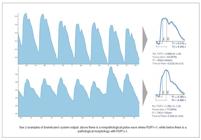
Since the clinical picture of HE is insidious and the recording horizon is relatively small for significant pathophysiological changes to occur in ICC, a well-adapted mean pulse morphology is sufficient. The protocol for selecting the time frame used to obtain these parameters was as follows: first, the trend of the P2/P1 relationship over time was identified and the intervals with the lowest confidence interval were prioritized. A graphical evaluation of all curves was then performed to exclude artifacts such as typical arterial pulses and discrepancies without clinical explanation. Therefore, those minutes were prioritized in which the pulse amplitude showed little variation and that did not belong to the first morphologies (period of adaptation and accommodation). Finally, the curve with the smallest confidence interval and the highest percentage of useful pulses was selected (Figure 3).
After contacting the participant, descriptive data were collected based on clinical history and information from the electronic medical record. These data were recorded on an electronic form for subsequent statistical analysis.
The following descriptive data were collected: medical record number; full name; date of birth; mother’s name; sex; education; date of hospitalization; comorbidities; medications in continuous use; medications for in-hospital use; whether there was a previous neurological event; social habits (alcoholism, smoking, and related burdens); family history; anthropometric measurements (weight, height); blood pressure; classification and etiology of liver disease; date and time of monitoring; participant’s mental status; HE classification; and clinical history of previous HE. With regard to laboratory tests, the following were collected: INR (most recent, maximum); prothrombin time; prothrombin activity; activated partial thromboplastin time; albumin; total bilirubin and fractions; GOT/AST; GPT/ALT; gamma-glutamyl transferase; alkaline phosphatase; serum creatinine. The most recent laboratory test results available, typically from the same or previous day, were recorded. The results were used to calculate the Model for End-Stage Liver Disease (MELD) score and Child-Pugh classification.
Data treatment
For the descriptive characteristics of the control group and the group with HE, the Shapiro-Wilk normality test was applied to the quantitative and ordinal qualitative variables, followed by the t-test for those with verified normality and the Mann-Whitney test for the others.
To evaluate whether the groups with and without HE show a statistically significant difference in ICC, assessed by the parameters P2/P1, TTP and P2/P1xTTP, a normality test was performed and the t-test or Mann-Whitney test was applied, depending on the best indication.
Finally, to evaluate the usefulness of ICP pulse morphology as a predictor of the presence of HE, diagnostic tests were performed estimating the sensitivity and specificity of each parameter (P2/P1 and TTP) over a spectrum of possibilities. By tabulating the sensitivity (true positive rate) by 1-specificity (false positive rate) for each threshold, the corresponding receiver operating characteristic (ROC) curve was found, which can be measured by the parameter area under the ROC curve (AUROC) - calculated by a non-parametric method analogous to the Willcoxon/Mann-Whitney test.
RESULTS
A total of 41 liver disease patients were evaluated, however, due to the poor quality of ICP monitoring, three participants had their ICPni retaken and the data of 1 participant was discarded. Thus, 20 liver disease patients who did not present clinical signs of HE (59.5±9.3 years; 70.0% male) were evaluated as a control group and, as a test group, 20 liver disease patients presenting a clinical picture indicative of HE (59.6±11.9 years; 65.0% male).
The groups had similar levels of education, but the group without HE practiced harmful social habits with greater prevalence and intensity. Alcoholism was a habit for 75% of participants without HE, while it was practiced by 60% of those with HE - the average alcohol load was 65 vs 7 doses/week, respectively. Smoking was practiced by 55% (average smoking history: 22.1 pack-years) of liver disease patients without HE, while 40% (average smoking history: 7.0 pack-years) of those with HE smoked. There were no other significant differences between the groups regarding demographic aspects and habits.
In terms of comorbidities, the groups had similar profiles. The prevalence of diagnosed cirrhosis, diabetes mellitus (DM), hypertension, and obesity for the group without HE compared to the group with HE [X%/X%] were: 90/70, 65/45, 40/40, and 5/15.
Regarding the etiology of liver disease, both groups had a higher prevalence of CLD (95%) compared to ALF (5%) at the time of enrollment. Participants with CLD were classified according to Child-Pugh: the prevalence of A, B and C [%/%/%] was 52.6/36.8/10.5 in those without HE and 0.0/52.6/47.4 in those with HE. The difference between MELD scores was also statistically significant (P=0.0322; power 86.5%): the mean score was 14.0±7.8 for the group without HE and 20.2±9.7 for the group with HE.
In addition, the etiologies of CLD often overlapped. The most common etiologies of CLD in the groups without and with HE were [X%/X%]: cirrhosis of alcoholic origin (57.9/36.8), non-alcoholic hepatic steatosis (26.3/36.8), viral hepatitis (21.5/15.8), hepatocellular carcinoma (15.8/5.3), autoimmune causes (5.3/15.8), hemochromatosis (0.0/10.5), primary biliary cirrhosis (5.3/5.3), other (0.0/10.5), andunknown causes (0.0/15.8).
After data collection, participants were matched for clinical characteristics. Although patients with HE tended to have lower systemic blood pressure, there were no statistically significant differences between the groups. In addition, since non-selective beta-blockers (BB) are used to control portal hypertension in chronic liver disease, it is worth noting that 50% of participants without HE were taking some type of BB continuously or in the hospital, while this number reached 65% among those with current HE.
Also, according to the most recent laboratory data available in the electronic medical records, the groups were statistically similar except for prothrombin activity (63.6±31.0% vs 46.3±13. 6%; P=0.0048), albumin (3.4±0.8g/dL vs 2.9±0.5g/dL; P=0.0253), and serum creatinine (0.9±0.3mg/dL vs 1.3±0.8mg/dL; P=0.0403), which were more critical in patients with current HE (Table 1).
Regarding mental status, among those without HE20, only one was lethargic and the rest were normal. Among those with HE20, 70% were lethargic, 10% were in delirium, 10% were obtunded, and 10% were in coma.
For the group with HE, the encephalopathy was classified, with 1 of the participants being type A (acute) and the rest19 being type C (chronic). Of the chronic cases, 47.4% were further subclassified as precipitated episodic HE (Ia), 10.5% as spontaneous episodic (Ib), 15.8% as recurrent episodic (Ic), 10.5% as mild persistent (IIa), and 15.8% as severe persistent (IIb). Considering types A and C, clinical factors predisposing to HE were identified in half (50%) of the participants with HE - the recurrent ones were: constipation, erysipelas, upper gastrointestinal bleeding, urinary tract infection, spontaneous bacterial peritonitis and drug intoxication.
Regarding ICPni monitoring, the average percentage of useful pulses for each group was 97.2±1.6% for the group without HE and 94.0±7.0% for the group with HE, indicating a statistically significant difference in the quality of monitoring (unpaired t-test: two-tailed P=0.048). This difference is partly explained by the myoclonus and underlying tremor that make up the clinical picture of HE.
When evaluating the P2/P1 ratio, the Shapiro-Wilk W test was significant (P=0.0123), indicating that the parameter does not follow a normal distribution. Comparative evaluation of the P2/P1 ratio using the Mann-Whitney test indicated that the groups were not statistically different (two-tailed P=0.2978). The median P2/P1 ratio for each group was 1.33 for the group without HE and 1.25 for the group with HE.
The mean and median TTP values for each group were 0.26±0.06 [0.25] for the group without EH and 0.22±0.07 [0.22] for the group with EH. The Shapiro-Wilk W test was significant (P=0.0237), indicating that the parameter did not follow a normal distribution. Comparative assessment of TTP using the Mann-Whitney test suggests a statistically significant difference between the groups (two-tailed P=0.0282; median difference=0.04). Furthermore, to control TTP by HR, since these variables are expected to be inversely correlated, it was found that HRxTTP has a statistically normal distribution (Shapiro-Wilk W: P=0.7531) and does not have a statistically significant difference (unpaired t-test: two-tailed P=0.2914) between the groups without and with HE.
The mean and median values of the P2/P1xTTP ratio for each group were 0.37±0.16 [0.34] for the group without HE and 0.28±0.14 [0.27] for the group with HE. The distribution is not statistically normal (Shapiro-Wilk W: P=0.0017). Comparative evaluation of the P2/P1xTTP relationships using the Mann-Whitney test suggests that the groups are not statistically different at the 5% significance level (P=0.0524), but since the P value is extremely close to 0.05, it is possible that this statistical difference will occur with a larger sample size and thus greater power of the test.
ICPni pulse morphologies also varied between the different clinical grades of HE stratified by WHC grades (Table 2). The composition of patients with HE was asymmetric for the mildest grades, with grade 1: 35%, grade 2: 45%, grade 3: 10%, and grade 4: 10%. The low sampling power does not allow to conclude statistically significant differences between grades.
In this context, the optimal cutoff points for P2/P1 and TTP were selected by analyzing the ROC curve (Figure 4). The P2/P1 parameter obtained an AUROC estimated by the Wilcoxon test of 0.5975, with a DeLong standard error of 0.0935. The selected threshold for P2/P1 was 1.31, with a sensitivity of 60% and a specificity of 75%. The TTP parameter obtained an AUROC estimated by the Wilcoxon test of 0.7013, with a DeLong standard error of 0.0857. The optimal threshold selected for TTP was 0.22, with a sensitivity of 90% and a specificity of 45%.

DISCUSSION
The pathophysiology of liver disease is not yet fully understood. From a hemodynamic perspective, CLD is recurrently associated with arterial hypotension and portal hypertension - often generating a state of hyperdynamic circulation. Also, insufficient protein synthesis leading to a decrease in colloid osmotic pressure, an increase in hydrostatic pressure due to a multiplication of vascular resistance in the liver, an increase in sympathetic tone, the load of pro-inflammatory cytokines and nitric oxide are some of the mechanisms that justify these changes.
In addition, cirrhosis - the final stage of CLD - is often associated with harmful habits such as alcoholism (cirrhosis of alcoholic origin) and obesity (non-alcoholic hepatic steatosis). Therefore, several liver diseases are comorbid with cardiovascular diseases such as DM and high BP. Each of these diseases has its own therapy, but antihypertensive therapy plays a special role from a hemodynamic point of view. In this context, non-selective beta-blockers to control portal hypertension are also added to the prescriptions - further compromising the maintenance of an adequate mean arterial BP to maintain CPP.
On the other hand, one of the systemic manifestations of liver disease, hepatic encephalopathy, still has ambiguous hemodynamic behavior. It is a syndrome with multiple pathophysiologies, whether metabolic encephalopathy (e.g., hyperammonemia), cerebral atrophy, cerebral edema, or a combination of these conditions. The medical literature correlates higher levels of HE with progressive increases in intracranial edema and hypertension, in addition to worsening CPP and greater impairment of neurotransmitter systems. The prevalence of comorbidities and the heterogeneous clinic make it difficult to have a clear pathophysiological explanation. In this sense, the classical mechanism of increased CPP in ICH, synthesized in Cushing’s triad, may be compromised in HE: the increase in nitric oxide levels and the dysfunction in the GABAergic and glutamate systems are some of the mechanisms that would justify this dysfunction in perfusion compensation. Thus, the dynamics of systemic blood pressure in a patient with HE cannot be clearly determined. ICP, which is closely correlated with arterial pressure because it is determined by extravasation from the choroid plexus, is also uncertain.
The present study investigated the association of ICP with the presence of HE and its different clinical grades. A diagnostic test was also performed to evaluate the accuracy of using non-invasive ICP morphology parameters as predictors of HE. A diagnostic test, if accurate, should provide status information and suggest management of a patient. However, in order to do this, the evaluated test must be compared to the gold standard, which should correctly assess the evaluated condition. This assessment can be challenging when the gold standard is clinical and passive of relative subjectivity. This is the case of HE: which represents subclinical states and prevalence correlated with other comorbidities.
The present study separates statistically significant differences in the morphological patterns of ICP between the groups evaluated, as well as trends in the parameters. The difference in P2/P1 ratio was not significant (Mann-Whitney: two-tailed P=0.2978); however, TTP proved to be a parameter with a significant difference between groups (Mann-Whitney: two-tailed P=0.0282; median difference = 0.04). A subsequent ROC curve analysis suggested P2/P1=1.31 (AUROC: 0.5975) and TTP=0.22 (AUROC: 0.7013) as optimal cutoff points, where the presence of HE in liver disease would be associated with parameters below these limits.
In addition, the clinical description of the study participants may provide clues for the management of these patients: factors predisposing to the HE crisis were identified in 50% of the participants with HE. All of these factors have a well-described prognostic value in the literature, a detailed specific management and are often treated with continuous prophylaxis.
Despite these findings, clinical heterogeneity and low sample power may be limiting factors for conclusions about the morphologic patterns of ICPni. Furthermore, normality standards for ICPni have not been described in the literature. New technologies may change these paradigms.
Regarding the method chosen to evaluate ICP and ICC, the brain4care® system has consolidated in the literature - several articles have been published since 2013 with applications in the most diverse fields24-34. For example, this technology has been used to assess ICC in dialysis patients with end-stage renal disease. Rickli et al. found a normalization of the P2/P1 ratio after dialysis32. More recently, Ballestero et al. found a significant non-linear relationship between ICPi and ICPni in the assessment of ICH. However, there was no significant association between compliance parameters (P2/P1, TTP and P2/P1xTTP) and ICH (ICP >22 mmHg) or neurological prognosis (measured by Glasgow scale). In the ROC curve analysis to evaluate ICH, the P2/P1 ratio was found to have a sensitivity/specificity/accuracy (%) of 100/62/64 for ICPi and 54/50/50 for ICPni34.
Thus, in line with the literature and the aforementioned studies, the growing applicability of non-invasive technologies in scientific investigations is evident.
CONCLUSION
The use of the brain4care® BWS device proved to be feasible in patients with liver disease without or with clinical signs of hepatic encephalopathy and was able to differentiate them. However, due to the small sample size, a larger case series, a better understanding of the intracerebral hydrodynamic picture in hepatic encephalopathy, and the need for longer-term clinical follow-up are required for definitive pathophysiological explanations.
ACKNOWLEDGMENTS
First, the authors would like to thank the study volunteers for their participation, whose idealism to contribute to research and for the benefit of their fellow human beings motivated them beyond their pain. The authors would also like to thank brain4care® for cordially providing the ICPni monitoring device and technical support for the research. Finally, the authors thank the liver transplant team at the institution for clinical access to medical records.
REFERENCES
-
1 Schulz M, Trebicka J. Acute-on-chronic liver failure: a global disease. Gut. [Internet]. 2022;71:5-6. Available from: https://gut.bmj.com/lookup/doi/10.1136/gutjnl-2020-323973
» https://doi.org/10.1136/gutjnl-2020-323973 -
2 Stravitz RT, Lee WM. Acute liver failure. The Lancet. [Internet]. 2019;394:869-81. Available from: http://dx.doi.org/10.1016/S0140-6736(19)31894-X
» https://doi.org/10.1016/S0140-6736(19)31894-X -
3 Datar S, Wijdicks EFM. Neurologic manifestations of acute liver failure. In: Handbook of Clinical Neurology. [Internet]. 1st ed. Elsevier BV. 2014:645-59. Available from: http://dx.doi.org/10.1016/B978-0-7020-4087-0.00044-9
» https://doi.org/10.1016/B978-0-7020-4087-0.00044-9 -
4 Maloney PR, Mallory GW, Atkinson JLD, Wijdicks EF, Rabinstein AA, Van Gompel JJ. Intracranial Pressure Monitoring in Acute Liver Failure: Institutional Case Series. Neurocrit Care. [Internet]. 2016;25:86-93. Available from: http://link.springer.com/10.1007/s12028-016-0261-y
» https://doi.org/10.1007/s12028-016-0261-y -
5 Kodali S, McGuire BM. Diagnosis and Management of Hepatic Encephalopathy in Fulminant Hepatic Failure [Internet]. Vol. 19, Clinics in Liver Disease. Elsevier Inc. 2015:565-76. Available from: http://dx.doi.org/10.1016/j.cld.2015.04.006
» https://doi.org/10.1016/j.cld.2015.04.006 -
6 Vilstrup H, Amodio P, Bajaj J, Cordoba J, Ferenci P, Mullen KD, et al. Hepatic encephalopathy in chronic liver disease: 2014 Practice Guideline by the American Association for the Study Of Liver Diseases and the European Association for the Study of the Liver. Hepatology. [Internet]. 2014;60:715-35. Available from: https://onlinelibrary.wiley.com/doi/10.1002/hep.27210
» https://doi.org/10.1002/hep.27210 -
7 Ferenci P, Lockwood A, Mullen K, Tarter R, Weissenborn K, Blei AT. Hepatic encephalopathy-Definition, nomenclature, diagnosis, and quantification: Final report of the Working Party at the 11th World Congresses of Gastroenterology, Vienna, 1998. Hepatology. [Internet]. 2002;35:716-21. Available from: http://doi.wiley.com/10.1053/jhep.2002.31250
» https://doi.org/10.1053/jhep.2002.31250 - 8 Ochoa-Sanchez R, Rose CF. Pathogenesis of Hepatic Encephalopathy in Chronic Liver Disease. J Clin Exp Hepatol. [Internet]. 2018;8:262-71. Available from: https://linkinghub.elsevier.com/retrieve/pii/S0973688318306480
-
9 Wijdicks EFM, Plevak DJ, Rakela J, Wiesner RH. Clinical and Radiologic Features of Cerebral Edema in Fulminant Hepatic Failure. Mayo Clin Proc. [Internet]. 1995;7:119-24. Available from: http://dx.doi.org/10.4065/70.2.119
» https://doi.org/10.4065/70.2.119 - 10 Frontera JA. Management of Hepatic Encephalopathy. Vol. 16, Current Treatment Options in Neurology. 2014;1-37.
-
11 Raschke RA, Curry SC, Rempe S, Gerkin R, Little E, Manch R, et al. Results of a protocol for the management of patients with fulminant liver failure*. Crit Care Med. [Internet]. 2008;36:2244-8. Available from: http://journals.lww.com/00003246-200808000-00004
» http://journals.lww.com/00003246-200808000-00004 -
12 Raghavan M, Marik PE. Therapy of Intracranial Hypertension in Patients With Fulminant Hepatic Failure. Neurocrit Care. [Internet]. 2006;4:179-89. Available from: http://link.springer.com/10.1385/NCC:4:2:179
» https://doi.org/10.1385/NCC:4:2:179 -
13 Bajaj JS, Cordoba J, Mullen KD, Amodio P, Shawcross DL, Butterworth RF, et al. Review article: the design of clinical trials in hepatic encephalopathy - an International Society for Hepatic Encephalopathy and Nitrogen Metabolism (ISHEN) consensus statement. Aliment Pharmacol Ther. [Internet]. 2011;33:739-47. Available from: https://onlinelibrary.wiley.com/doi/10.1111/j.1365-2036.2011.04590.x
» https://doi.org/10.1111/j.1365-2036.2011.04590.x -
14 Lidofsky SD, Bass NM, Prager MC, Washington DE, Read AE, Wright TL, et al. Intracranial pressure monitoring and liver transplantation for fulminant hepatic failure. Hepatology. [Internet]. 1992;16:1-7. Available from: https://onlinelibrary.wiley.com/doi/10.1002/hep.1840160102
» https://doi.org/10.1002/hep.1840160102 - 15 Blei AT, Olafsson S, Webster S, Levy R. Complications of intracranial pressure monitoring in fulminant hepatic failure. The Lancet. [Internet]. 1993;341:157-8. Available from: https://linkinghub.elsevier.com/retrieve/pii/014067369390458S
-
16 Rabelo NN, Silveira Chaves PH, Gonçalves de Sena Barbosa M, Frigieri G. Is It Possible to Monitor the Wave Form with Noninvasive Methods? World Neurosurg. [Internet]. 2021;152:231-2. Available from: https://doi.org/10.1016/j.wneu.2021.06.050
» https://doi.org/10.1016/j.wneu.2021.06.050 -
17 Kirkness CJ, Mitchell PH, Burr RL, March KS, Newell DW. Intracranial Pressure Waveform Analysis: Clinical and Research Implications. Journal of Neuroscience Nursing. [Internet]. 2000;32:271-7. Available from: http://journals.lww.com/01376517-200010000-00007
» http://journals.lww.com/01376517-200010000-00007 -
18 Frigieri G, Yukie Hayashi C, Nunes Rabelo N, Brasil S. Intracranial Pressure Waveform: History, Fundamentals and Applications in Brain Injuries. In: Advancement and New Understanding in Brain Injury [Internet]. IntechOpen. 2021;1-15. Available from: https://www.intechopen.com/books/advancement-and-new-understanding-in-brain-injury/intracranial-pressure-waveform-history-fundamentals-and-applications-in-brain-injuries
» https://www.intechopen.com/books/advancement-and-new-understanding-in-brain-injury/intracranial-pressure-waveform-history-fundamentals-and-applications-in-brain-injuries -
19 Zakrzewska AP, Placek MM, Czosnyka M, Kasprowicz M, Lang EW. Intracranial pulse pressure waveform analysis using the higher harmonics centroid. Acta Neurochir (Wien). [Internet]. 2021;163:3249-58. Available from: https://doi.org/10.1007/s00701-021-04958-1
» https://doi.org/10.1007/s00701-021-04958-1 -
20 Brasil S, Solla DJF, Nogueira R de C, Jacobsen Teixeira M, Malbouisson LMS, Paiva WS. Intracranial Compliance Assessed by Intracranial Pressure Pulse Waveform. Brain Sci. [Internet]. 2021 Jul 23;11(8):971. Available from: https://www.mdpi.com/2076-3425/11/8/971
» https://www.mdpi.com/2076-3425/11/8/971 -
21 Mascarenhas S, Vilela GHF, Carlotti C, Damiano LEG, Seluque W, Colli B, et al. The New ICP Minimally Invasive Method Shows That the Monro-Kellie Doctrine Is Not Valid. 2012;114:3-6. Available from: http://www.springerlink.com/index/10.1007/978-3-7091-0956-4
» http://www.springerlink.com/index/10.1007/978-3-7091-0956-4 -
22 Vilela GHF, Cabella B, Mascarenhas S, Czosnyka M, Smielewski P, Dias C, et al. Validation of a New Minimally Invasive Intracranial Pressure Monitoring Method by Direct Comparison with an Invasive Technique. In: Acta Neurochirurgica, Supplementum. [Internet]. 2016;97-100. Available from: https://link.springer.com/10.1007/978-3-319-22533-3_19
» https://link.springer.com/10.1007/978-3-319-22533-3_19 -
23 Cabella B, Vilela GHF, Mascarenhas S, Czosnyka M, Smielewski P, Dias C, et al. Validation of a New Noninvasive Intracranial Pressure Monitoring Method by Direct Comparison with an Invasive Technique. In: Acta Neurochirurgica, Supplementum. [Internet]. Springer-Verlag Wien. 2016;93-6. Available from: https://link.springer.com/10.1007/978-3-319-22533-3_18
» https://link.springer.com/10.1007/978-3-319-22533-3_18 -
24 Galdino GAM, Moura-Tonello SCG, Linares SN, Milan-Mattos JC, Spavieri Jr DL, Oliveira SM, et al. Intracranial compliance in type 2 diabetes mellitus and its relationship with the cardiovascular autonomic nervous control. Brazilian Journal of Medical and Biological Research. [Internet]. 2022;55:1-12. Available from: http://www.scielo.br/scielo.php?script=sci_arttext&pid=S0100-879X2022000100660&tlng=en
» http://www.scielo.br/scielo.php?script=sci_arttext&pid=S0100-879X2022000100660&tlng=en -
25 Ballestero MFM, Frigieri G, Cabella BCT, de Oliveira SM, de Oliveira RS. Prediction of intracranial hypertension through noninvasive intracranial pressure waveform analysis in pediatric hydrocephalus. Child’s Nervous System. [Internet]. 2017;33:1517-24. Available from: http://link.springer.com/10.1007/s00381-017-3475-1
» http://link.springer.com/10.1007/s00381-017-3475-1 -
26 Bollela VR, Frigieri G, Vilar FC, Spavieri Jr DL, Tallarico FJ, Tallarico GM, et al. Noninvasive intracranial pressure monitoring for HIV-associated cryptococcal meningitis. Brazilian Journal of Medical and Biological Research. [Internet]. 2017;50. Available from: http://www.scielo.br/scielo.php?script=sci_arttext&pid=S0100-879X2017000900501&tlng=en
» http://www.scielo.br/scielo.php?script=sci_arttext&pid=S0100-879X2017000900501&tlng=en -
27 Brasil S, Renck AC, Taccone FS, Fontoura Solla DJ, Tomazini BM, Wayhs SY, et al. Obesity and its implications on cerebral circulation and intracranial compliance in severe COVID‐19. Obes Sci Pract. [Internet]. 2021;7:751-9. Available from: https://onlinelibrary.wiley.com/doi/10.1002/osp4.534
» https://doi.org/10.1002/osp4.534 -
28 Brasil S, Taccone FS, Wayhs SY, Tomazini BM, Annoni F, Fonseca S, et al. Cerebral Hemodynamics and Intracranial Compliance Impairment in Critically Ill COVID-19 Patients: A Pilot Study. Brain Sci. [Internet]. 2021;11:874. Available from: https://www.mdpi.com/2076-3425/11/7/874
» https://www.mdpi.com/2076-3425/11/7/874 - 29 da Silveira D, Rabelo NN, de Sena Barbosa MG, Frigeri G, Vellosa JCR. Intracranial pressure and laboratory parameters in high- And low-risk pregnant women. Surg Neurol Int. 2021;12:1-7.
- 30 Frigieri G, Andrade RAP, Dias C, Spavieri DL, Brunelli R, Cardim DA, et al. Analysis of a non-invasive intracranial pressure monitoring method in patients with traumatic brain injury. In: Acta Neurochirurgica, Supplementum. Springer-Verlag Wien. 2018;107-10.
-
31 Nagai Ocamoto G, Spavieri Junior DL, Matos Ribeiro JA, Frigieri Vilela GH, Catai AM, Russo TL. Noninvasive Intracranial Pressure Monitoring in Chronic Stroke Patients with Sedentary Behavior: A Pilot Study. In: Acta Neurochirurgica, Supplementum. [Internet]. Springer Science and Business Media Deutschland GmbH. 2021;55-8. Available from: https://link.springer.com/10.1007/978-3-030-59436-7_12
» https://link.springer.com/10.1007/978-3-030-59436-7_12 -
32 Rickli C, Kalva DC, Frigieri GH, Schuinski AFM, Mascarenhas S, Vellosa JCR. Relationship between dialysis quality and brain compliance in patients with end-stage renal disease (ESRD): a cross-sectional study. Sao Paulo Medical Journal. [Internet]. 2022;140:398-405. Available from: http://www.scielo.br/scielo.php?script=sci_arttext&pid=S1516-31802022000300398&tlng=en
» http://www.scielo.br/scielo.php?script=sci_arttext&pid=S1516-31802022000300398&tlng=en -
33 Rojas SSO, Ordinola AAM, Veiga VC, Souza JM de. The use of a noninvasive intracranial pressure monitoring method in the intensive care unit to improve neuroprotection in postoperative cardiac surgery patients after extracorporeal circulation. Rev Bras Ter Intensiva. [Internet]. 2021;33:469-76. Available from: http://criticalcarescience.org.br/artigo/detalhes/0103507X-33-3-18
» http://criticalcarescience.org.br/artigo/detalhes/0103507X-33-3-18 -
34 Ballestero M, Dias C, Gomes ICN, Grisi LS, Cardoso RAM, Júnior ELZ, et al. Can a new noninvasive method for assessment of intracranial pressure predict intracranial hypertension and prognosis? Acta Neurochir (Wien). [Internet]. 2023;165:1495-503. Available from: https://link.springer.com/10.1007/s00701-023-05580-z
» https://link.springer.com/10.1007/s00701-023-05580-z