
 Myrrhone exert cardioprotective effect against isoproterenol-induced myocardial injury in rats via alteration of NF-κB and Bax/Bcl-2/caspase-3 signaling
ABSTRACT
Purpose: To examine the cardioprotective effects of myrrhone against isoproterenol (ISO)-induced myocardial injury in rats.
Methods: Myocardial injury was induced in the rats through subcutaneous administration of ISO (85 mg/kg). The body weight, heart weight, electrocardiogram (ECG), cardiac, antioxidant, electrolyte, membrane-bound enzymes, antioxidant, cytokines, and inflammatory parameters were estimated. The mRNA expression of inflammatory parameters was estimated in the cardiac tissue.
Results: Myrrhone treatment significantly (p < 0.001) altered ECG parameters, body weight, heart weight, and heart weight/body weight ratio. Myrrhone significantly (p < 0.001) improved the level of 5-hydroxytryptamine (5-HT) and suppressed the level of cardiac parameters like creatine kinase-MB, creatine kinase, lactate dehydrogenase, cardiac troponin I, and cardiac troponin T. It also suppressed the level of hepatic parameters such as aspartate aminotransferase, and alanine transaminase; altered the electrolyte, membrane-bound enzymes, and antioxidant parameters. Myrrhone treatment significantly (p < 0.001) altered the level of cytokines such as tumor necrosis factor-α (TNF-α), interleukin (IL)-10, IL-1β, IL-17, and IL-6; inflammatory parameters like cyclooxygenase-2, prostaglandin, inducible nitric oxide synthetase, nuclear factor kappa-light-chain enhancer of activated B cells; matrix metalloproteinases such as 2, and 9; apoptosis parameters viz., Bcl-2-associated X protein (Bax), B-cell lymphoma 2 protein (Bcl-2), and caspase-3 parameters. Myrrhone treatment significantly (p < 0.001) altered the mRNA expression IL-6, IL-1β, IL-10, TNF-α, Bcl-2, caspase-3, Bax, and caspase-9.
Conclusion: Myrrhone exhibited the cardioprotective effect against ISO-induced myocardial injury in rats via alteration of kappa-light-chain enhancer of activated B cells (NF-κB) and Bax/Bcl-2/caspase-3 signaling.
Key words
Myocardial Reperfusion Injury; Inflammation; Matrix Metalloproteinases; Apoptosis; Oxidative Stress
Introduction
It is widely known that the heart is a vital organ, and any dysfunction in its tissue can have significantly effects on other organs1. Myocardial injury is a major cause of morbidity and mortality in both developing and developed countries2,3. Myocardial injury, commonly known as a heart attack, is a serious medical condition that occurs when the blood supply to portion of the heart tissue is blocked. This obstruction prevents oxygen and nutrients from reaching the heart muscle, resulting in tissue damage or death4. Plaque accumulation in the coronary arteries is the source of this occlusion, which can result in either total or partial arterial obstruction5. There is a mismatch between the demands of the heart for oxygenated blood and the ability of the coronary arteries to meet that demand resulting from coronary artery blockage, which is the root cause of myocardial injury6. The heart has limited capacity for anaerobic metabolism and cannot compensate the lack of oxygen, blood, and the necessary nutrients during a myocardial damage. This inadequacy tends to produce a pathologic change followed by congestive heart failure4.
However, myocardial injury condition can induce the various symptoms such as myocardial fibrosis and hypertrophy. Previous reports suggest that apoptosis, oxidative stress, and inflammatory reactions are involved in the mechanism of myocardial injury7. Previous research indicates that isoproterenol (ISO)-induced oxidative stress is mediated by free radicals or reactive oxygen species (ROS). This is evidenced by a significant increase in tissue lipid peroxidation (LPO) and a reduction in antioxidant parameters. Antioxidants play a crucial role in protecting against myocardial injury. It is believed that any phytoconstituent or compound with potent antioxidant activity can safeguard cardiac tissue from myocardial injury7,8.
During the myocardial injury, increased production of cytokines leads to tissue infiltration by inflammatory cells. Inflammatory cytokines can accelerate myocardial injury, often occurring alongside severe congestive heart diseases9. During the myocardial injury, neutrophils infiltrate the infarcted area, exacerbating injury to myocardial cells by triggering the production of cytokines, proteolytic enzymes, and chemokines10. Ultimately, this cascade leads to increased ROS generation and heightened oxidative stress. Elevated levels of cytokines, ROS, and chemokines can lead to impaired organ function. Cytokines, particularly tumor necrosis factor-α (TNF-α), enhance neutrophil migration into the ischemic area of infarcted myocardial tissue11. Interleukin (IL)-6 and IL-1β involved in the inflammatory response to stress in the myocardial tissue. In myocardial injury condition, adverse reactions are classified based on the activation of cellular signaling molecules, including nuclear factor kappa-light-chain enhancer of activated B cells (NF-κB). NF-κB is a well-known key regulator during inflammatory and apoptotic conditions12. During myocardial injury, NF-κB phosphorylation triggers an intracellular signaling cascade, leading to the induction of inflammatory cytokines such as TNF-α, IL-1β, and IL-6, along with other inflammation-related proteins. These changes contribute to the pathophysiology associated with myocardial injury9.
ISO (1-3,4-dihydroxyphenyl-2-isopropylamino ethanol hydrochloride) is a synthetic catecholamine and β-adrenoceptor agonist, which is commonly used in the treatment of ventricular bradycardia, bronchial asthma, glaucoma, allergic emergencies and finally induces the cardiac arrest7,13. Therefore, a high dose of ISO induces myocardial stress by depleting the energy stored in cardiomyocytes, ultimately leading to infarct-like necrosis and cellular injury. Following autooxidation, ISO generates a large amount of free radicals, altering the endogenous antioxidant levels in the tissues7. The subcutaneous administration of ISO induces the myocardial injury in the rodent. The acute hemodynamic and electrocardiographic changes observed in ISO-induced myocardial injury closely resemble those seen in patients experiencing myocardial injury4,14. ISO is a well-established model with high validity, reproducibility, and low mortality compared to other rodent models.
The lock-and-key concept describes how allopathic Western drugs work, focusing on a specific metabolic or signaling system. However, the inability to effectively treat diseases often stems from the involvement of multiple molecular mechanisms13. Many synthetic drugs have been investigated for their efficacy against myocardial injury, but unfortunately, most of them exhibit side effects, and in some cases, they can even lead to patient’s mortality. These synthetic drugs manifest various undesirable effects that pose serious health risks15. Currently, researchers are directing their efforts toward studying plant extracts and their phytoconstituents as potential cardioprotective agents against myocardial injury injuries16.
Myrrhone (naphtho2,1-b.furan-6(7H)-one, 8,9-dihydro-1,5,8-trimethyl-, (8R)-) is isolated from Commiphora myrrha tree17,18. It is a common Chinese herbal medicine with various physiological properties, which play a crucial role in the liver damage and renal cancer19. Myrrhone exhibited the antioxidant and anti-inflammatory potential against various diseases. In this experimental study, we investigated the cardioprotective effect against ISO-induced myocardial injury in rats and explored the underlying mechanism.
Methods
Experimental rats
Swiss Wistar rats (12–14 weeks old, weighing 200 ± 20 g, male) were used in this study. They were maintained under standard laboratory conditions (temperature 22 ± 5°C, relative humidity = 65%, 12/12-h dark/light cycle) and provided with a standard chow pellet diet and water ad libitum. The rats were acclimatized to the laboratory environment for seven days prior to the experimental study. The entire study was conducted in accordance with the institution’s Ethical Committee Guidelines.
Toxicant and tested drug preparation
Subcutaneous administration of ISO (85 mg/kg) was used to induce the myocardial injury20. The tested drug (myrrhone) was administered to the rats as an oral suspension, prepared by dissolving the drug in 1% carboxymethyl cellulose (CMC).
Experimental group
The rats were randomly divided into following groups (n = 6). The groups are presented in Fig. 1. The ISO was administered to the experimental rats for two successive days as per the previous reported method with minor modification4,20.

Electrocardiography recording
At the end of the experimental study (30 days), the rats were anesthetized with urethane (125 mg/kg, b.w.) and given a stabilization period of 30 minutes before recording electrocardiography20. Electrocardiography (ECG) parameters were recorded using an ECG coupler and a student’s physiograph. Briefly, electrodes made from 26-gauge hypodermic needles were attached to both the front and hind paws. One precordial lead was used and placed in a position corresponding to V4 in humans. The QT interval, RR interval, corrected QT interval, QRS interval, and heart rate were estimated.
Blood and plasma sample preparation
At the end of the study, the rats were anesthetized with ketamine (80 mg/kg) to collect blood samples from the retro-orbital plexus. The blood samples were collected in dry test tubes and allowed to coagulate at room temperature for 30 minutes. After coagulation, the samples were centrifuged for 10 minutes at 5,000 rpm.
Cardiac parameters
Cardiac parameters such as cardiac troponin T (cTnT) (Cat. No. E-EL-H0503, Elabscience), creatine kinase-MB (CK-MB) (Cat. No. E1343Hu, BT Lab), creatine kinase (CK) (MBS703319, MyBioSource), cardiac troponin I (cTnI) (Cat. No. ab200016, Abcam), and lactate dehydrogenase (LDH) (Cat. No. E-EL-H6004, Elabscience) were estimated using enzyme-linked immunosorbent assay (ELISA) kits, following the manufacturer’s instructions.
Cardiac 5HT, serum 5HT and cardiac total Ca2+
The level of serum and cardiac 5-HT (Cat. No. KLU0048, Krishgen GENLISA) were estimated using the ELISA kits following the manufacture’s instruction.
Lipid parameters
The level of lipid parameters like triglycerides (TG), total cholesterol (TC), and high-density lipoprotein cholesterol (HDL-C) were estimated using the ELISA kits following the manufacture’s instruction. The level of low-density lipoprotein cholesterol (LDL-C) and very low density lipoprotein cholesterol (VLDL-C) were estimated using the previous formula of Friedewald et al.21.
Hepatic parameters
A previously reported method was used for the estimation of hepatic parameters with minor modifications7. Briefly, 100-µL serum sample was mixed into the 100 mM L-aspartate (0.5 mL) and 100 mM α-oxoglutarate in phosphate buffer at pH = 7.4 and left for incubation at room temperature (37°C) for 30 min and mix the 0.5-mL 2,4 dinitrophenylhydrazine and re-incubated at room temperature against serum free reagent (blank sample). The enzyme activity was assessed by employing a standard curve based on commercially available standards, and the results were expressed in international units per liter (IU/L).
Electrolytes and membrane-stabilizing enzyme
The electrolytes such as potassium (K+), sodium (Na+), calcium (Ca2+), and chlorine (Cl-) (Cat. No 2114) were determined using an automated electrolyte analyzer based on the ion selective electrode technology. Briefly, the blood samples were collected in all group rats and allowed to clot and centrifuged for 10 min at 3,000 rpm to collect the serum. The serum samples were then analyzed as the manufacture’s instruction (Roche Diagnostics, United States of America).
The membrane stabilizing enzymes like Mg2+ATPase, Na+/K+ ATPase, and Ca2+ATPase were determined by the spectrophotometrically via determining the inorganic phosphate (Pi) liverbated during the ATP hydrolysis using the previous report method with minor modification22,23. Briefly, we prepared the tissue homogenates in a suitable ice-cold buffer and centrifuged them at 10,000 rpm for 5 min to obtain the microsomal fraction. The reaction mixture contained the appropriate level of Tris HCl buffer, NaCl, MgCl2, CaCl2, and KCl, depending on the specific enzyme being determined. ATP was added in the reaction mixture to initiate the reaction and was incubated at room temperature (37°C) for defined time. For the termination of the reaction, adding the cold trichloroacetic acid (TCA), the level of Pi generated was quantified. Finally, the enzyme activities were presented as µmol Pi liberated per mg protein per hour.
Inflammatory cytokines, apoptosis, MMP, and oxidative stress parameters
Inflammatory cytokines such as TNF-α (Cat. No. NBP1-91170, Novus Biologicals), IL-1β (Cat. No. EKF57064-96T, Biomatik), IL-17 (Cat. No. ELH-IL17A-1, RayBiotech), IL-6 (Cat. No. ELH-IL6-1, RayBiotech), and IL-10 (Cat. No. 950.060.192, Medix Biochemica); inflammatory parameters like cyclooxygenase-2 (COX-2) (Cat. No. EH125RB, Thermo Fisher Scientific), prostaglandin (PGE2) (Cat. No. 514010, Cayman Chemical), inducible nitric oxide synthetase (iNOS) (Cat. No. EEL035, Thermo Fisher Scientific), and NF-κB (Cat. No. 85-86081-11, Thermo Fisher Scientific); apoptosis marker viz., B-cell lymphoma 2 protein (Bcl-2) (Cat. No. BMS244-3, Thermo Fisher Scientific), Bax (Cat. No. EEL030, Thermo Fisher Scientific), caspase-3 (Cat. No. KHO1091, Thermo Fisher Scientific); oxidative stress parameters, including catalase (CAT) (Cat. No. A007-1-1, Nanjing Jiancheng Bioengineering Institute, Nanjing), superoxide dismutase (SOD) (Cat. No. 19160, Cayman Chemical), glutathione peroxidase (GPx) (Cat. No. CGP1, Sigma-Aldrich), reduced glutathione (GSH) (Cat. No. CS0260, Sigma-Aldrich), and malondialdehyde (MDA) (Cat. No. MAK085, Sigma-Aldrich); and MMP parameters such as MMP-2 (Cat. No. MMP200, Thermo Fisher Scientific) and 9 (Cat. No. KHC3081, Thermo Fisher Scientific) were estimated using the ELISA’s instruction.
Quantitative real-time reverse transcription polymerase chain reaction
Total RNA from the hearts of the experimental rats was isolated with the Trizol reagent as the manufacturer’s instructions (Invitrogen, United States of America). The first strand of cDNA was synthesized by reverse transcription (20 μL) with a random primer by using total 2 µg of RNA. After a denaturing step of 1 min at 95°C, several polymerase chain reaction (PCR) cycles resulted in the amplification of β-actin. Primer list is shown in Table 1.
Statistical analysis
For statistical analysis, GraphPad Prism software was utilized. Dunnett’s T-test was used after a one-way analysis of variance (ANOVA), and p < 0.05 was deemed statistically significant.
Results
Electrocardiography parameters
ECG parameters of myocardial injury rats induced by ISO are significantly different from those of control animals. In particular, the QRS complex, HR, QT interval, R-R intervals, and the corrected QT interval (QTc) were severely affected (Fig. 2a–2e). These alterations in ECG characteristics are a manifestation of the myocardial dysfunction and resultant electrical derangements of myocardial injury. ISO-induced myocardial injury model is commonly used to investigate the pathophysiology of myocardial injury and estimation the therapeutic interventions. Myrrhone treatment significantly (p < 0.001) reduced the ECG levels, indicating the possible cardioprotective activity of myrrhone. The modulation of ECG parameters by myrrhone demonstrates its competence to normalize the cardiac electrophysiological abnormalities induced by myocardial injury and possibly enhance the cardiac performance.
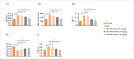
Effect of myrrhone on the electrocardiogram parameters of normal and isoproterenol (ISO)-induced myocardial injury group rats. (a) QRS, (b) heart rate (HR), (c) QT, (d) R-R and (e) QTc. Data are expressed as mean ± standard error of the mean (n = 6 for each group).
Body weight, heart weight and heart weight/body weight ratio
Body weight (BW), heart weight (HW), and HW/BW ratio are indispensable physiological parameters for medical and biological studies. Body mass is a rough index of an individual’s size and general nutritional condition. HW correlates with the size and status of this essential organ. The HW/BW ratio is a derived index that corrects HW for differences in body size and facilitates comparability between smaller and larger individuals or groups. ISO-induced myocardial injury rats showed increased HW (Fig. 3a) and decreased BW (Fig. 3b). Myrrhone treatment significantly (p < 0.001) reduced HW and increased BW. In the ISO-induced myocardial injury group, rats exhibited an increased HW/BW ratio (Fig. 3c), which was significantly reduced (p < 0.001) by myrrhone treatment.

Effect of myrrhone on the body weight (BW), heart weight (HW) and HW/BW ratio of normal and isoproterenol (ISO)-induced myocardial injury group rats. (a) Heart weight, (b) body weight, and (c) HW/BW ratio. Data are expressed as mean ± standard error of the mean (n = 6 for each group).
Cardiac parameters
ISO group rats exhibited the boosted level of LDH (Fig. 4a), CK (Fig. 4b), CK-MB (Fig. 4c), CTnI (Fig. 4d), and CTnT (Fig. 4e). These are enzymes that usually enter the blood stream following damage or stress to cardiac muscle cells. Myrrhone treatment significantly (p < 0.001) repressed the level of cardiac parameters.

Effect of myrrhone on the biochemical parameters of normal and isoproterenol (ISO)-induced myocardial injury group rats. (a) lactate dehydrogenase (LDH), (b) creatine kinase (CK), (c) CK-MB, (d) cardiac troponin I (cTnI), and (e) cardiac troponin T (cTnT). Data are expressed as mean ± standard error of the mean (n = 6 for each group).
Electrolytes
Electrolyte imbalance is a well-recognized consequence of myocardial injury, with significant implications for both cardiac function and overall physiological homeostasis. Myocardial injury was induced in rats with ISO, and it was found that concentrations of K+, Na+, Cl-, and Ca2+ underwent remarkable changes during the progress of myocardial injury. ISO induced myocardial injury rats showed the altered level of K+ (Fig. 5a), Na+ (Fig. 5b), Cl- (Fig. 5c), and Ca2+ (Fig. 5d). Myrrhone treatment significantly (p < 0.001) modulated the electrolyte level.

Effect of myrrhone on the electrolyte levels of normal and isoproterenol (ISO)-induced myocardial injury group rats. (a) K+, (b) Na+, (c) Cl-, and (d) Ca2+. Data are expressed as mean ± standard error of the mean (n = 6 for each group).
Membrane-bound enzyme
The membrane-bound enzymes play a crucial role in maintaining electrochemical gradients across cell membranes, regulating intracellular ion concentrations, and facilitating various physiological processes. The decrease in their activity suggests compromised cellular energy metabolism and impaired ion transport mechanisms, which are characteristic features of myocardial damage during myocardial injury. The level of Na+K+ ATPase (Fig. 6a), Mg2+ ATPase (Fig. 6b), and Ca2+ ATPase (Fig. 6c) reduced in the ISO-induced myocardial injury group rats and myrrhone treatment significantly (p < 0.001) boosted the level of membrane-bound enzymes. This restoration of enzyme activity suggests that myrrhone possesses cardioprotective properties, potentially through its ability to stabilize cell membranes, improve energy metabolism, and restore ion homeostasis.

Effect of myrrhone on the membrane bound enzyme and 5-HT parameters of normal and isoproterenol (ISO)-induced myocardial injury group rats. (a) Na+K+ ATPase, (b) Mg2+ ATPase, (c) Ca2+ ATPase, (d) 5-HT in serum, and (e) 5-HT in tissue. Data are expressed as mean ± standard error of the mean (n = 6 for each group).
5-hydroxytryptamine
The findings suggested that reduced ISO-induced myocardial injury in rats may be accompanied by reduced levels of 5-hydroxytryptamine (HT) in serum and tissue. Such decrease in 5-HT level may be involved in the pathogenesis of myocardial injury and could influence the cardiac function, platelet aggregation and vascular tone. ISO induced myocardial injury group rats showed the suppressed level of 5-HT in the serum (Fig. 6d) and tissue (Fig. 6e), and myrrhone treatment significantly (p < 0.001) boosted the level in serum and tissue. The substantial rise of 5-HT level induced by myrrhone treatment may have great implications for cardiac recovery and function post-myocardial injury and may contribute to improve cardiovascular outcomes via better regulation of heart rate, blood pressure, and platelet function.
Hepatic parameters
ISO-induced myocardial injury group rats showed the enhanced level of hepatic parameters like aspartate aminotransferase (AST) (Fig. 7a), and alanine transaminase (ALT) (Fig. 7b). These enzymes are a common indicator of liver function or liver injury, and their elevation in the blood is indicative of hepatocellular injury or liver damage. The increase in these indices in the ISO-induced myocardial injury group may reflect secondary effects of myocardial injury on liver function, potentially associated with hemodynamic changes or a systemic inflammatory response resulting from cardiac damage in the rat model. Myrrhone ability to decrease the levels of AST and ALT may indicate that it might attenuate liver function and integrity during cardiac stress, perhaps through anti-inflammatory or antioxidant potentials.

Effect of myrrhone on the level of hepatic parameters of normal and isoproterenol (ISO)-induced myocardial injury group rats. (a) aspartate aminotransferase (AST), and (b) alanine transaminase (ALT) in serum. Data are expressed as mean ± standard error of the mean (n = 6 for each group).
Lipid parameters
ISO group rats exhibited the altered level of lipid parameters like TC (Fig. 8a), TG (Fig. 8b), high-density lipoprotein (HDL) (Fig. 8c), low-density lipoprotein (LDL) (Fig. 8d), and very low-density lipoprotein (VLDL) (Fig. 8e). Such alterations of the lipid profile reflect the disturbed lipid metabolism and may be linked to an elevated cardiovascular risk profile. Myrrhone treatment significantly (p < 0.001) altered the level of lipid parameters. Modulation of these lipid parameters by myrrhone may be useful in treatment of lipid-related disorders or as cardioprotective agent.
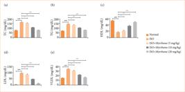
Effect of myrrhone on the lipid parameters of normal and isoproterenol (ISO)-induced myocardial injury group rats. (a) Total cholesterol (TC), (b) triglycerides (TG), (c) high-density lipoprotein (HDL), (d) low-density lipoprotein (LDL), and (e) very low-density lipoprotein (VLDL). Data are expressed as mean ± standard error of the mean (n = 6 for each group).
Oxidative stress parameters
ISO-induced myocardial injury group rats showed the altered levels of SOD (Fig. 9a), MDA (Fig. 9b), GPx (Fig. 9c), CAT (Fig. 9d), and GSH (Fig. 9e), and myrrhone treatment significantly (p < 0.001) modulated the level of oxidative stress parameters.
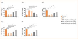
Effect of myrrhone on the antioxidant parameters of normal and isoproterenol (ISO)-induced myocardial injury group rats. (a) superoxide dismutase (SOD), (b) malondialdehyde (MDA), (c) glutathione peroxidase (GPx), (d) catalase (CAT), and (e) reduced glutathione (GSH). Data are expressed as mean ± standard error of the mean (n = 6 for each group).
Cytokines
ISO-induced myocardial injury group rats exhibited the altered level of cytokines like IL-1β (Fig. 10a), TNF-α (Fig. 10b), IL-6 (Fig. 10c), IL-10 (Fig. 10d), and IL-17 (Fig. 10e). Modifications of cytokine responses are consistent with those of the post-myocardial injury inflammatory reaction. The deregulation of these cytokines is involved in the pathophysiology of myocardial injury, leading to tissue injury, the remodeling of the heart and dysfunction. Myrrhone treatment significantly (p < 0.001) restored the level of inflammatory cytokines. This indicates that myrrhone has strong anti-inflammatory activity over inflammation in myocardial injury.
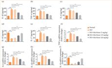
Effect of myrrhone on the inflammatory cytokines in tissue and inflammatory cytokines mRNA expression of normal and isoproterenol (ISO)-induced myocardial injury group rats. (a) Interleukin (IL)-1β, (b) tumor necrosis factor (TNF)-α, (c) IL-6, (d) IL-10, (e) IL-17, (f) TNF-α, (g) IL-6, (h) IL-1β and (i) IL-10. Data are expressed as mean ± standard error of the mean (n = 6 for each group).
ISO-induced myocardial injury group rats showed altered mRNA expression of cytokines such as TNF-α (Fig. 10f), IL-6 (Fig. 10g), IL-1β (Fig. 10h), and IL-10 (Fig. 10i), and myrrhone treatment significantly (p < 0.001) modulated the mRNA expression.
Inflammatory parameters
The myocardial injury group rats exhibited the increased levels of various inflammatory cytokines, which suggested a severe inflammatory response after suffering from myocardial injury. ISO-induced myocardial injury group rats showed the increased level of PGE2 (Fig. 11a), COX-2 (Fig. 11b), iNOS (Fig. 11c), NF-κB (Fig. 11d), and C-reactive protein (Fig. 11f). These inflammatory mediators are critical for the advancement of myocardial injury and subsequent repair following injury. Myrrhone treatment significantly (p < 0.001) suppressed the level of inflammatory parameters. The ability of myrrhone to reduce several inflammatory mediators at a time suggests that myrrhone could serve as a treatment for post-infarct inflammation and possibly overall cardiac outcomes.

Effect of myrrhone on the inflammatory parameters of normal and isoproterenol (ISO)-induced myocardial injury group rats. (a) Prostaglandin (PGE2), (b) cyclooxygenase-2 (COX-2), (c) inducible nitric oxide synthetase (iNOS), (d) kappa-light-chain enhancer of activated B cells (NF-κB), and (e) C-reactive protein. Data are expressed as mean ± standard error of the mean (n = 6 for each group).
Apoptosis parameters
ISO-induced myocardial injury group rats demonstrated the altered level of Bcl-2 (Fig. 12a), Bax (Fig. 12b), and caspase-3 (Fig. 12c). They reflect a disturbance in the equilibrium between pro-apoptotic and anti-apoptotic factors, which may result in enhanced cell death in the myocardium. Myrrhone treatment significantly (p < 0.001) restored the level of apoptosis parameters. This normalization of apoptotic equilibrium may be responsible for diminution of death of cardiac-myocytes and normalization of myocardial function, suggesting that the myrrhone possesses potential therapeutic implications for myocardial injury and other related cardiac disorders.
ISO-induced myocardial injury group rats showed the altered mRNA expression of Bax mRNA expression (Fig. 12d), Bcl-2 mRNA expression (Fig. 12e), caspase-3 mRNA expression (Fig. 12f), and caspase-9 mRNA expression mRNA expression (Fig. 12g), and myrrhone treatment significantly (p < 0.001) modulated the mRNA expression.

Effect of myrrhone on the apoptosis parameters and mRNA expression of normal and isoproterenol (ISO)-induced myocardial injury group rats. (a) B-cell lymphoma 2 protein (Bcl-2), (b) Bcl-2-associated X protein (Bax), (c) caspase-3, (d) Bcl-2 mRNA, (e) Bax mRNA, (f) caspase-3 mRNA, and (g) caspase-9 mRNA. Data are expressed as mean ± standard error of the mean (n = 6 for each group).
MMP
ISO-induced myocardial injury group rats exhibited the boosted level of MMP-9 (Fig. 13a), and MMP-2 (Fig. 13b). This MMP upregulation is a typical constituent of the pathologic remodeling process after myocardial damage. The elevated MMP activity has a role in the disruption of structural proteins in the heart, resulting in the adverse cardiac remodeling, ventricular dilation, and heart failure. Myrrhone treatment significantly (p < 0.001) suppressed the MMP level.

Effect of myrrhone on the MMP level of normal and isoproterenol (ISO)-induced myocardial injury group rats. (a) MMP-9, and (b) MMP-2. Data are expressed as mean ± standard error of the mean (n = 6 for each group).
Discussion
Myocardial injury causes permanent damage to heart tissue and increases the mortality rate among affected individuals24,25. The pathogenesis of myocardial injury involves several processes, such as cardiac injury, oxidative stress, contractile dysfunction, calcium overload, and cell death through apoptosis, necrosis, or both4. The ISO-induced myocardial injury model is a well-established and non-invasive approach for reliably evaluating the cardioprotective potential of various therapeutic agents4,26.
Previous reports suggested that the secretion of 5-HT begins during the acute inflammation, cardiac ischemia, and tissue injury, primarily from platelets27. During these conditions, 5-HT secreted from the platelets4. Thrombus formation, platelet aggregation, coronary artery spasms, and smooth muscle cell contraction all are triggered by the secretion of additional 5-HT from the dense granules of platelets after it binds to 5-HT2A receptors27,28. The myocardial interstitium accumulates 5-HT, which contributes to cardiac cellular damage through various pathways, including 5-HT2A and 5-HT2B receptors, as well as 5-HT uptake transporters4,28.
A heart attack, also known as myocardial injury, occurs when blood flow to part of the heart is blocked long enough that part of the heart muscle is damaged or dies. Often the blockage is due to a buildup of fat, cholesterol, and other substances that creates a plaque in the coronary arteries20. When blood flow is disrupted, the affected heart tissue is deprived of oxygen and nutrients, leading to cell injury and death. This can cause significant damage to the heart muscle, impairing its ability to pump blood effectively5,7,14. When blood flow is restored, an inflammatory reaction and oxidative stress are initiated, which play a crucial role in damaging cells and tissues. During myocardial injury, various cardiac biomarkers such as CK-MB, LDH, cTnT, and cTnI are secreted into the blood1,13. Elevated levels of these biomarkers indicate myocardial necrosis (death of heart muscle cells) and cell membrane injury. These biomarkers are used to estimate, diagnose, and monitor the severity of myocardial injury. An abnormally high level of CK-MB during myocardial injury is considered a specific biomarker for assessing myocardial injury. CK-MB and CK levels significantly increase within 3–4 hours after myocardial injury7,29. LDH is an enzyme found in many tissues throughout the body, including the heart. CK-MB and LDH are also used as biomarkers for diagnose the myocardial injury. cTnI is a protein found in heart muscle and is highly specific for myocardial injury. Cardiac troponins are the preferred biomarkers for diagnosing acute myocardial injury30,31. Elevated levels indicate heart muscle damage and are used to assess the severity of the injury. cTnT, like cTnI, is a protein specific to the heart muscle and is a critical marker for myocardial injury. cTnT is used alongside cTnI to diagnose myocardial injury and evaluate the extent of heart muscle damage7,30. During myocardial injury, the heart muscle begins to sustain damage, leading to the secretion of these enzymes into the blood. The myrrhone treatment ameliorate the cardiac enzymes, which suggest the cardioprotective effect.
Na+, Cl-, K+, and Ca2+ are crucial electrolytes involved in the proper functioning of the heart. Their roles in myocardial injury are significant due to their influence on various physiological processes, including cellular signaling, muscle contraction and fluid balance32,33. Na+ is essential for generating action potentials in cardiac cells, facilitating electrical conduction, and maintaining osmotic balance. Chloride helps to maintain the electrical neutrality and osmotic balance within cells. It also participates in the regulation of cell volume and pH. During myocardial injury, sodium influx into cardiac cells increases the level of Cl-, leading to cellular swelling and potentially contributing to cell death. Increased intracellular sodium can also lead to calcium overload through the sodium-calcium exchanger, exacerbating injury33–36. Changes in Cl- levels can influence the acid-base balance and contribute to arrhythmias during myocardial injury. Chloride shifts can affect cellular excitability and exacerbate damage in ischemic conditions. K+ is critical for repolarization of cardiac cells after a potential action. It helps maintain resting membrane potential and overall cardiac rhythm. During myocardial injury condition, potassium can leak out of damaged cells, leading to hyperkalemia in the extracellular space. This can cause arrhythmias and impair cardiac function. Additionally, hypokalemia can occur, further destabilizing cardiac electrical activity. Calcium is vital for excitation-contraction coupling in cardiac muscle cells. It triggers contraction by binding to troponin and facilitates the release of neurotransmitters. Calcium overload is a major factor in myocardial injury. Excessive calcium influx into cells can activate proteolytic enzymes, disrupt mitochondrial function, and lead to cell death. This calcium overload can result from ischemia-reperfusion injury, in which reintroduction of blood supply after ischemia leads to a surge in calcium influx32,33,37. Myrrhone treatment effectively restored the level of electrolyte levels towards the normal physiological ranges, which indicate that electrolytes (sodium, calcium, potassium, and chloride) are the indicative of the impaired cardiac function and systemic imbalance. Myrrhone treatment enhanced the level of electrolyte, suggesting the corrective effect on ISO-induced electrolyte disturbances.
ISO, through its sympathetic stimulation, can cause the dysregulation of lipid metabolism and increase the serum lipid levels, thereby contributing to a higher risk of coronary heart disease and myocardial injury13. It may stimulate the biosynthesis of lipids in the liver. An increase in acyl coenzyme A (acyl CoA) levels can lead to higher lipid synthesis. Elevated production of glycerol can contribute to triglyceride synthesis and accumulation38,39. Dysregulation of lipid metabolism is a significant factor in the development of myocardial injury. ISO is a synthetic catecholamine that mimics the effects of the sympathetic nervous system by stimulating beta-adrenergic receptors7. Its cardiovascular effects include increased heart rate, cardiac output, and myocardial oxygen consumption. Previous reports have shown that ISO administration leads to elevated levels of serum lipids, including LDL, TC, VLDL, and TG13,38. High levels of LDL and VLDL and low levels of HDL are associated with an increased risk of coronary heart disease and myocardial injury13. ISO administration induced the significant dyslipidemia, evidence via enhanced the serum levels of LDL, TC, TG, and VLDL and reduced the HDL. Myrrhone treatment considerably restore these alterations, restoring lipid profile balance. The lipid lowering effect of myrrhone treatment suggests its potential role to mitigate ISO-induced atherogenic risk and also contributing to its overall cardioprotective mechanism.
The overproduction of ROS, combined with depleted antioxidant defenses, leads to oxidative stress, triggering a series of pathological events that cause significant functional and structural damage to cardiomyocytes26,40. During myocardial injury, there is a massive production of ROS, particularly due to the autooxidation of ISO. These ROS can attack various molecules, with a particular affinity for polyunsaturated fatty acids in cell membranes. ROS target polyunsaturated fatty acids, leading to the formation of peroxyl radicals. These radicals initiate a chain reaction of lipid peroxidation, a key pathogenic event in myocardial necrosis. The resulting lipid hydroperoxides damage cardiac cell membranes and other cellular components4. The pathogenesis of myocardial injury involves a complex interplay of oxidative stress and lipid peroxidation. Excessive production of ROS leads to the peroxidation of membrane lipids, resulting in cellular damage and necrosis1. MDA serves as a critical biomarker for oxidative stress and lipid peroxidation in myocardial injury. Elevated MDA levels indicate increased lipid peroxidation and oxidative stress. The presence of high MDA levels in the ISO-treated group confirms the occurrence of oxidative stress in myocardial injury. GSH is a crucial non-enzymatic antioxidant that helps neutralize ROS. In myocardial injury, GSH levels drop significantly due to excessive ROS production. This depletion of GSH further exacerbates oxidative stress and cellular damage1,41,42. In the current study, the ISO administration considerably enhanced the MDA level and reduced the level of GPx, CAT, SOD, and GSH activities, suggesting the increased oxidative stress and myrrhone treatment significantly restored the levels of these parameters. The alteration in the level of antioxidant parameters suggest the potent free radical scavenging effect and antioxidant capacity. The antioxidant nature of myrrhone contribute to its cardioprotective potential against ISO induced myocardial injury in rats.
Myocardial injury is often accompanied by excessive generation of inflammatory cytokines, which exacerbates tissue damage13. IL-1β induces intense inflammatory reaction, which could induce tissue injury due to the enhancement of neutrophil invasion and other inflammatory factors. It reflects the degree of severity and highly correlates with the inflammatory level. Myocardial injury has been associated with raising levels of IL-1β, which complements cardiac damage via contributing to the inflammatory cascade. Several inflammatory reactions such as neutrophil development and mobilization are IL-6 dependent. Cardiopathies contribute to increased IL-6, its high concentration enhancing inflammatory and toxic effects, while suppressing catalase activity13,43. TNF-α is another key cytokine involved in inflammation. Its production is regulated by NF-κB, a transcription factor central to the inflammatory response. TNF-α exacerbates tissue damage and dysfunction in myocardial injury. Under normal conditions, NF-κB is kept inactive in the cytoplasm by inhibitory proteins called IκB. In response to inflammatory stimuli, IκB is phosphorylated and degraded, releasing NF-κB44–46. This allows NF-κB to translocate to the nucleus, in which it binds to promote sequences of target genes and induces the transcription of inflammatory cytokines such as IL-1β, IL-6, and TNF-α. Activation of the NF-κB pathway is a critical step in the inflammatory response during myocardial injury. It leads to the production of various inflammatory cytokines, amplifying the inflammatory response and contributing to tissue damage13,44. ISO considerably activated the NF-κB in the cardiac tissue, which further enhanced the production of inflammatory cytokines. Myrrhone treatment significantly ameliorated NF-κB activation, as evidenced via reduced the inflammatory response. The anti-inflammatory action directly contributes to enhanced the myocardial integrity and function.
Apoptosis is of great importance in the ISO-induced myocardial injury, which is mainly related to the imbalance of pro-apoptotic and anti-apoptotic signaling pathways. Increased expression of Bax and caspase-3 and decreased Bcl-2 expression induce cardiomyocyte apoptosis. The results of the present study showed that treatment with ISO up-regulated expression of Bax and caspase-3, and down-regulated expression of Bcl-2, indicating a promoted apoptosis. Reportedly, myrrhone treatment apparently revoked the pattern, resulting in inhibitions of Bax and caspase-3 and induction of Bcl-2 expression to restore the apoptotic equilibrium. These results emphasize the anti-apoptotic efficacy of myrrhone and indicate that its cardioprotective properties are, at least in part, through the maintenance of cardiomyocyte viability via the regulation of the Bax/Bcl-2/caspase-3 signaling pathway.
Conclusion
The present study showed that myrrhone has a potent cardioprotective effect against ISO-induced myocardial injury in rats. Myrrhone treatment significantly ameliorated the ECG and physiological parameters, restored the antioxidant enzymes to their regular molecules, and ameliorated the cardiac and hepatic markers and the correcting the electrolytic imbalances. Moreover, it alleviated inflammatory and apoptotic reactions, as evidenced by a reduced expression of pro-inflammatory cytokines and pro-apoptotic markers, as well as increased anti-inflammatory and anti-apoptotic factors. Inhibiting NF-κB activation and regulating the Bax/Bcl-2/Caspase-3 signaling pathway are likely the molecular mechanisms responsible for myrrhone’s protective effects. These results indicate that myrrhone could be a potential drug for preventing or treating myocardial injury induced by oxidative stress, inflammation, and apoptosis.
Acknowledgements
Not applicable.
-
Research performed at Xi’an No.5 Hospital, Xi’an, China.
-
Funding
Not applicable.
Data availability statement
The data will be available upon request to the corresponding author.
References
-
1 Zheng XH, Liu CP, Hao ZG, Wang YF, Li XL. Protective effect and mechanistic evaluation of linalool against acute myocardial ischemia and reperfusion injury in rats. RSC Adv. 2017;7(55):34473–81. https://doi.org/10.1039/C7RA00743D
» https://doi.org/10.1039/C7RA00743D -
2 Clementino Neto J, Paulino ET, Rodrigues AKBF, da Silva JCG, Bernardino AC, dos Santos Oliveira JM, Nascimento TG, Oliveira WS, Santos JCC, Smaniotto S, Ribeiro EAN. Cardioprotective effect of hydroalcoholic extract of Brazilian red propolis against isoproterenol-induced myocardial infarction in rats. Phytomedicine Plus. 2022;2(1):100190. https://doi.org/10.1016/j.phyplu.2021.100190
» https://doi.org/10.1016/j.phyplu.2021.100190 -
3 Li J, Zhao Y, Zhou N, Li L, Li K. Dexmedetomidine attenuates myocardial ischemia-reperfusion injury in diabetes mellitus by inhibiting endoplasmic reticulum stress. J Diabetes Res. 2019;2019:7869318. https://doi.org/10.1155/2019/7869318
» https://doi.org/10.1155/2019/7869318 -
4 Ahmed MI, Abdelrazek HMA, Moustafa YM, Alshawwa SZ, Mobasher MA, Abdel-Wahab BA, Abdelgawad FE, Khodeer DM. Cardioprotective effect of flibanserin against isoproterenol-induced myocardial infarction in female rats: role of cardiac 5-HT2A receptor gene/5-HT/Ca2+ pathway. Pharmaceuticals (Basel). 2023;16(4):502. https://doi.org/10.3390/ph16040502
» https://doi.org/10.3390/ph16040502 -
5 Shodieva GR, Ruziyeva AA, Nizamov BU, Sharipov AM. Acute myocardial infarction. Int J Curr Res Rev. 2020;12:91-5. http://doi.org/10.31782/IJCRR.2020.121419
» https://doi.org/10.31782/IJCRR.2020.121419 -
6 Madigan M, Atoui R. Therapeutic use of stem cells for myocardial infarction. Bioengineering (Basel). 2018;5(2):28. https://doi.org/10.3390/bioengineering5020028
» https://doi.org/10.3390/bioengineering5020028 -
7 Zhang X, Zhao Y, Zhao X, Zhang J, Diao J, Jia S, Feng P, Yu P, Cheng G. Anti-inflammatory, cardioprotective effect of gypenoside against isoproterenol-induced cardiac remodeling in rats via alteration of inflammation and gut microbiota. Inflammopharmacology. 2023;31(5):2731–50. https://doi.org/10.1007/s10787-023-01307-9
» https://doi.org/10.1007/s10787-023-01307-9 -
8 Panda S, Kar A, Biswas S. Preventive effect of Agnucastoside C against Isoproterenol-induced myocardial injury. Sci Rep. 2017;7:16146. https://doi.org/10.1038/s41598-017-16075-0
» https://doi.org/10.1038/s41598-017-16075-0 -
9 Zhang B, Wang H, Yang Z, Cao M, Wang K, Wang G, Zhao Y. Protective effect of alpha-pinene against isoproterenol-induced myocardial infarction through NF-κB signaling pathway. Hum Exp Toxicol. 2020;39(12):1596–606. https://doi.org/10.1177/0960327120934537
» https://doi.org/10.1177/0960327120934537 -
10 Khoury MK, Yang H, Liu B. Macrophage biology in cardiovascular diseases. Arterioscler Thromb Vasc Biol. 2021;41(2):E77–81. https://doi.org/10.1161/atvbaha.120.313584
» https://doi.org/10.1161/atvbaha.120.313584 -
11 El Kazzi M, Rayner BS, Chami B, Dennis JM, Thomas SR, Witting PK. Neutrophil-mediated cardiac damage after acute myocardial infarction: Significance of defining a new target cell type for developing cardioprotective drugs. Antioxid Redox Signal. 2020;33(10):689–712. https://doi.org/10.1089/ars.2019.7928
» https://doi.org/10.1089/ars.2019.7928 -
12 Zhu C, Li W, Wang X, Xue J, Zhao L, Song Y, Zhou T, Zhang M. Thiopental sodium loaded solid lipid nano-particles attenuates obesity-induced cardiac dysfunction and cardiac hypertrophy via inactivation of inflammatory pathway. Drug Deliv. 2020;27(1):1188–200. https://doi.org/10.1080/10717544.2020.1803449
» https://doi.org/10.1080/10717544.2020.1803449 -
13 Cheng G, Zhang J, Jia S, Feng P, Chang F, Yan L, Gupta P, Wu H. Cardioprotective effect of gossypin against myocardial ischemic/reperfusion in rats via alteration of oxidative stress, inflammation and gut microbiota. J Inflamm Res. 2022;15:1637–51. https://doi.org/10.2147/jir.s348883
» https://doi.org/10.2147/jir.s348883 -
14 Khalifa AA, El Sokkary NH, Elblehi SS, Diab MA, Ali MA. Potential cardioprotective effect of octreotide via NOXs mitigation, mitochondrial biogenesis and MAPK/Erk1/2/STAT3/NF-kβ pathway attenuation in isoproterenol-induced myocardial infarction in rats. Eur J Pharmacol. 2022;925:174978. https://doi.org/10.1016/j.ejphar.2022.174978
» https://doi.org/10.1016/j.ejphar.2022.174978 -
15 Mladěnka P, Applová L, Patočka J, Costa VM, Remiao F, Pourová J, Mladěnka A, Karlíčková J, Jahodář L, Vopršalová M, Varner KJ, Štěrba M; TOX-OER and CARDIOTOX Hradec Králové Researchers and Collaborators. Comprehensive review of cardiovascular toxicity of drugs and related agents. Med Res Rev. 2018;38(4):1332–403. https://doi.org/10.1002/med.21476
» https://doi.org/10.1002/med.21476 -
16 Qin L, Liu H, Wang J, Wang W, Zhang L. Crocetin exerts a cardio-protective effect on mice with coxsackievirus b3-induced acute viral myocarditis. J Oleo Sci. 2021;70(8):1115–24. https://doi.org/10.5650/jos.ess21100
» https://doi.org/10.5650/jos.ess21100 -
17 Sun Z, Zhan X. Myrrhone inhibits the progression of hepatic fibrosis by regulating the abnormal activation of hepatic stellate cells. J Biochem Mol Toxicol. 2022;36(11):e23177. https://doi.org/10.1002/jbt.23177
» https://doi.org/10.1002/jbt.23177 -
18 Greve HL, Kaiser M, Kaiser M, Schmidt TJ. Investigation of antiplasmodial effects of terpenoid compounds isolated from myrrh. Planta Med. 2020;86(9):643–54. https://doi.org/10.1055/a-1157-9463
» https://doi.org/10.1055/a-1157-9463 -
19 Yu Q, Tian L, Zhang J. Chemoprotective effect of myrrhone against diethylnitrosamine and ferric nitrile induced renal cancer via alteration of HO-1/Nrf2 and TRL4/NF-κB signaling pathway. Dokl Biochem Biophys. 2025;520:130–43. https://doi.org/10.1134/S160767292460091X
» https://doi.org/10.1134/S160767292460091X -
20 Kharadi GB, Patel KJ, Purohit BM, Baxi SN, Tripathi CB. Evaluation of cardioprotective effect of aqueous extract of Allium cepa Linn. bulb on isoprenaline-induced myocardial injury in Wistar albino rats. Res Pharm Sci. 2016;11(5):419–27. https://doi.org/10.4103/1735-5362.192494
» https://doi.org/10.4103/1735-5362.192494 - 21 Friedewald WT, Levy RI, Fredrickson DS. Estimation of the concentration of low-density lipoprotein cholesterol in plasma, without use of the preparative ultracentrifuge. Clin Chem. 1972;18:499–502.
-
22 Young DS. Effects of drugs on clinical laboratory tests. Ann Clin Biochem. 1997;34(Pt 6):579–81. https://doi.org/10.1177/000456329703400601
» https://doi.org/10.1177/000456329703400601 -
23 Young DS, Thomas DW, Friedman RB, Pestaner LC. Effects of drugs on clinical laboratory tests. Clin Chem. 1972;18(10):1041–303. https://doi.org/10.1093/clinchem/18.10.1041
» https://doi.org/10.1093/clinchem/18.10.1041 -
24 Mc Namara K, Alzubaidi H, Jackson JK. Cardiovascular disease as a leading cause of death: how are pharmacists getting involved. Integr Pharm Res Pract. 2019;8:1–11. https://doi.org/10.2147/iprp.s133088
» https://doi.org/10.2147/iprp.s133088 -
25 Akhmerov A, Parimon T. Extracellular vesicles, inflammation, and cardiovascular disease. Cells. 2022;11(14):2229. https://doi.org/10.3390/cells11142229
» https://doi.org/10.3390/cells11142229 -
26 Chen Y, Peng L, Shi S, Guo G, Wen H. Boeravinone B alleviates gut dysbiosis during myocardial infarction-induced cardiotoxicity in rats. J Cell Mol Med. 2021;25(13):6403–16. https://doi.org/10.1111/jcmm.16620
» https://doi.org/10.1111/jcmm.16620 -
27 Duerschmied D, Canault M, Lievens D, Brill A, Cifuni SM, Bader M, Wagner DD. Serotonin stimulates platelet receptor shedding by tumor necrosis factor-alpha-converting enzyme (ADAM17). J Thromb Haemost. 2009;7(7):1163–71. https://doi.org/10.1111/j.1538-7836.2009.03476.x
» https://doi.org/10.1111/j.1538-7836.2009.03476.x -
28 Soslau G. Cardiovascular serotonergic system: Evolution, receptors, transporter, and function. J Exp Zool A Ecol Integr Physiol. 2022;337(2):115–27. https://doi.org/10.1002/jez.2554
» https://doi.org/10.1002/jez.2554 -
29 Ren T, Cao G, Han X, Tan F, Chen Q, Yang S, Zhang H. Cardioprotective effect of naringenin against myocardial ischemia-reperfusion injury via alteration of apoptotic signaling pathway. Pak J Zool. 2022;54(5):2285–94. https://doi.org/10.17582/journal.pjz/20200104090131
» https://doi.org/10.17582/journal.pjz/20200104090131 -
30 Aydin S, Ugur K, Aydin S, Sahin I, Yardim M. Biomarkers in acute myocardial infarction: Current perspectives. Vasc Health Risk Manag. 2019;15:1–10. https://doi.org/10.2147/vhrm.s166157
» https://doi.org/10.2147/vhrm.s166157 -
31 Sychyov IV, Eryomkina TY, Madonov KS, Pushkina YA, Kupriyanov YY, Makarova YA, Goncharova LN. MicroRNA as potential marker of acute myocardial ischemia. Med Alphabet. Med Alphabet. 2020;13–6. https://doi.org/10.33667/2078-5631-2020-28-13-16
» https://doi.org/10.33667/2078-5631-2020-28-13-16 -
32 Fhadil S, Wright P. Electrolytes in cardiology. Pharm J. 2015;294(7849):181–4. https://doi.org/10.1211/PJ.2015.20067712
» https://doi.org/10.1211/PJ.2015.20067712 - 33 Hansoti RC, Shah VV, Anklesaria CM. The electrocardiogram in the tetralogy of Fallot. A study of seventeen cases. Indian Heart J Teach Ser.1965; 17(3):229-33.
-
34 Kyndiah A, Dipalo M, Molazemhosseini A, Viola FA, Modena F, Iachetta G, Zorn NF, Berger FJ, Zaumseil J, Caironi M, De Angelis F. Direct recording of action potentials of cardiomyocytes through solution processed planar electrolyte-gated field-effect transistors. Sens Actuators B Chem. 2023;393:134227. https://doi.org/10.1016/j.snb.2023.134227
» https://doi.org/10.1016/j.snb.2023.134227 -
35 Macdonald JE, Struthers AD. What is the optimal serum potassium level in cardiovascular patients. J Am Coll Cardiol. 2004;43(2):155–61. https://doi.org/10.1016/j.jacc.2003.06.021
» https://doi.org/10.1016/j.jacc.2003.06.021 -
36 Kibert JL, Franck JB, Dietrich NM, Quffa LH, Franck AJ. Impact of a pharmacy-cardiology collaborative management program during initiation of antiarrhythmic drugs. J Am Coll Clin Pharm. 2020;3(1):30–5. https://doi.org/10.1002/jac5.1143
» https://doi.org/10.1002/jac5.1143 -
37 Aslam S, Afzal M, Kousar R, Waqas A, Gilani SA. The assessment of nurses’ knowledge and practices about fluid and electrolytes monitoring and administration among cardiac surgery patients: a case of Punjab Institute of Cardiology. Int J Appl Sci Biotechnol. 2017;5(2):208–15. https://doi.org/10.3126/ijasbt.v5i2.17626
» https://doi.org/10.3126/ijasbt.v5i2.17626 -
38 Jeon YG, Kim YY, Lee G, Kim JB. Physiological and pathological roles of lipogenesis. Nat Metab. 2023;5(5):735–59. Physiological and pathological roles of lipogenesis. Nat Metab. https://doi.org/10.1038/s42255-023-00786-y
» https://doi.org/10.1038/s42255-023-00786-y -
39 Schultz JR, Tu H, Luk A, Repa JJ, Medina JC, Li L, Schwendner S, Wang S, Thoolen M, Mangelsdorf DJ, Lustig KD, Shan B. Role of LXRs in control of lipogenesis. Genes Dev. 2000;14(22):2831–8. https://doi.org/10.1101/gad.850400
» https://doi.org/10.1101/gad.850400 -
40 Wong ZW, Thanikachalam PV, Ramamurthy S. Molecular understanding of the protective role of natural products on isoproterenol-induced myocardial infarction: A review. Biomed Pharmacother. 2017;94:1145–56. https://doi.org/10.1016/j.biopha.2017.08.009
» https://doi.org/10.1016/j.biopha.2017.08.009 -
41 Jin HB, Yang YB, Song YL, Zhang YC, Li YR. Protective roles of quercetin in acute myocardial ischemia and reperfusion injury in rats. Mol Biol Rep. 2012;39(12):11005–9. https://doi.org/10.1007/s11033-012-2002-4
» https://doi.org/10.1007/s11033-012-2002-4 -
42 Jiang WL, Zhang SM, Tang XX, Liu HZ. Protective roles of cornuside in acute myocardial ischemia and reperfusion injury in rats. Phytomedicine. 2011;18(4):266–71. https://doi.org/10.1016/j.phymed.2010.07.009
» https://doi.org/10.1016/j.phymed.2010.07.009 -
43 Penna C, Andreadou I, Aragno M, Beauloye C, Bertrand L, Lazou A, Falcão-Pires I, Bell R, Zuurbier CJ, Pagliaro P, Hausenloy DJ. Effect of hyperglycaemia and diabetes on acute myocardial ischaemia–reperfusion injury and cardioprotection by ischaemic conditioning protocols. Br J Pharmacol. 2020;177(23):5312–35. https://doi.org/10.1111/bph.14993
» https://doi.org/10.1111/bph.14993 -
44 Smit SE, Manirafasha C, Marais E, Johnson R, Huisamen B. Cardioprotective function of green rooibos (Aspalathus linearis) extract supplementation in ex vivo ischemic prediabetic rat hearts. Planta Med. 2022;88(1):62–78. https://doi.org/10.1055/a-1239-9236
» https://doi.org/10.1055/a-1239-9236 -
45 Cheng XF, He ST, Zhong GQ, Meng JJ, Wang M, Bi Q, Tu RH. Exosomal HSP90 induced by remote ischemic preconditioning alleviates myocardial ischemia/reperfusion injury by inhibiting complement activation and inflammation. BMC Cardiovasc Disord. 2023;23(1):58. https://doi.org/10.1186/s12872-023-03043-y
» https://doi.org/10.1186/s12872-023-03043-y -
46 Pan X, Zhang X, Wu X, Zhao Y, Li Y, Chen Z, Hu Y, Cao X. Association between non-high-density lipoprotein cholesterol to high-density lipoprotein cholesterol ratio and obstructive sleep apnea: a cross-sectional study from NHANES. 2024;23(1):209. Lipids Health Dis. https://doi.org/10.1186/s12944-024-02195-w
» https://doi.org/10.1186/s12944-024-02195-w
Edited by
-
Section editor:
Eiji Kobayashi https://orcid.org/0000-0001-8617-3778

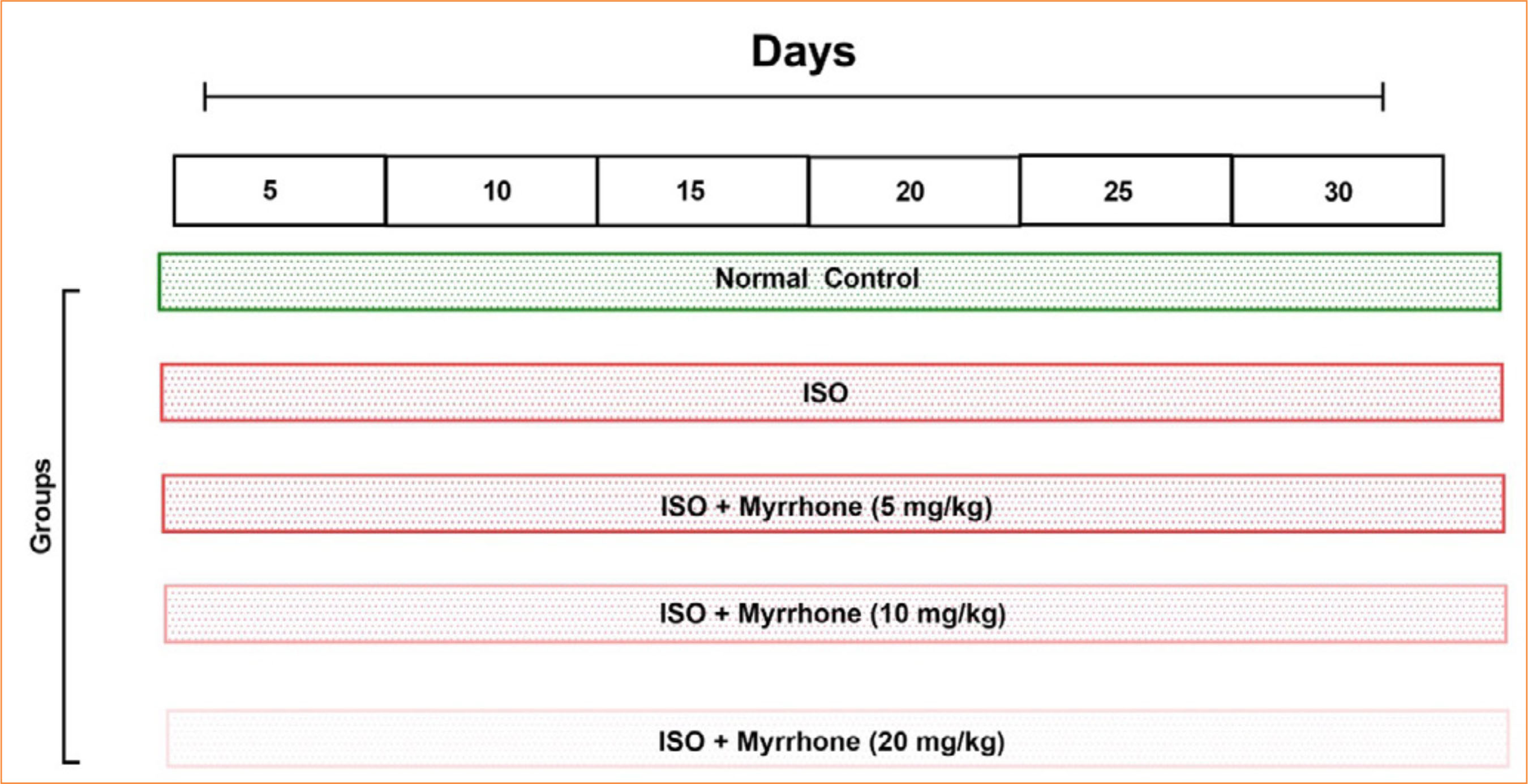 ISO: isoproterenol. Source: Elaborated by the authors.
 #p < 0.05 as compared to normal; *p < 0.05 as compared to ISO control analyzed by one-way analysis of variance followed by Dunnett’s T test. Source: Elaborated by the authors.
 #p < 0.05 as compared to normal; *p < 0.05 as compared to ISO control analyzed by one-way analysis of variance followed by Dunnett’s T test. Source: Elaborated by the authors.
 #p < 0.05 as compared to normal; *p < 0.05 as compared to ISO control analyzed by one-way analysis of variance followed by Dunnett’s T test. Source: Elaborated by the authors.
 #p < 0.05 as compared to normal; *p < 0.05 as compared to ISO control analyzed by one-way analysis of variance followed by Dunnett’s T test. Source: Elaborated by the authors.
 #p < 0.05 as compared to normal; *p < 0.05 as compared to ISO control analyzed by one-way analysis of variance followed by Dunnett’s T test. Source: Elaborated by the authors.
 #p < 0.05 as compared to normal; *p < 0.05 as compared to ISO control analyzed by one-way analysis of variance followed by Dunnett’s T test. Source: Elaborated by the authors.
 #p < 0.05 as compared to normal; *p < 0.05 as compared to ISO control analyzed by one-way analysis of variance followed by Dunnett’s T test. Source: Elaborated by the authors.
 #p < 0.05 as compared to normal; *p < 0.05 as compared to ISO control analyzed by one-way analysis of variance followed by Dunnett’s T test. Source: Elaborated by the authors.
 #p < 0.05 as compared to normal; *p < 0.05 as compared to ISO control analyzed by one-way analysis of variance followed by Dunnett’s T test. Source: Elaborated by the authors.

 #p < 0.05 as compared to normal; *p < 0.05 as compared to ISO control analyzed by one-way analysis of variance followed by Dunnett’s T test. Source: Elaborated by the authors.
