
 Microsurgical education for medical students: systematic review
ABSTRACT
Purpose: To perform a systematic review about microsurgical training for medical students.
Methods: A systematic search was conducted in several databases. Screening was performed by two independent reviewers based on predetermined criteria, following PRISMA 2020 guidelines.
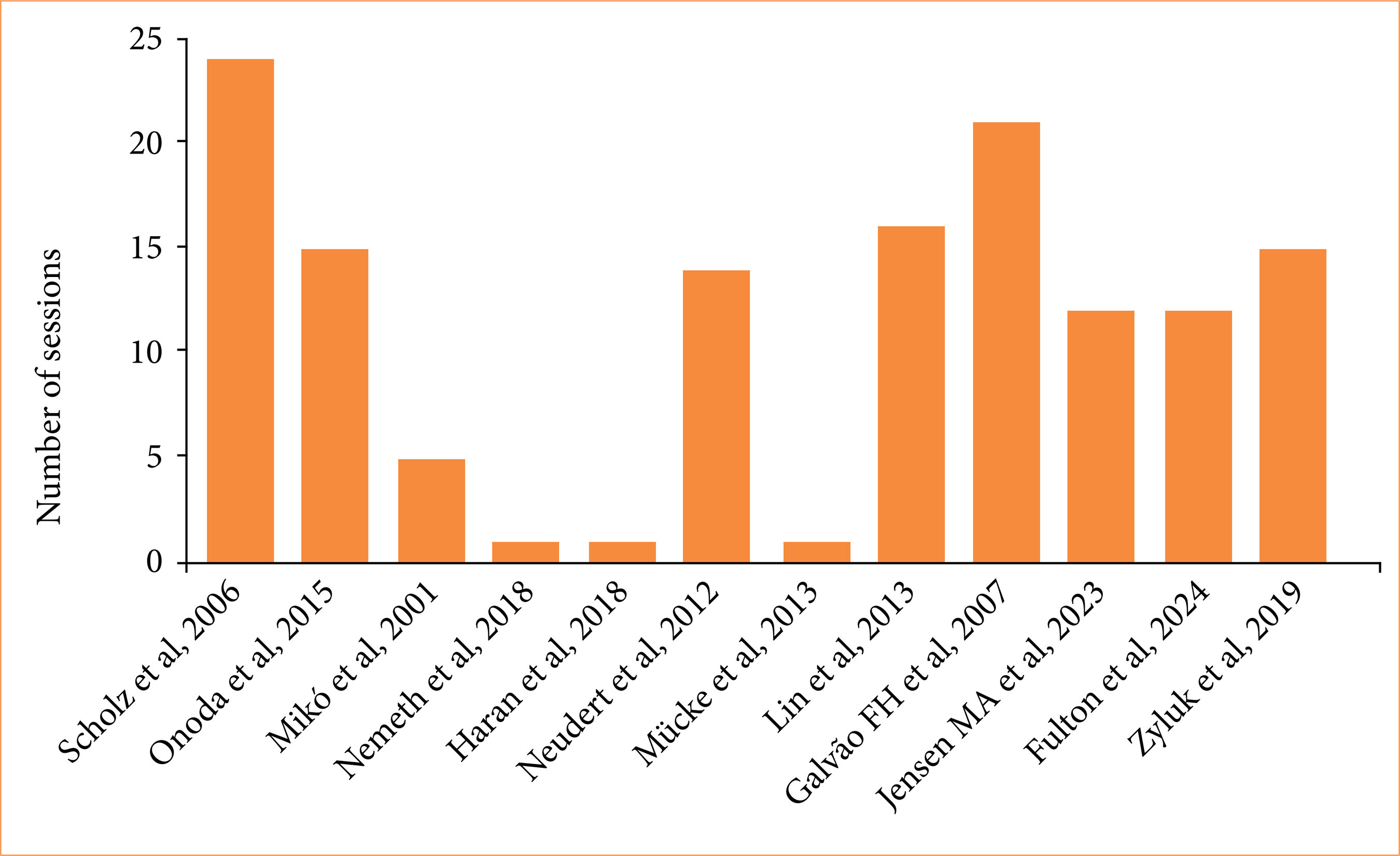
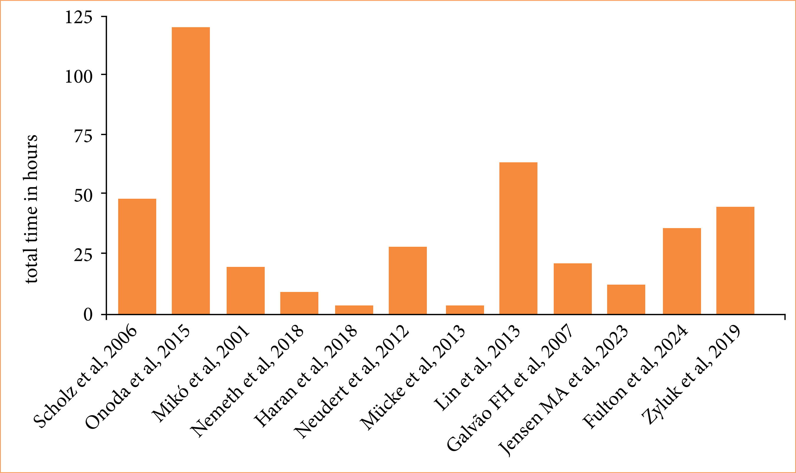
Results: A total of 433 articles were obtained. After screening, 40 were included. Teaching methods were heterogeneous and restricted to a few countries. There are specific methods to assess microsurgery training, general surgical activities, learning curve and anxiety. The most used materials were surgical gloves, silicone tubes, chicken thighs and rats. Animals were not essential in early stages of education. There was a variation between one and 24 sessions regarding teaching frequency and between 3 and 120 hours of teaching period. The average age of students was 23.7, and 67.5% were men. Teaching groups had an average of 5.8 students per group. Physical activity and anxiety had negative effect on microsurgical ability gain, while caffeine had positive effect. Alcohol and sleep deprivation had no significant effect.
Conclusion: Students with adequate training could achieve performance similar to that obtained by experts. Microsurgical education is realistic during medical graduation and may enable better career choices. More investigations are needed to improve this training and expand knowledge to other medical schools.
Key words
Microsurgery; Models, Theoretical; Rats; Education, Medical
Introduction
Microsurgery emerged in the 20th century and nowadays represents the pinnacle of medical precision1. In clinical practice, microsurgery plays a fundamental role expanding treatments for complex diseases involving neurosurgery2, plastic surgery3, hand surgery4, ophthalmology5, among others. Another critical application of this procedure is translational scientific research using small animals such as rodents, which fosters the development of innovative therapies6.
The teaching of surgery is a highly complex process involving difficult evaluation methods of skill acquisition. It often requires extensive training, sophisticated instruments and expensive surgical sutures. Research on microsurgical education helps allocate tasks based on skills, reduces future losses from inability, monitors the training costs and assesses student ability gain and learning curves7. Inadequate training is associated with increased surgical complications and high treatment costs. Therefore, surgical education is a public health issue8.
Due to its complexity, microsurgical training typically occurs at later stages of surgical carrier, with scant opportunities for medical students9. However, its increasing importance prospects that microsurgery will be gradually integrated into medical education following the same path as regular surgery in the past10.
Nevertheless, there is a lack of review articles assessing microsurgical training for medical students. Thus, the objective of this manuscript was to perform a systematic review about method of microsurgical training for medical students.
Methods
This systematic review was conducted according to PRISMA 2020 guidelines and registered in the PROSPERO system, with the identification number CRD420251169243. The data search was performed by evaluating the PubMed, Virtual Health Library (BVS), Embase, Scopus, and Web of Science databases, using the following terms: “(teaching OR education) AND (microsurgery) AND (medical students)”, without filters or period limits.
Eligibility criteria
The inclusion criteria were all articles that evaluate the teaching of microsurgery applied to medical students. Exclusion criteria include specialist-focused training, absence of microsurgery training, absence of medical students in the training, and lack of reference to the educational process.
Data selection
From the articles found, the selection was carried out by two independent reviewers, who compared the results, jointly resolving any discrepancies with the aid of the Rayyan program, using title and abstract analysis.
Data collection
The complete reading of the articles was carried out by the same two reviewers. They also worked independently in this phase, collecting specific data from the articles on predefined topics and highlighting other relevant information. A study design table was created for greater transparency and to make it easier to find the original articles (Table 1). The predefined analysis points included materials used, teaching methodology, number of students per group, duration of teaching, sociodemographic information, influence of non-surgical factors on the learning curve, training evaluation methods, and comparison between medical students, residents, and groups of experienced surgeons. From each study, all results compatible with each expected outcome domain were collected.
Risk of bias assessment and data synthesis
Risk of bias assessment was performed independently by two reviewers using the Critical Appraisal Skills Program (CASP) tool for qualitative studies (Table 2). Disagreements were resolved by consensus. Qualitative data were presented descriptively, and quantitative analyses were expressed as simple and weighted averages.
Results
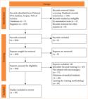
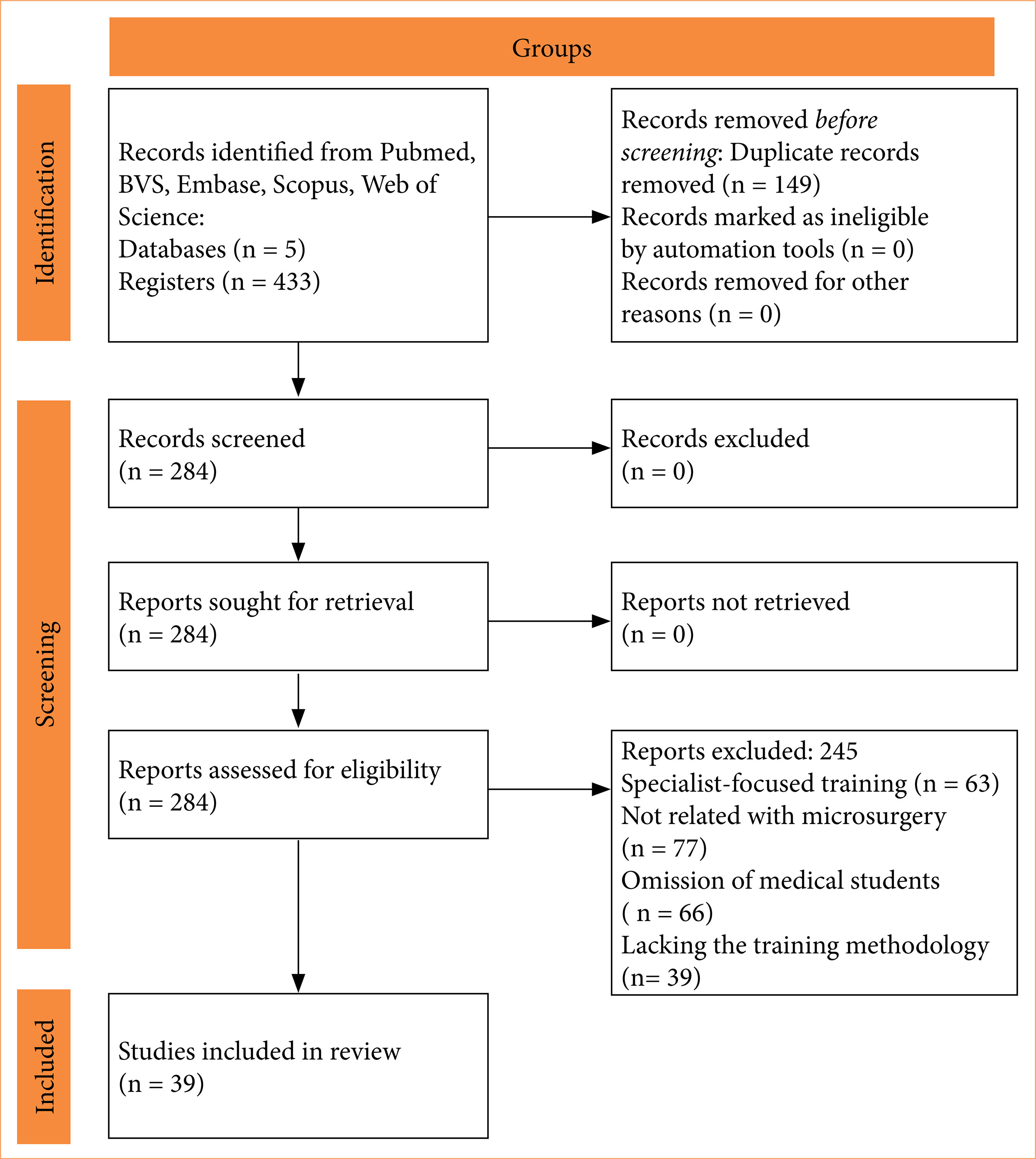
A total of 433 articles were assessed, 114 from PubMed, 27 from BVS, 140 from Embase, 75 from Scopus, and 77 from Web of Science, with the last consultation on November 22nd, 2024. A total of 149 duplicates were excluded, lasting 284 articles. From these 284 articles, 244 were excluded, 63 due to specialist-focused training, 77 not related with microsurgery topic, 65 for omitting medical students and 39 for lacking the methodology of the training. In the end, 40 articles were included in this survey (Fig. 1).
Most of the articles present microsurgery as elective courses/subjects11–20 or as part of research projects21–32. In one article, the institution included microsurgical training as a mandatory subject33.
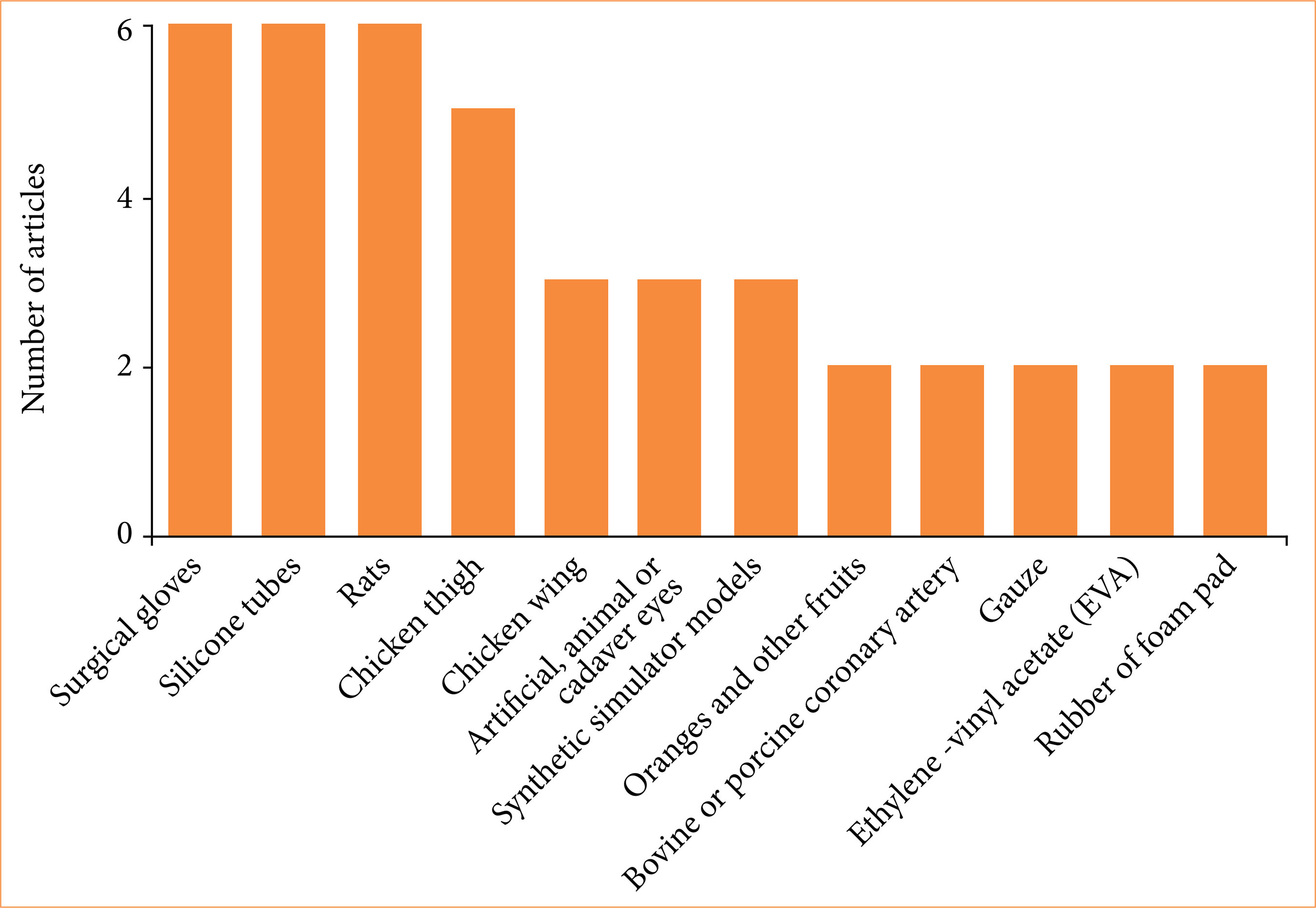
Various materials were employed for the training, with emphasis on the surgical glove21,22,27,29,33,34, silicone tubes13,20,24,31,32,35, rats11,13,18,34–36 and chicken thighs25,29,33,37,38 (Fig. 2). Other materials less used included pig skin12, newspaper33, goat or canine sciatic nerve21, pig spleen23, and pig dura mater28.

Materials used to teach microsurgery to medical students and the number of times each was described in the articles.
Kinshoku et al.18 advocates for the use of already deceased rats for medical student training, with the cadavers originating from prior practices in other health-related courses and researches. This procedure allows the rational use of animals and provides training closer to real-life scenarios.
Galvão et al.34 introduced a training of intestinal transplantation technique in rats for medical students and observed that most students abandoned the training before completion of the investigation. This result was attributed to the high complexity of the procedure, lack of time, and frustration. Only 5% of the students were able to complete the total procedure of intestinal transplantation technique in the end of the training.
Two articles20,39 mention the use of smartphone magnification in locations with limited access to bench microscopes. However, its application for medical students32 had poor results, which was explained by the failure to convert the learned skills to the operating microscope.
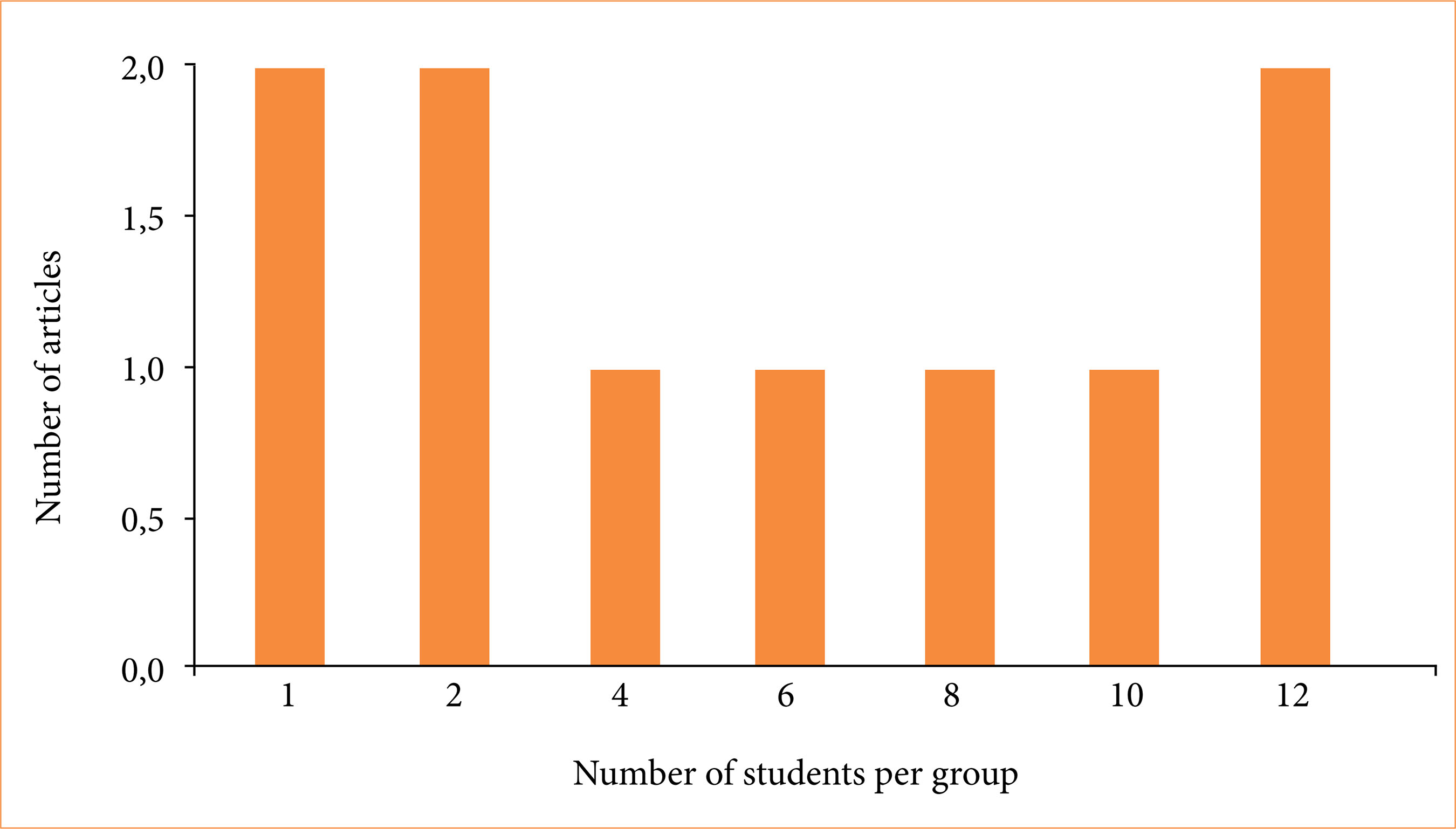
The number of students per teaching group was recorded (Fig. 3). Beier et al.37 highlight “peer-assisted learning” with excellent results. In this review, the average was 5.8 students per teaching group.

Number of students per teaching group in microsurgery and the number of times it was applied.
Regarding teaching time, information on the number of sessions (Fig. 4) and total training time in hours (Fig. 5) was recorded.

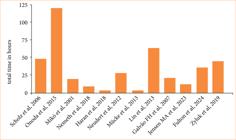
Regarding skill retention, one study30 compared suture accuracy after course of 8 hours in a single day with other group that had four sessions of 2 hours each for one month and observed that both groups had comparable skills, but the group with spaced-out lessons for one month performed significantly better in microsurgical suturing.
Regarding skill retention based on the distribution of teaching over time, one study analyzed suture accuracy one month after an 8-hour course. One group completed all 8 hours in a single day, while the other group had four sessions of 2 hours each. At the end of the course, both groups had comparable skills. However, after one month, the group with spaced-out lessons performed significantly better in microsurgical suturing30.
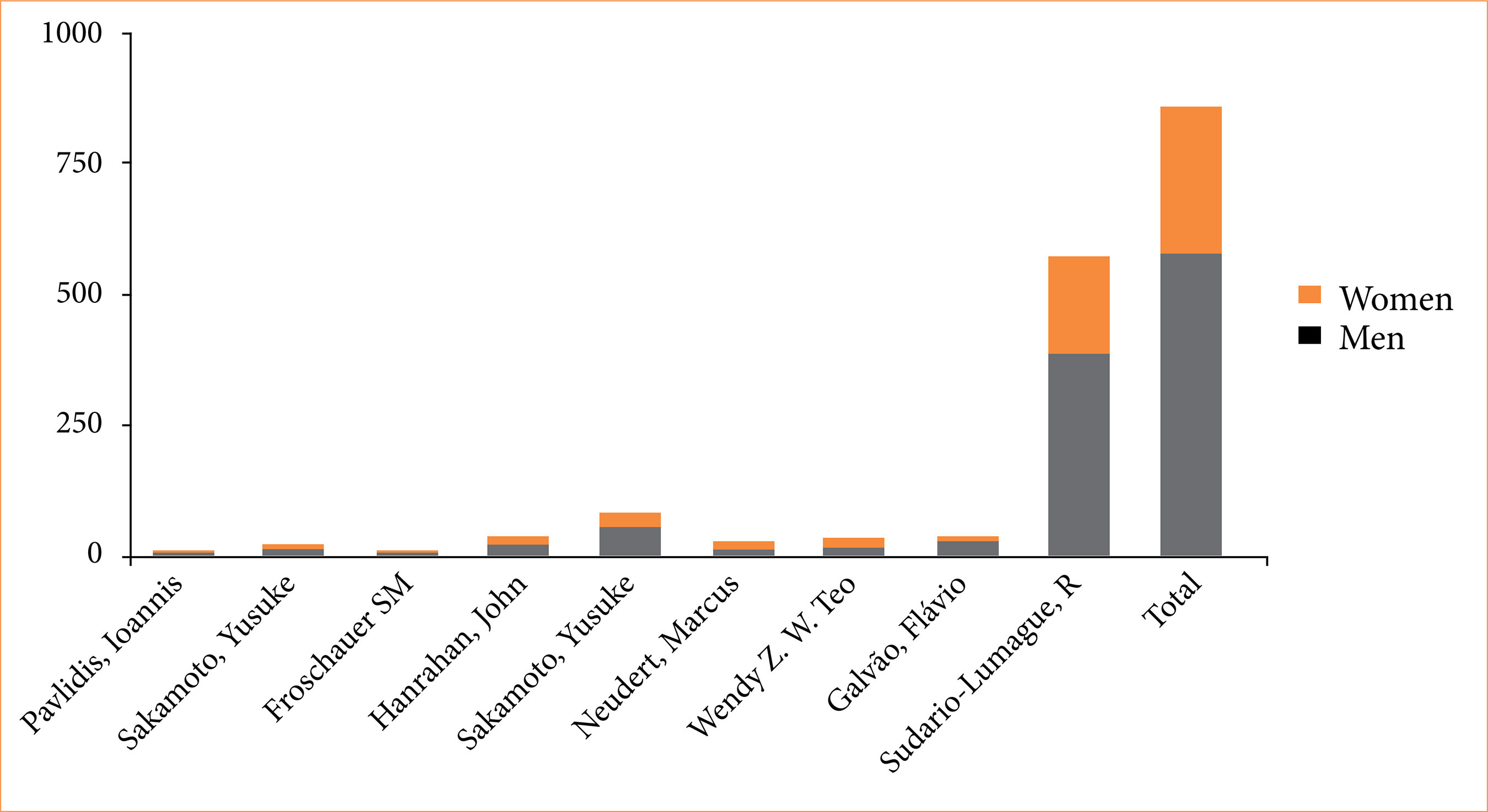
The average age of participants in six articles was 23.7 years. Gender stratification was cited in nine articles, 67.5% of the participants were men and 32.5% were women (Fig. 6). Sudario-Lumague et al.27 compared microsurgical performance between genders and demonstrated no significant difference.
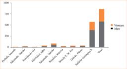
Gender stratification among the students participating in each article and among all articles in this research.
Two articles21,40 concluded that younger individuals acquire motor skills more easily than older individuals, what encourages early teaching.
Al Omran et al.26 compared the effects of regular physical activity on microsurgical performance, finding that high levels of physical activity in medical students are associated with slower anastomosis and an increased number of movements for the same task. However, this does not apply to specialists. Zyluk et al.41 found a positive effect of caffeine and a negative effect of physical exercise on microsurgical performance shortly before the task. A small dose of alcohol taken before the task showed little performance effect.
Hanrahan et al.28 evaluated the role of hand tremors and anxiety in students’ microsurgical skills and observed that overall performance decreased with a higher subjective perception of anxiety. However, greater physiological tremor and higher anxiety in objective tests were not associated with a decline in microsurgical performance.
Micko et al.42 studied the effects of sleep deprivation in medical students and neurosurgery residents to simulate a night shift. The results showed that performance scores significantly increased and time remained stable in both groups, but both groups had worse performance.
Pavlidis et al.22 affirmed that manual skills are facilitated by the absence of strong sympathetic stimuli, through the elimination of potential stressors in the context of informal education. High levels of stress during surgical training trigger fight-or-flight responses and high error rates. This article, as well as the one by Fulton et al.43, concluded that the early introduction of microsurgery during medical school, which has a lighter routine compared to residency, reduces exogenous stress and improves skills.
Regarding the method for evaluating students’ performance, time measurements25 were used. A reproducible scale was employed in the article by Sudario-Lumague et al.27. The Stanford Microsurgery and Resident Training (SMaRT) scale was used in one article25. SmaRT is an established and well-known scale with nine evaluation categories, each rated from 1 to 5, resulting in a score ranging from 9 to 45. The Nagoya University Microvascular Anastomosis Assessment System was also used in one article24.
The global rating scale, objective structured assessment of technical skill (OSATS), University of Bergen assessment tool31, McGill global rating scale44, and direct observation of procedural skills28 appeared in one article each, except for the OSATS scale, which was cited in two articles. All of these are tools used to measure performance in practical and technical skills, being more comprehensive and not specific to microsurgery.
The Swedish occupational fatigue inventory32 evaluates work-related fatigue. The surgical task load index32 measures perceived workload during surgical procedures, an adaptation of the NASA task load index22. State trait anxiety inventory22 and workload in traffic control systems28 were utilized to assess anxiety.
Microsurgery teaching was directly linked to medical specialties and influenced students’ ambitions toward surgery as a future career13,29,31. In four articles12,14–16, the course offering was related to ophthalmology; in three44–46, to otorhinolaryngology; and in one37, to plastic surgery. These activities highlight young talent and sparks interest in microsurgery and the specialty.
Microsurgical performance comparisons between students and more experienced groups such as residents and surgeons conclude that students’ results are very similar to those of more experienced individuals, sometimes even better13,24,29,36,47,48.
Discussion
The principles of microsurgical teaching, according to Mikó et al.11, were: activity, synchronization, video assistance, self-control, individualization, and analysis. Also, three areas of focus have been mentioned: safety training, risk management, and technical guidance. These areas are especially important when handling animals, using sharp materials and contributing to translational research35. To meet these principles, animals are not necessary at the initial microsurgical teaching and should be offered later if there is a genuine desire for specialization in microsurgery. This approach reduces animal suffering, respects bioethical considerations, and lessens the stress on students of handling live animals21–23,37. Inert models bear little resemblance to clinical situations, so they should be used as an introduction to microsurgical training with animals38.
The ethylene vinyl acetate model is practical, reproducible, portable, extremely cheap, readily available and allows teaching both two and three-dimensional suturing techniques17. The chicken thigh or wing model allows handling nerves, veins and arteries, in addition to enabling more complex procedures. However, chicken thigh and wing models’ limitations include absence of blood flow and inability to replicate thrombotic phenomena38. The technique taught should not be too complex in order to maintain a balance between students’ difficulty and level of instruction, ensuring lessons without significant frustration34.
The content regarding microsurgery for medical students is heterogeneous in purpose, time, teaching method, teacher-to-student ratio, and investment. Despite a reasonable number of reports, they are limited to a few developed countries and research centers of excellence, such as Germany, France, England, Japan, the United States, Canada, Brazil, Romania, and Hungary.
Practical learning with mentor feedback is superior to self-directed theoretical learning through instructional videos49. Teaching groups should be small, so each student can be closely monitored, with their mistakes corrected moment by moment, preventing the acquisition of poor manual habits. The course should be short, allowing intensive training, but spread over more than one session, so that each session is not exhausting, ensuring maximum learning with better retention.
We believe that learning this skill is feasible during medical school, where there is greater ease and availability of time to acquire technical skills. Thus, when students begin training during medical school, they gain the potential to achieve superior skills during residency practice33. Another reason for teaching microsurgery for medical students is their involvement in surgical research, which is valuable for research method35.
Finally, anxiety is an endogenous stress factor for nowadays’ medical students. The self-perception of anxiety undermines the student’s self-confidence and is even more harmful than its somatic effects, such as the intensification of physiological tremors. Thus, maximizing learning should be achieved through the reduction of stress factors.
In our Laboratory of Universidade de São Paulo Medical School, inspired by the legendary Professor Robert Zhen Zhong (in memoriam), we involve medical students in our research program of scientific initiation using rats as microsurgical models. For bioethical reasons, before performing surgery in animals, the students perform courses for animal use and microsurgery training using the latex glove simulator, in two weekly sessions for four weeks, with peer-assisted learning. In these sessions, the students are required to perform three continuous sutures with the extension of 3 cm on the upper face of the glove during 1 hour. After that training and other specific education, the students may participate in our experiments involving innovative translational microsurgical animal research that include intestinal transplantation34, Cuff-Glue sutureless anastomosis10, multivisceral transplantation50,51, anorectal transplantation52, hepatic ischemia-reperfusion experiments53, among others.
Conclusion
Microsurgical education for medical students is possible, feasible, and expandable. However, much still needs to be researched and developed, especially regarding the expansion of this knowledge to other countries and medical reference centers.
Acknowledgements
Not applicable.
-
Research performed at Scientific Initiation Program in the Department of Gastroenterology, Faculdade de Medicina of the Universidade de São Paulo, São Paulo (SP), Brazil. Tutor: Prof. Dr. Flávio Henrique Ferreira Galvão.
-
Funding
Not applicable.
-
Declaration of use of artificial intelligence tools
We declare that we have not used artificial intelligence tools.
Data availability statement
All dataset were generated or analyzed in the current study.
References
-
1 Mavrogenis AF, Markatos K, Saranteas T, Ignatiadis I, Spyridonos S, Bumbasirevic M, Georgescu AV, Beris A, Soucacos PN. The history of microsurgery. Eur J Orthop Surg Traumatol. 2019;29(2):247–54. https://doi.org/10.1007/s00590-019-02378-7
» https://doi.org/10.1007/s00590-019-02378-7 - 2 Willenberg E, Krumbholz S, Berge G. Möglichkeiten und Grenzen der Mikrochirurgie in der. Z Arztl Fortbild. 1981;75:674–5.
-
3 Mendoza JM, Chi JJ. Reconstruction of animal bite injuries to the head and neck. Curr Opin Otolaryngol Head Neck Surg. 2019;27(5):407–12. https://doi.org/10.1097/MOO.0000000000000564
» https://doi.org/10.1097/MOO.0000000000000564 -
4 Beris AE, Lykissas MG, Korompilias AV, Mitsionis GI, Vekris MD, Kostas-Agnantis IP. Digit and hand replantation. Arch Orthop Trauma Surg. 2010;130:1141–7. https://doi.org/10.1007/s00402-009-1021-7
» https://doi.org/10.1007/s00402-009-1021-7 -
5 Roper-Hall MJ. Microsurgery in ophthalmology. Br J Ophthalmol. 1967;51(6):408–14. https://doi.org/10.1136/bjo.51.6.408
» https://doi.org/10.1136/bjo.51.6.408 -
6 Aitman T, Dhillon P, Geurts AM. A RATional choice for translational research? Dis Model Mech. 2016;9(10):1069–72. https://doi.org/10.1242/dmm.027706
» https://doi.org/10.1242/dmm.027706 - 7 Rabinovich I, Kabanova SA, Lozhkevich IIu. The role of the Russian Surgery Research Center, Russian Academy of Medical Sciences, in the postgraduate training of medical staff. Vestn Ross Akad Med Nauk. 2002;5:51–4. https://pubmed.ncbi.nlm.nih.gov/12094754/
-
8 Fallah PN, Jayaram A, Hauser BM. Moving the needle on global surgery education in the US. J Surg Educ. 2021;78(6):1780–2. https://doi.org/10.1016/j.jsurg.2021.04.006
» https://doi.org/10.1016/j.jsurg.2021.04.006 -
9 Pinto LOAD, Silva RC, Bentes LGB, Bacelar HPH, Kietzer KS. Low-fidelity simulation models in urology resident’s microsurgery training. Acta Cir Bras. 2023;38:e386523. https://doi.org/10.1590/acb386523
» https://doi.org/10.1590/acb386523 -
10 Waisberg DR, Galvão FH, De Castro Galvão R, Chaib E, D’Albuquerque LA. Intestinal transplantation using cuff-glue sutureless technique for microanastomosis in rats. Microsurgery. 2011;31(7):584–5. https://doi.org/10.1002/micr.20924
» https://doi.org/10.1002/micr.20924 -
11 Mikó I, Bráth E, Furka I. Basic teaching in microsurgery. Microsurgery. 2001;21(4):121–3. https://doi.org/10.1002/micr.1021
» https://doi.org/10.1002/micr.1021 -
12 Müller A, Pfeiffer N, Schmidt F, Prokosch V. Ein mikrochirurgisches Wetlab für Studierende steigert das Interesse an der. Ophthalmologe. 2020;117(11):1087–91. https://doi.org/10.1007/s00347-020-01079-5
» https://doi.org/10.1007/s00347-020-01079-5 -
13 Onoda S, Kimata Y, Sugiyama N, Tokuyama E, Matsumoto K, Ota T, Thuzar M. Analysis of 10-year training results of medical students using the microvascular research center training program. J Reconstr Microsurg. 2016;32(5):336–41. https://doi.org/10.1055/s-0035-1568884
» https://doi.org/10.1055/s-0035-1568884 -
14 Liu EY, Li B, Hutnik CML. Canadian ophthalmic microsurgery course: an innovative spin on wet lab-based surgical education. Can J Ophthalmol. 2016;51(5):315–20. https://doi.org/10.1016/j.jcjo.2016.04.021
» https://doi.org/10.1016/j.jcjo.2016.04.021 -
15 Deuchler S, Scholtz J, Ackermann H, Seitz B, Koch F. Implementation of microsurgery simulation in an ophthalmology clerkship in Germany: a prospective, exploratory study. BMC Med Educ. 2022;22(1):599. https://doi.org/10.1186/s12909-022-03634-x
» https://doi.org/10.1186/s12909-022-03634-x -
16 Cole J, Chen TA, Ahmad T, Parikh N. Ophthalmic microsurgery lab for medical students: enhancing learner intrinsic motivation and comfort with microsurgery. J Acad Ophthalmol. 2021;13(2):e234–41. https://doi.org/10.1055/s-0041-1740067
» https://doi.org/10.1055/s-0041-1740067 -
17 Bastos EM, Silva RD. Proposal of a synthetic ethylene-vinyl acetate bench model for surgical foundations learning: suture training. Acta Cir Bras. 2011;26(2):149–52. https://doi.org/10.1590/s0102-86502011000200014
» https://doi.org/10.1590/s0102-86502011000200014 -
18 Kinshoku MR, Rodriguez CAL, Fidalgo RS, Duran CCG, Leme PLS, Duarte IS. Uso racional de modelos animais para pesquisa e ensino de microcirurgia. Rev Col Bras Cir. 2012;39(5):414–7. https://doi.org/10.1590/S0100-69912012000500013
» https://doi.org/10.1590/S0100-69912012000500013 -
19 Lin D, Lewis T, Mobley M, Burnett B, Tunstall RG, Abrahams PH. Small is beautiful: a practical course in microsurgery for medical students. The FASEB J. 2013;27:lb24. https://doi.org/10.1096/fasebj.27.1_supplement.lb24
» https://doi.org/10.1096/fasebj.27.1_supplement.lb24 -
20 Jensen MA, Bhandarkar AR, Riviere-Cazaux C, Bauman MMJ, Wang K, Carlstrom LP, Graffeo CS, Spinner RJ. The early bird gets the worm: introducing medical students to microsurgical technique via a low-cost, user-friendly, reusable simulation system. World Neurosurg. 2022;167:89–94. https://doi.org/10.1016/j.wneu.2022.08.100
» https://doi.org/10.1016/j.wneu.2022.08.100 -
21 Scholz M, Mücke T, Hölzle F, Schmieder K, Engelhardt M, Pechlivanis I, Harders AG. A program of microsurgical training for young medical students: are younger students better? Microsurgery. 2006;26(6):450–5. https://doi.org/10.1002/micr.20269
» https://doi.org/10.1002/micr.20269 -
22 Pavlidis I, Zavlin D, Khatri A, Wesley A, Panagopoulos G, Echo A. Absence of stressful conditions accelerates dexterous skill acquisition in surgery. Sci Rep. 2019;9:1747. https://doi.org/10.1038/s41598-019-38727-z
» https://doi.org/10.1038/s41598-019-38727-z -
23 Maluf Junior I, da Silva AB, Groth AK, Lopes MA, Kurogi AS, Freitas Rda S, Tomasich FD. An alternative experimental model for training in microsurgery. Rev Col Bras Cir. 2014;41(1):72–4. https://doi.org/10.1590/s0100-69912014000100014
» https://doi.org/10.1590/s0100-69912014000100014 -
24 Sakamoto Y, Okamoto S, Shimizu K, Araki Y, Hirakawa A, Wakabayashi T. Comparative prospective study of microvascular anastomosis training by self-learning or with expert instruction. World Neurosurg. 2018;118:e818–24. https://doi.org/10.1016/j.wneu.2018.07.061
» https://doi.org/10.1016/j.wneu.2018.07.061 -
25 Froschauer SM, Holzbauer M, Kwasny O, Kastner P, Schnelzer RF, Hopfner M, Duscher D. Effect of music on the efficiency of performing a microsurgical arterial anastomosis: a prospective randomized study. J Hand Microsurg. 2020;15(1):13–7. https://doi.org/10.1055/s-0040-1716992
» https://doi.org/10.1055/s-0040-1716992 -
26 Al Omran Y, Kostusiak M, Myers S, Ghanem AM. Effects of habitual physical activity on microsurgical performance. Br J Oral Maxillofac Surg. 2016;54(9):1025–7. https://doi.org/10.1016/j.bjoms.2016.01.008
» https://doi.org/10.1016/j.bjoms.2016.01.008 -
27 Sudario-Lumague R, Chiang YC, Lin TS. Gender comparison of medical student microsurgical skills in a laboratory model. J Reconstr Microsurg. 2018;34(5):359–62. https://doi.org/10.1055/s-0038-1626694
» https://doi.org/10.1055/s-0038-1626694 -
28 Hanrahan J, Sideris M, Pasha T, Tsitsopoulos PP, Theodoulou I, Nicolaides M, Georgopoulou EM, Kombogiorgas D, Bimpis A, Papalois A. Hands train the brain-what is the role of hand tremor and anxiety in undergraduate microsurgical skills? Acta Neurochir. 2018;160(9):1673–9. https://doi.org/10.1007/s00701-018-3609-6
» https://doi.org/10.1007/s00701-018-3609-6 -
29 Mücke T, Borgmann A, Ritschl L, Kesting MR, Loeffelbein DJ, Wolff K-D. Microvascular training of medical students and surgeons: a comparative prospective study. J Craniomaxillofac Surg. 2013;41(8):e187–90. https://doi.org/10.1016/j.jcms.2013.01.017
» https://doi.org/10.1016/j.jcms.2013.01.017 -
30 Teo WZW, Dong X, Yusoff SKBM, De SD, Chong AKS. Randomized controlled trial comparing the effectiveness of mass and spaced learning in microsurgical procedures using computer aided assessment. Sci Rep. 2021;11:2810. https://doi.org/10.1038/s41598-021-82419-6
» https://doi.org/10.1038/s41598-021-82419-6 -
31 Almeland SK, Lindford A, Sundhagen HP, Hufthammer KO, Strandenes E, Svendsen HL, Guttormsen AB, Hansson E. The effect of microsurgical training on novice medical students’ basic surgical skills—a randomized controlled trial. Eur J Plastic Surg. 2020;43:459–66. https://doi.org/10.1007/s00238-019-01615-w
» https://doi.org/10.1007/s00238-019-01615-w -
32 Jensen MA, Bhandarkar AR, Bauman MMJ, Riviere-Cazaux C, Wang K, Carlstrom LP, Graffeo CS, Spinner RJ. The LazyBox educational intervention trial: can longitudinal practice on a low-fidelity microsurgery simulator improve microsurgical skills? Cureus. 2023;15(11):e49675. https://doi.org/10.7759/cureus.49675
» https://doi.org/10.7759/cureus.49675 -
33 Nemeth N, Miko I, Furka I. Experiences with basic microsurgical training programs and skill assessment methods at the University of Debrecen, Hungary. Acta Cir Bras. 2018;33(9):842–52. https://doi.org/10.1590/s0102-865020180090000013
» https://doi.org/10.1590/s0102-865020180090000013 -
34 Galvão FH, Bacchella T, Machado MC. Teaching intestinal transplantation in the rat for medical student. Microsurgery. 2007;27(4):277–81. https://doi.org/10.1002/micr.20355
» https://doi.org/10.1002/micr.20355 -
35 Zhang F, Dorsett-Martin W, Fischer K, Angel M, Buncke HJ, Lineaweaver WC. Role of medical students in microsurgery research. J Reconstr Microsurg. 2001;17(2):89–93. https://doi.org/10.1055/s-2001-12696
» https://doi.org/10.1055/s-2001-12696 -
36 Climov M, Măciuceanu Zarnescu MB, Stefănescu A, Zamfirescu D, Lascăr I. Learning curve in hemifacial transplantation in rats. Chirurgia (Bucur). 2013;108(2):234–40. https://doi.org/10.1097/01.prs.0000400392.01661.40
» https://doi.org/10.1097/01.prs.0000400392.01661.40 -
37 Beier JP, Horch RE, Boos AM, Taeger CD, Breuer G, Arkudas A. Etablierung und evaluation eines mikrochirurgie – kurses für medizinstudierende. Handchir Mikrochir Plast Chir. 2015;47(6):400–7. https://doi.org/10.1055/s-0041-108196
» https://doi.org/10.1055/s-0041-108196 -
38 Couceiro J, Ozyurekoglu T, Sanders S, Tien H. Microsurgical training regimen with nonliving chicken models. Microsurgery. 2013;33(3):251–2. https://doi.org/10.1002/micr.22060
» https://doi.org/10.1002/micr.22060 -
39 Bhandarkar AR, Jensen MA, Riviere-Cazaux C, Bauman MMJ, Wang K, Graffeo CS, Spinner RJ. Countertop microsurgery for medical students: letter to the editor regarding “lazy glass microsurgical trainer: a frugal solution for microsurgical training”. World Neurosurg. 2021;147:241. https://doi.org/10.1016/j.wneu.2020.11.082
» https://doi.org/10.1016/j.wneu.2020.11.082 -
40 Haran C, Brunger N, Pumphrey O, Simpson K, Roberts J, Kejriwal N, Parkinson G, Mccormack D, El-Gamel A. Microsurgical dexterity tuition of students and house surgeons: a necessary and worthwhile investment. Heart, Lung and Circulation. 2018;27(Suppl.3):S516. https://doi.org/10.1016/j.hlc.2018.04.043
» https://doi.org/10.1016/j.hlc.2018.04.043 -
41 Zyluk A, Czernikiewicz K, Antoniak J, Abramczyk U. An analysis of the effect of selected factors on microsurgical performance. Handchir Mikrochir Plast Chir. 2022;54(5):442–6. https://doi.org/10.1055/a-1380-3922
» https://doi.org/10.1055/a-1380-3922 -
42 Micko A, Knopp K, Knosp E , Wolfsberger S. Microsurgical performance after sleep interruption: a neurotouch simulator study. World Neurosurg. 2017;106:92–101. https://doi.org/10.1016/j.wneu.2017.06.142
» https://doi.org/10.1016/j.wneu.2017.06.142 -
43 Fulton M, Donnelly DT, Nkana ZH, Jung S, Zeng W, Dingle AM. The impact of early exposure to microsurgery training on undergraduates: a pilot course. WMJ. 2024;123(5):368–73. https://wmjonline.org/123no5/fulton/
» https://wmjonline.org/123no5/fulton/ -
44 Kaplan DJ, Vaz-Guimaraes F, Fernandez-Miranda JC, Snyderman CH. Validation of a chicken wing training model for endoscopic microsurgical dissection. Laryngoscope. 2015;125(3):571–6. https://doi.org/10.1002/lary.24977
» https://doi.org/10.1002/lary.24977 -
45 Neudert M, Kluge A, Beleites T, Kemper M, Zahnert T. Microsurgical skills training with a new tympanoplasty model. Otol Neurotol. 2012;33(3):364–70. https://doi.org/10.1097/mao.0b013e318238081c
» https://doi.org/10.1097/mao.0b013e318238081c -
46 Ilgner J, Park JJ-H, Westhofen M. Teaching microsurgery to undergraduate medical students by means of high-definition stereo video microscopy: the Aachen “skills lab” experience. Proceedings of SPIE. 2008;6803:680313. https://doi.org/10.1117/12.763867
» https://doi.org/10.1117/12.763867 -
47 Furka I, Brath E, Nemeth N, Miko I. Learning microsurgical suturing and knotting techniques: comparative data. Microsurgery. 2006;26(1):4–7. https://doi.org/10.1002/micr.20201
» https://doi.org/10.1002/micr.20201 -
48 von Reibnitz D, Weinzierl A, Grünherz L, Giovanoli P, Lindenblatt N. Learning curve of robotic assisted microsurgery in surgeons with different skill levels: a prospective preclinical study. J Robot Surg. 2024;18(1):353. https://doi.org/10.1007/s11701-024-02114-2
» https://doi.org/10.1007/s11701-024-02114-2 -
49 Sakamoto Y, Okamoto S, Shimizu K, Araki Y, Hirakawa A, Wakabayashi T. Hands-on simulation versus traditional video-learning in teaching microsurgery technique. Neurol Med Chir. 2017;57(5):238–45. https://doi.org/10.2176/nmc.oa.2016-0317
» https://doi.org/10.2176/nmc.oa.2016-0317 -
50 Galvão FH, Waisberg DR, Cruz RJ Jr, Chaib E, D’Albuquerque LAC. Modified multivisceral transplantation in the rat. Transplantation. 2013;96(2):e3–e4. https://doi.org/10.1097/tp.0b013e3182981167
» https://doi.org/10.1097/tp.0b013e3182981167 -
51 Galvao FHF, Araki J, Fonseca ABS, Cruz RJ Jr, Lanchotte C, Waisberg DR, Chaib E, Nacif LS, Traldi MCC, de Mello EB, Andraus W, Carneiro-D’Albuquerque L. Multivisceral transplantation of pelvic organs in rats. Front Surg. 2023;10:1086651. https://doi.org/10.3389/fsurg.2023.1086651
» https://doi.org/10.3389/fsurg.2023.1086651 -
52 Galvão FH, Waisberg DR, Seid VE, Costa AC, Chaib E, Baptista RR, Capelozzi VL, Lanchotte C, Cruz RJ, Araki J, D’Albuquerque LC. Allogeneic anorectal transplantation in rats: technical considerations and preliminary results. Sci Rep. 2016;6:30894. https://doi.org/10.1038/srep30894
» https://doi.org/10.1038/srep30894 -
53 Pantanali CA, Rocha-Santos V, Kubrusly MS, Castro IA, Carneiro-D’Albuquerque LA, Galvão FH. The protective effect of nutraceuticals on hepatic ischemia-reperfusion injury in wistar rats. Int J Mol Sci. 2023;24(12):10264. https://doi.org/10.3390/ijms241210264
» https://doi.org/10.3390/ijms241210264
Edited by
-
Section editor:
Cristina Camargo https://orcid.org/0000-0002-3134-0003

 Source: Elaborated by the authors.
 Source: Elaborated by the authors.
 Source: Elaborated by the authors.
 Source: Elaborated by the authors.
 Source: Elaborated by the authors.
 Source: Elaborated by the authors.