Elkafas
et al.6
|
2020 |
10 Eker rats (Long Evans; Tsc2 Ek /+) |
2 groups |
Transgenic animals + Hormonal |
Not specified |
Accumulation of DNA damage (γH2AX)* and reduced expression of repair proteins (RAD50, MRE11, NBS1) in myometrial stem cells*
|
| Yang et al.7
|
2023 |
10 Eker rats (Long Evans; Tsc2 Ek /+) |
2 groups |
Transgenic animals + Hormonal |
Not specified |
Fibrosis** and inflammatory infiltration of macrophages (CD68+) associated with hypomethylation of the Stat3 gene*
|
Yasong
et al.9
|
2016 |
50 Sprague Dawley rats |
5 groups |
Hormonal |
Not specified |
Thickening of the smooth muscle layer with cellular disarray, polymorphic nuclei and large mitochondria**; overexpression of MMP-2 and reduction of TIMP-2*
|
| Ju and Xiao10
|
2016 |
60 Wistar rats |
6 groups |
Hormonal |
Not specified |
Macroscopic thickening of the uterine horns, smooth muscle hyperplasia with cellular disarray and degeneration*; overexpression of ER, PR and Bcl-2 with reduction of Bax |
| Liu et al.11
|
2017 |
C57B6 mice |
4 groups |
Hormonal |
Not specified |
Significant thickening of the smooth muscle layer with cellular disarray and pleomorphic nuclei**; overexpression of Bcl-2 and reduction of Bax and Caspase-3*
|
| Li et al.12
|
2019 |
Wistar rats |
5 groups |
Hormonal |
Not specified |
Uterine hyperplasia evidenced by a significant increase in the uterine coefficient and cervical diameter*; overexpression of Bcl-2, Akt, p-Akt and increased NOS activity, concomitant with a reduction in Bax and Caspase-3*
|
| Li et al.13
|
2019 |
40 Sprague Dawley rats |
4 groups |
Hormonal |
Not specified |
Uterine hyperplasia evidenced by a significant increase in the uterine coefficient, transverse diameter and smooth muscle thickness*; overexpression of TGF-β3 and MMP-11 proteins, increased arachidonic acid and lysophosphatidylcholine associated with inflammation and abnormal angiogenesis*
|
| Lin et al.14
|
2019 |
ICR (CD-1) mice |
3 groups |
Hormonal |
Not specified |
Smooth muscle hyperproliferation characterized by a significant increase in the thickness of the myometrial and endometrial layers, with cellular hypertrophy and irregular arrangement of muscle fibers*; overexpression of PCNA, ER-α, and PR*
|
| Yu et al.15
|
2019 |
30 Sprague Dawley rats |
6 groups |
Hormonal |
Not specified |
Distorted and sparse nuclei with a "smudged" chromatin pattern and poorly defined contours, associated with a significant increase in extracellular matrix (ECM) thickness**; overexpression of genes linked to ECM formation (Lcn2, Aqp8) and proliferation, with dysregulation of MAPK, PPAR, Notch, and TGF-β pathways*
|
| Lin et al.16
|
2020 |
70 specific pathogen-free rats |
7 groups |
Hormonal |
Not specified |
Hypertrophy and hyperplasia of smooth muscle cells**; intense endometrial proliferation and eosinophil infiltration, with the presence of hemorrhagic damage*; overexpression of PCNA, ER-α, ER-β, PR, Survivin, and Bcl-2, with a reduction in Bax and Caspase-3*
|
| Olowofolahan et al.17
|
2020 |
24 Wistar rats |
4 groups |
Hormonal |
Not specified |
Disorganization of tissue architecture and a significant increase in fibroblast density*; overexpression of ER-α and β-catenin concomitant with loss of E-cadherin, suggesting activation of the Wnt pathway and epithelial-mesenchymal transition |
Yamei
et al.18
|
2021 |
140 Sprague Dawley rats |
10 groups |
Hormonal |
Not specified |
Thickening of the smooth muscle layer with cellular disarray and intense inflammatory infiltration**; overexpression of hormone receptors (ER-α, ER-β, PR)*
|
Yousefi
et al.19
|
2021 |
84 Wistar rats |
2 groups |
Hormonal |
Surgery + macroscopic analysis |
Thickening of the uterine wall with disorganization of smooth muscle cells and extensive fibrosis**; increased expression and activity of MMP-2 and MMP-9, and oxidative stress (elevation of MDA and reduction of antioxidants SOD, CAT, GSH)*
|
Zhang
et al.20
|
2021 |
27 Sprague Dawley rats |
3 groups |
Hormonal |
Not specified |
Focal hypertrophy and hyperplasia of smooth muscle cells with hyaline degeneration and vacuolization of epithelial cells*; increased tissue concentrations of E2, P, ER, and PR, in addition to increased plasma viscosity |
| Yu et al.15
|
2019 |
30 Sprague Dawley rats |
6 groups |
Hormonal |
Not specified |
Distorted and sparse nuclei with a "smudged" chromatin pattern and poorly defined contours, associated with a significant increase in extracellular matrix (ECM) thickness**; overexpression of genes linked to ECM formation (Lcn2, Aqp8) and proliferation, with dysregulation of MAPK, PPAR, Notch, and TGF-β pathways*
|
| Lin et al.16
|
2020 |
70 specific pathogen-free rats |
7 groups |
Hormonal |
Not specified |
Hypertrophy and hyperplasia of smooth muscle cells**; intense endometrial proliferation and eosinophil infiltration, with the presence of hemorrhagic damage*; overexpression of PCNA, ER-α, ER-β, PR, Survivin, and Bcl-2, with a reduction in Bax and Caspase-3*
|
| Olowofolahan et al.17
|
2020 |
24 Wistar rats |
4 groups |
Hormonal |
Not specified |
Disorganization of tissue architecture and a significant increase in fibroblast density*; overexpression of ER-α and β-catenin concomitant with loss of E-cadherin, suggesting activation of the Wnt pathway and epithelial-mesenchymal transition |
Yamei
et al.18
|
2021 |
140 Sprague Dawley rats |
10 groups |
Hormonal |
Not specified |
Thickening of the smooth muscle layer with cellular disarray and intense inflammatory infiltration**; overexpression of hormone receptors (ER-α, ER-β, PR)*
|
Yousefi
et al.19
|
2021 |
84 Wistar rats |
2 groups |
Hormonal |
Surgery + macroscopic analysis |
Thickening of the uterine wall with disorganization of smooth muscle cells and extensive fibrosis**; increased expression and activity of MMP-2 and MMP-9, and oxidative stress (elevation of MDA and reduction of antioxidants SOD, CAT, GSH)*
|
Zhang
et al.20
|
2021 |
27 Sprague Dawley rats |
3 groups |
Hormonal |
Not specified |
Focal hypertrophy and hyperplasia of smooth muscle cells with hyaline degeneration and vacuolization of epithelial cells*; increased tissue concentrations of E2, P, ER, and PR, in addition to increased plasma viscosity |
| Chen et al.21
|
2022 |
48 Sprague Dawley rats |
6 groups |
Hormonal |
Surgical evaluation of three rats from each group |
Thickening of smooth muscle with abnormally shaped/sized nuclei, disordered arrangement, and intense infiltration of inflammatory cells in the myometrium**; overexpression of Med12 protein and activation of the Wnt/beta-Catenin pathway, promoting cell proliferation |
| Qiu et al.22
|
2022 |
40 Sprague Dawley rats |
5 groups |
Hormonal |
Not specified |
Hypertrophy of smooth muscle cells with thickening of the muscle layer and extensive deposition of fibrous connective tissue between muscle bundles**; overexpression of IDO1, PI3KCA, and AKT1, inhibition of PTEN and apoptosis/autophagy markers (Caspase-9, LC-3), in addition to systemic immunosuppression (increased Tregs)*
|
| Shi et al.23
|
2023 |
36 Sprague Dawley rats |
6 groups |
Hormonal |
Histological evaluation of one rat from each group |
Hyperplasia and disarray of smooth muscle cells with hypertrophic and dense nuclei, associated with inflammatory infiltration of neutrophils in the lamina propria*; overexpression of miR-21-5p and activation of the PI3K/AKT pathway (increased p-PI3K and p-AKT) concomitant with reduced PTEN*
|
| Yuan et al.24
|
2023 |
108 Sprague Dawley rats |
9 groups |
Hormonal |
Surgery + macroscopic analysis of two rats per group |
Smooth muscle layer hyperplasia with cellular disarray, enlarged nuclei, connective tissue hyperplasia, and inflammatory infiltration**; elevated serum levels of E2, P, IL-6, and TNF-alpha and tissue overexpression of ER-alpha and PR*
|
| Zeng et al.25
|
2023 |
6 Sprague Dawley rats |
2 groups |
Hormonal |
Histopathological analysis of the model group |
Thickening of the smooth muscle layer with focal and multiple hyperplasia, cellular hypertrophy, disordered arrangement, and connective tissue hyperplasia (fibrosis), associated with severe inflammatory infiltration**; overexpression of COX-2 and Bcl-2 concomitant with reduced Bax and elevated serum levels of E2 and P*
|
| Gao et al.26
|
2024 |
48 Sprague Dawley rats |
6 groups |
Hormonal |
Not specified |
Smooth muscle hyperplasia (thickening) with disordered fiber arrangement, indistinct cell boundaries, and evident inflammatory infiltration**; overexpression of Bcl-2 concomitant with reduction of Bax and Caspase-3, in addition to systemic and tissue elevation of E2, P and ER |
| Yang et al.27
|
2023 |
196 Kunming mice |
14 groups |
Hormonal |
Not specified |
Hypertrophy and thickening of the smooth muscle layer with disordered arrangement of fibers, focal hyperplasia of the endometrium and infiltration of lymphocytes and eosinophils*; elevation of serum levels of E2, P, TNF-alpha and NOS activity, in addition to tissue overexpression of ER and PR*
|
| Ezejiofor and Okoroafor28
|
2022 |
21 Wistar rats |
3 groups |
Monosodium glutamate |
Not specified |
Densely compacted fusiform fibrous tissue and multifocal tumor cells in the endometrium*; significant elevation of serum levels of E2 and P, in addition to markers of oxidative stress (increased MDA, reduced SOD, CAT, GSH) and renal insufficiency (increased urea and creatinine); |
Imade
et al.29
|
2024 |
25 Sprague Dawley rats |
5 groups |
Monosodium glutamate |
Not specified |
Thick bundles of spindle-shaped smooth muscle fibers arranged in a disordered and crisscrossed manner, invading the endometrial glands and stroma*; increased serum levels of total cholesterol and E2, indicating hormonal and lipid dysregulation associated with the tumor*
|
Kayode
et al.30
|
2021 |
24 Wistar rats |
4 groups |
Monosodium glutamate |
Not specified |
Severe hyperplasia of endometrial spindle cells, disrupting the normal architecture of connective tissue*; elevated serum levels of E2, FSH, LH and total cholesterol*; high oxidative stress (elevated MDA and reduced SOD and CAT)*
|
Oyebode
et al.31
|
2020 |
28 Wistar rats |
4 groups |
Monosodium glutamate |
Not specified |
Intense deposition of collagenous connective tissue in the myometrium and ovarian stroma, associated with a higher density of spindle fibroblasts**; elevated serum levels of E2, progesterone, total cholesterol, and triglycerides*
|
Mamoon
et al.32
|
2021 |
33 Wistar rats |
7 groups |
Monosodium glutamate + Hormonal |
Ultrasound with Dopplerfluxometry |
Increased diameter and blood flow on Doppler ultrasound; proliferation of smooth muscle cells with excessive collagen deposition**; elevated serum levels of E2, progesterone (P4) and its receptors (ER, PGR), in addition to hemodynamic changes in the uterine arteries |
| Lin et al.33
|
2017 |
24 Wistar rats |
3 groups |
Ischemic injury |
Not specified |
Thickening of the myometrium and atrophy of the endometrium, with disarray of muscle fibers and significant accumulation of collagen**; overexpression of TGF-β, mast cells and activation of the Wnt/beta-Catenin pathway, promoting transdifferentiation of myofibroblasts*
|
| Zhao et al.35
|
2018 |
Sprague Dawley rats |
5 groups |
Hormonal + external stimulation |
Not specified |
Focal hyperplasia of smooth muscle, hyaline and vacuolar degeneration, disordered arrangement and inflammatory infiltration**; elevated serum P and increased tissue levels of E2, P, ER, and PR*; NOTE: the group subjected only to stress (without hormones) did not develop tumor nodules, only functional alterations |
| Feng et al.36
|
2021 |
72 Sprague Dawley rats |
9 groups |
Hormonal + external stimulation |
Not specified |
Disarray of smooth muscle cells, indistinct cell boundaries and focal degeneration/necrosis**; overexpression of fibroblast activating protein (FAP) and TGF-beta, promoting excessive deposition of collagen I and activation of the AKT/ERK proliferation pathway |
Abdelaziz
et al.37
|
2016 |
18 naked mice |
3 groups |
ELT-3 + Hormonal injection |
Physical examination + Digital caliper |
Increased cell proliferation*; overexpression of extracellular matrix genes (TGF-beta) and angiogenesis (VEGF, IGF-1), in addition to Bcl-2 and PCNA*
|
| Bar-Joseph et al.38
|
2020 |
14 specific pathogen-free rats |
2 groups |
ELT-3 + Hormonal injection |
Digital caliper |
Evident vascularization in the capsule and dependence on estrogen and VEGF for growth*; expression of estrogen receptors (alpha and beta), PEDF receptors (PNPLA2 and laminin) and VEGF production by tumor cells*
|
| Chen et al.39
|
2019 |
10 nude mice (Foxn1nude) |
2 groups |
ELT3 injection |
Digital caliper |
High cell proliferation (PCNA overexpression) and intense extracellular matrix deposition (high levels of fibronectin and alpha-SMA), associated with an anti-apoptotic profile (reduced Bax/Bcl-2 ratio) |
Halder
et al.40
|
2014 |
24 athymic nude mice |
3 groups |
ELT-3 + Hormonal injection |
Physical examination |
High cellularity and intense extracellular matrix deposition (fibronectin and type i collagen overexpression)*; high cell proliferation (elevated PCNA, Mcm2 and Cyclin D1) and activation of critical signaling pathways, including TGF-beta, Wnt/beta-Catenin*; high expression of hormone receptors (ERα, PR-A, PR-B)*
|
| Lin and Wu41
|
2022 |
14 BALB/C mice |
2 groups |
ELT3 injection |
Digital caliper |
High cell proliferation rate (elevated PCNA)*; constitutive activation of the AKT/mTORC1 survival and proliferation pathway and resistance to apoptosis (low levels of cleaved Caspase-3)*
|
| Lin et al.42
|
2020 |
Not specified |
2 groups |
ELT3 injection |
Not specified |
High rate of cell proliferation (PCNA overexpression) and constitutive activation of the Shh/Gli1 signaling pathway (elevated levels of Sonic Hedgehog and Glioma-associated oncogene homolog 1 proteins)*
|
| Rhee et al.44
|
2007 |
6 rabbits |
1 group |
VX2 injection |
Magnetic resonance imaging + Ultrasound with Doppler flowmetry |
Intense hypervascularization confirmed by angiography and enhancement on MRI; extensive central necrosis surrounded by viable tumor cells in the periphery**; NOTE: this is a cancer model used as a hemodynamic and not biological substitute for leiomyoma |
Borahay
et al.47
|
2015 |
20 immunodeficient mice NOG (NOD/Shi-scid/IL-2Rγnull) |
2 groups |
Xenograft + Hormonal |
Digital caliper + Ultrasound |
High rate of cell proliferation (high expression of the Ki67 marker) |
Corachán
et al.48
|
2020 |
48 NOD-SCID mice |
6 groups |
Xenograft + Hormonal |
Digital caliper + PET/CT |
Dense arrangements of smooth muscle fibers surrounded by an abundant collagenous extracellular matrix (ECM)**; rlevated expression of E2 and P receptors, in addition to active cell proliferation markers (Ki67 and PCNA) and high deposition of fibrotic proteins, specifically collagen I, fibronectin and PAI-1, regulated by the TGF-beta signaling pathway |
Huang
et al.49
|
2016 |
83 SCID mice |
7 groups >> 3 |
Xenograft + Hormonal |
Not specified |
Tumor nodules adherent to the peritoneum with visible neovascularization; bundles of smooth muscle cells, significant increase in microvascular density and high VEGF expression**; upregulation of E2 and P receptors, high proliferative index evidenced by Ki67 nuclear staining*
|
| Lee et al.50
|
2014 |
10 NOG mice (NOD+Shi / scid+IL-2Rγnull) |
2 groups |
Xenograft + Hormonal |
Digital caliper + Physical examination |
Bundles of spindle-shaped smooth muscle cells and organized collagenous extracellular matrix*; stable expression of proliferative markers (Ki67 and PCNA) and regulation of cell cycle (Cyclin D1) and anti-apoptotic (Bcl-2) proteins*
|
| Li et al.51
|
2022 |
24 NOD-SCID mice |
4 groups |
Xenograft + Bisphenol A |
Digital caliper |
Morphology of solid nodules with proliferation dependent on activation of the XBP1/ITGA2 molecular axis*; upregulation of XBP1 and ITGA2, accompanied by activation of the PI3K/AKT signaling pathway (increased p-PI3K and p-AKT) and acceleration of the cell cycle*
|
| Nair et al.52
|
2019 |
17 immunodeficient NOD-SCID mice |
3 groups |
Xenograft + Hormonal |
Not specified |
Bundles of spindle-shaped smooth muscle cells and significant collagen deposition in the extracellular matrix**; strong basal expression of proliferation markers (PCNA and Ki67) and maintenance of functional hormone receptors (ER and PR)*
|
| Sousa et al.53
|
2015 |
6 Wistar rats |
2 groups |
Xenograft + Hormonal + immunosuppression |
Surgery + macroscopic analysis |
High graft survival rate, evidenced by the absence of necrosis and presence of active neovascularization; maintenance of the fibrotic component of the extracellular matrix, with 66.7٪ of peritoneal cases presenting grade III fibrosis and predominantly mild lymphocytic inflammatory infiltrate, without signs of cellular atypia or mitoses**
|
Suzuki
et al.54
|
2018 |
39 BALB/C nu/nu mice |
12 groups |
Xenograft + Hormonal |
Digital caliper |
Fascicular architecture of smooth muscle cells and collagen-rich extracellular matrix, with evidence of peripheral growth mediated by synthetic/dedifferentiated cells*; overexpression of ER-alpha, ER-beta and PR receptors, activation of proliferation pathways (Ki67 and IGF2)*
|
Zakaria
et al.55
|
2020 |
24 naked mice |
4 groups >> 5 |
Xenograft |
Not specified |
Dense cellular organization with pleomorphic and hyperchromatic nuclei, presenting a disorganized fascicular arrangement typical of smooth muscle tumors with a high proliferative index*; induction of cell survival pathways, with a low rate of spontaneous apoptosis and maintenance of mitochondrial membrane integrity |
| Koohestani et al.56
|
2016 |
43 NOD-scid IL2Rynull mice |
6 groups |
Xenograft + Hormonal |
Surgery on two rats per group |
Dense bundles of smooth muscle cells and significant deposition of extracellular matrix**; elevated basal cell proliferation index (Ki67 positive), functional expression of ER and PR, marked presence of fibrotic proteins (collagen I and III) and expression of pro-fibrotic factors (TGF-beta, LOX3 and dermatopontin)*
|
Qiang
et al.57
|
2014 |
NOD-scid IL2Rynull mice |
Not specified |
Xenograft + Hormonal |
Not specified |
Excessive accumulation of rigid ECM, composed predominantly of dense collagen I and III fibers, preserving the morphology of smooth muscle cell bundles**; consistent down-regulation of miR-29b, resulting in overexpression of pro-fibrotic target genes, including COL1A1 and COL3A1, in addition to maintaining stable expression of functional hormone receptors (ER and PR)*
|
Fritsch
et al.58
|
2015 |
SCID mice |
36 groups |
Xenograft + Hormonal |
Not specified |
Intertwined bundles of fusiform smooth muscle cells and abundant deposition of disorganized collagenous ECM**; persistent expression of ER and PR, specific structural markers of smooth muscle (desmin and smooth muscle alpha-actin)*; high rates of active cell proliferation, evidenced by BrdU incorporation and Ki67 nuclear labeling*
|
| Laik-Schandelmaier et al.59
|
2017 |
83 Guinea pigs |
1 group |
Spontaneous |
Surgery on 64 animals |
Bundles and whorls of spindle-shaped smooth muscle cells, endometrial adenomas, and malignant mixed Müllerian tumors*; molecular pathogenesis is strongly associated with a state of persistent hyperestrogenism, suggested by the massive coexistence of cysts in the rete ovarii and cystic follicles, which promote a continuous proliferative environment in the myometrium and endometrium |
| Mozzachio et al.60
|
2004 |
106 potbellied pigs |
1 group |
Spontaneous |
Not specified |
Firm, whitish, and often multifocal masses, with a fasciculated or spiral arrangement on the cut surface, ranging from microscopic nodules (2 mm) to voluminous masses (45 kg) located predominantly in the uterine horns*; intertwined bundles of well-differentiated leiomyocytes amidst an ECM with variable collagen deposition, which may exhibit hyaline degeneration and cystic cavitations*; strong cytoplasmic immunoreactivity for smooth muscle actin and low mitotic rates |
| Berry et al.61
|
2006 |
50 hens |
1 group |
Spontaneous |
Not specified |
Regions of active growth with prominent staining in connective tissue nuclei and greater proliferative intensity at the interface between normal tissue and tumor*; significant overexpression of ER and PR, in addition to a marked increase in PCNA and Bcl-2 |
Machado
et al.62
|
2012 |
263 hens |
1 group |
Spontaneous |
Not specified |
Intertwined fascicles of spindle cells (arrangement in "thread ball" pattern) with abundant eosinophilic cytoplasm, elongated nuclei and absence of significant mitotic figures, showing hyaline degeneration and intense collagen deposition in the extracellular matrix*; smooth muscle phenotype (SMA and Desmin positive) accompanied by upregulation of ER-alpha and PR, increased BCL2 and elevated mRNA levels of profibrotic factors such as TGFB3 and type I collagen*
|
| Silva et al.63
|
2006 |
1 chimpanzee |
1 group |
Spontaneous |
Not specified |
Whitish, firm and coalescent nodules that merge with the original myometrium*; well-differentiated neoplastic leiomyocyte bundles arranged in various directions, with abundant eosinophilic cytoplasm and elongated nuclei with dense chromatin, surrounded by moderately abundant and well-vascularized connective tissue*; strong and diffuse nuclear staining for ER and PR, accompanied by intense cytoplasmic reactivity for smooth muscle alpha-actin and low proliferative activity (few cells positive for MIB-1/Ki67) |
Videan
et al.64
|
2011 |
195 chimpanzees |
1 group |
Spontaneous |
Physical examination + Transabdominal or transrectal ultrasound |
Nodular masses detectable by ultrasound, laparoscopy or necropsy, frequently associated with clinical signs of excessive bleeding and anemia |
| Simmons and Mattison65
|
2011 |
217 monkeys rhesus |
2 groups |
Spontaneous |
Not specified |
Solid tumor masses in the myometrium*; intertwined bundles of spindle-shaped cells and occasional morphological variants, such as epithelioid leiomyoma or the presence of intratumoral neurilemmomas |
| Long et al.66
|
2010 |
1 Squirrel Monkey |
1 group |
Spontaneous |
Physical examination + X-ray + Ultrasound + Exploratory laparotomy |
Intertwined bundles of smooth muscle cells with sparse fibrous stroma, exhibiting elongated cells with poorly defined borders, "cigar-shaped" nuclei with single, small nucleoli, and an extremely low mitotic rate |

 Induced and spontaneous uterine leiomyomas in animal models: a scoping review
 Thumbnail
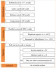
 Source: Elaborated by the authors.