
 Induced and spontaneous uterine leiomyomas in animal models: a scoping review
ABSTRACT
Purpose: To map the experimental models of induced and spontaneous uterine leiomyomas (UL) in animals, describing induction techniques, evaluation methods, advantages, limitations, and research gaps.
Methods: We conducted a systematic review of studies published between 2004 and 2024 with in-vivo animal experiments reporting UL induction or spontaneous occurrence. Data were extracted on animal species, sample size, induction methods, confirmation techniques, and reported advantages or disadvantages.
Results: A total of 56 studies were included. Rats and mice were the most frequently used species. Induction methods comprised hormonal administration, genetic predisposition, chemical exposure (monosodium glutamate), ischemic injury, external stimulation, cell transplantation, xenotransplantation, and spontaneous development. A critical finding that emerged was that 60.7% of the studies failed to report confirmation of UL induction. Among the 22 studies that reported confirmation, caliper measurement was the most frequently used technique. This widespread lack of methodological rigor significantly undermines the credibility and reproducibility of many published models.
Conclusion: This scoping review highlights not only the heterogeneity of UL animal models and the lack of standardized induction and evaluation protocols, but also identifies a critical gap in induction validation reporting that compromises the foundation of preclinical research in this field.
Key words
Leiomyoma; Models, Animal; Scoping Review
Introduction
Uterine leiomyomas (UL) are one of the most common gynecological diseases, affecting women of childbearing age, causing abnormal uterine bleeding, symptoms related to increased uterine volume, and fertility problems, significantly reducing the quality of life of patients and resulting in high costs for the health system1.
Although the ideal pharmacotherapy for treating UL has not been developed yet, nonsteroidal anti-inflammatory drugs, hormonal agents, and gonadotropin-releasing hormone (GnRH) agonists are currently the first line of treatment. In cases of symptomatic uterine fibroids that do not respond to medical treatment, invasive procedures such as uterine artery embolization, high-intensity focused ultrasound, myomectomy, or hysterectomy may be necessary, resulting in more costs, trauma, and risks to patients with UL, justifying the search for new clinical treatments2.
Preclinical research is essential in this context, starting with identifying the need for a new drug to treat a specific disease or improve an existing treatment. At this stage, scientists investigate compounds with therapeutic potential through laboratory studies, molecular screening, and computational modeling3. The promising compounds are tested in cultured cells and animal models to assess efficacy and safety4. Promising drugs undergo rigorous testing to assess their toxicity in different organs and systems. If positive results are obtained, the development of the pharmaceutical form (tablets, capsules, or injections) and appropriate dosage begin, stages that precede clinical trials5.
Despite the crucial role of animal models in preclinical research, current literature on UL models exhibits significant heterogeneity in induction techniques, confirmation protocols, and evaluation methods, with a conspicuous lack of standardization across studies. This fragmentation hinders the ability of researchers to compare findings, select appropriate models for specific research objectives, identify best practices, and critically assess methodological quality, ultimately compromising the translational value of published research. To address this knowledge gap, a comprehensive mapping of available experimental models, their induction strategies, and validation approaches is urgently needed to guide researchers in model selection and promote standardization within the field.
Based on these premises, this study aimed to investigate the induction or spontaneous growth, evaluation methods, and advantages and disadvantages of UL in animal models, as well as to contribute to future research on clinical treatments of the disease.
Methods
Study design and registration
This study was conducted as a scoping review, guided by the Preferred Reporting Items for Systematic Reviews and Meta-Analyses extension for Scoping Reviews (PRISMA-ScR).
To ensure transparency, reproducibility, and to avoid duplication of efforts, the protocol was prospectively registered in the International Prospective Register of Systematic Reviews (PROSPERO), under ID No. CRD42025587878 on January 8, 2025.
Research question
Using the Population, Concept, Context (PCC) framework, the guiding questions were: what experimental animal models of UL have been described in the literature?; and what are their induction methods, evaluation techniques, advantages, and limitations?
-
Population (P): in-vivo animal models;
-
Concept (C): induced or spontaneous UL;
-
Context (C): experimental studies published between 2004 and 2024.
Eligibility criteria
The analysis included randomized and observational in-vivo studies reporting UL induction or spontaneous occurrence in animals published between 2004 and 2024. Reviews, meta-analyses, case reports, conference abstracts, commentaries, books, and grey literature were excluded. Duplicate records and studies without full-text access were removed.
Sources and search strategy
The search was conducted between November 2024 and February 2025 in the PubMed, Embase, Scopus, and Scientific Electronic Library Online (SciELO) databases. The search terms included were: “induced,” “leiomyoma,” “myoma,” “uterine fibroids,” “smooth muscle tumors,” and “animal model,” combined with Boolean operators AND and OR (Table 1).
Data collection process
Initially, the titles were analyzed, and the selected studies were extracted from each platform in .cvs format. Duplicates were removed, titles and abstracts were read, and those not meeting the eligibility criteria were removed. Then, the eligible studies were thoroughly evaluated. Two examiners were involved in the search, selection, and complete analysis of the articles. The data were compared, and a third examiner was consulted in case of disagreement or divergence.
Data registration
A database was created in table format using Microsoft Excel 2019. Data were extracted into a structured table including study design, animal species, sample size, induction method, confirmation technique, and reported advantages or disadvantages.
Synthesis of results
Findings were summarized descriptively and organized into categories of induction methods (hormonal, genetic, chemical, ischemic, external stimulation, transplantation, xenotransplantation, and spontaneous occurrence), as well as the mechanism for evaluating the induction proof, among the studies that reported on it.
Results
Study selection
After applying the inclusion criteria, it was found that, among the 2,802 articles in the search, 1,047 were duplicates, and 1,691 were excluded after reading the title and abstract. Furthermore, through careful analysis of the remaining 64 articles, 56 eligible studies were identified (Fig. 1).
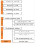
Flow diagram of evidence retrieval and selection according to Preferred Reporting Items for Systematic Reviews and Meta-Analyses extension for Scoping Reviews (PRISMA-ScR) guidelines.
Synthesis of results
A total of 56 studies were included in this scoping review, comprising diverse animal models and methods of UL induction or spontaneous occurrence. Among those, 1,078 rats, 570 mice, 83 guinea pigs, 106 potbellied pigs, 313 hens, 196 chimpanzees, 217 rhesus macaques, and a single squirrel monkey were evaluated. Most studies (n = 37) used hormonal induction as a stand-alone method (n = 19) or combined with other techniques (n = 18), such as xenotransplantation or cellular implantation, followed by xenograft models (n = 10), transgenic or genetically predisposed animals (n = 2), monosodium glutamate (MSG) administration (n = 5), ischemic injury (n = 1), and external stimulation (n = 2), and seven studies investigated spontaneous UL development in non-human primates, pigs, and poultry. The majority (n = 34) did not report how experimental UL induction was confirmed. Among the studies that did report it (n = 22), the use of a digital caliper was the most frequently applied technique (n = 9) (Table 2).
Induced rodent models, particularly those using hormonal administration or MSG, stand out for their accessibility, low cost, and ease of handling, making them suitable for initial pharmacological screenings and basic mechanistic studies, despite limitations imposed by the lack of standardized protocols and the occurrence of adverse systemic effects that hinder direct extrapolation to human clinical practice. In contrast, specific models such as ischemic injury and VX2 tumor in rabbits serve specific investigative niches, respectively focused on the pathogenesis of fibrosis and the development of interventional radiology techniques (Table 3).
In terms of translational potential, xenotransplantation models and spontaneously occurring models (especially in non-human primates and swine) are superior, as they replicate the tissue architecture and hormonal responses observed in women. Xenotransplantation is consolidating as a robust tool for preclinical efficacy testing of new drugs directly in human tissue, although it requires complex infrastructure to maintain immunodeficient animals. Spontaneous models, while ideal for understanding the natural history of the disease due to high anatomical and genetic similarity, face significant ethical barriers and difficulties in access and standardization, which restrict their large-scale application (Table 3).
Discussion
This scoping review mapped the available evidence on induced and spontaneous UL in animal models. The included studies revealed a wide diversity of species, induction methods, and evaluation techniques, reflecting the complexity of reproducing UL pathophysiology in experimental settings.
Hormonal in transgenic animals
Elkafas et al.6 and Yang et al.7 used Eker rats (Long Evans; Tsc2 Ek/+), transgenic rats lineage genetically predisposed to develop UL and renal carcinomas due to mutations in the Tuberous Sclerosis 2 gene, to establish the model. To accelerate the development of the UL, 10 µg of diethylstilbestrol, a synthetic hormone derived from estrogen, was administered daily subcutaneously on days 10, 11, and 12 after birth, since this is a critical phase for the development of UL. The rats were euthanized after five months of life for evaluation.
Genetic predisposition combined with hormone administration mimics the endocrine and genetic factors associated with the development of UL in humans, allowing the investigation of specific molecular mechanisms, such as the interaction between genetic mutations and hormonal stimuli, offering a highly relevant model for translational studies6,7.
Hormonal alone
Preclinical and clinical reports indicate that estradiol (E2) and progesterone (P) levels may serve as a key to the development and growth of UL8. The hormone-induced model alone was reported by 19 studies9–27.
Yasong et al.9 initially applied estrogen (0.5 mg/kg) intraperitoneally daily for four weeks in Wistar rats and then added progesterone at the same dose for five days, highlighting a progressive combined regimen.
One interesting fact about this model is the use of different dosages, routes of administration, and duration of estrogen and progesterone use. Ju and Xiao10 and Li et al.12 administered, in rats, diethylstilbestrol intramuscularly (IM) at a concentration of 2 mg/mL, with a dose of 0.02 mL daily for 30 days, while Yousefi et al.19 administered estradiol benzoate (0.2 mg/kg) IM, twice a week, for eight weeks. Olowofolahan et al.17 chose to administer estradiol valerate (3 mg/kg) by gavage daily for 12 weeks. In Kunming mice, Yang et al.27 divided the animals into three model groups and used estradiol benzoate (EB) in increasing doses of 0.3 (group 1), 0.6 (group 2), and 0.9 mg/kg (group 3), administered IM for periods of 15, 30, and 45 days, respectively. In this study, animals that received EB at a dose of 0.3 mg/kg for 15 days were the most successful in induction, presenting significant changes (p < 0.01) in both uterine coefficient and smooth muscle thickness compared to the control group.
The administration of estrogen associated with progestogens appears to be more effective in the development of hormone-induced UL when compared with estrogen alone and has been used in different protocols adapted to their experimental needs9,11,13–16,18,20–26.
Li et al.13 used Sprague-Dawley rats and administered estradiol benzoate (0.5 mg/kg) IM on Mondays, Wednesdays, and Fridays for 12 weeks, and at week 13, introduced progesterone (5 mg/kg) diluted with estradiol benzoate, maintaining this regimen until week 15. Similarly, Yamei et al.18 administered intramuscular progesterone (1 mg/kg) on Mondays, Wednesdays, and Fridays, complemented with EB (2 mg/kg) applied on Tuesdays, Thursdays, and Saturdays for eight weeks.
Adding this approach, Yu et al.15 employed diethylstilbestrol (0.167 mg/kg) by gavage daily and progesterone (1 mg/kg) IM once a week for 20 weeks, indicating that the route of administration can vary depending on the objective of the experiment. Similarly, Yuan et al.24 applied estradiol benzoate (0.5 mg/kg) intraperitoneally daily and progesterone (4 mg/kg) weekly for five weeks, followed by an additional one-week period with both hormones at the same dose and route of administration. Zeng et al.25 administered estradiol benzoate (0.5 mg/kg) intramuscularly three times a week for eight weeks and, subsequently, introduced progesterone (4 mg/kg) intraperitoneally twice a week for another four weeks, demonstrating a phased induction model.
Zhang et al.20 applied diethylstilbestrol (1.35 mg/kg) by gavage daily and progesterone (1 mg) intramuscularly for five weeks. Gao et al.26 administered estradiol benzoate (0.5 mg/kg) IM, combined with progesterone (1 mg/kg) daily for nine weeks, highlighting a simple and continuous protocol. Chen et al.21 followed a split regimen, using estradiol benzoate (0.5 mL/kg) IM for eight weeks, followed by the introduction of progesterone (0.25 mL/kg) intraperitoneally, twice a week, for four weeks. The study by Lin et al.14, which used Institute of Cancer Research (ICR) mice (CD-1), started with diethylstilbestrol (0.4 mg/kg) by daily gavage for four weeks, and then this compound was associated with medroxyprogesterone 17-acetate (5 mg/kg) by the same route for another four weeks.
Lin et al.16 followed a simple and continuous protocol, using daily injections of estradiol benzoate (0.2 mg) and progesterone (0.2 mg) for 60 days in specific pathogen-free (SPF) mice. Qiu et al.22 applied estradiol benzoate (0.5 mg/kg) daily intraperitoneally and progesterone (4 mg/kg) weekly for 10 weeks in Sprague Dawley rats, adding both hormones simultaneously in the last five days. Still dealing with hormonal induction, Shi et al.23 administered estradiol benzoate (0.5 mg/kg) IM for 35 days and progesterone (1 mg) daily, managing to induce the model in only 14 days, evidencing the efficiency of their approach.
Finally, Liu et al.11 chose to use C57B6 mice, which received intramuscular injections of estradiol benzoate at 0.05 mg/100 g three times a week for 12 weeks. After this period, the animals began receiving intramuscular progesterone injections twice weekly for another four weeks.
The main advantage of hormonal methods is their simplicity compared to surgical or genetic approaches. Administration of estrogen alone or in combination with progestogen allows precise control of doses and exposure time, in addition to not requiring invasive procedures. This factor reduces stress and the risk of direct complications and is less aggressive. The costs associated with hormones are relatively low, and the technique requires only basic training from the team in administering substances orally, IM, or intraperitoneally.
The limitations of this model are due to the prolonged use of high doses of hormones that can cause undesirable systemic effects, such as metabolic, hepatic, or renal changes; the development of UL may vary between individuals, requiring a larger number of animals to ensure an adequate sample; the lack of confirmation before starting the experimental research, in most of the studies evaluated; this model may not fully reflect the etiological complexity of UL in humans, which also involve genetic, inflammatory and environmental factors; and finally, there is no standardization regarding the type, associations, doses, route of administration, and time of use of the hormones used.
Monosodium glutamate
MSG, a non-essential amino acid commonly used as a flavor enhancer in processed foods, was used alone to induce UL by increasing cholesterol and consequently elevating estrogen and progesterone levels, leading to uterine hyperplasia and development of UL, with daily doses of 200 mg/kg administered orally for 30 days in Wistar or Sprague Dawley rats28–31.
Mamoon et al.32 used a different scheme in Wistar rats, administering MSG 200 mg/kg in the first week, 400 mg/kg in the second week, and 600 mg/kg from the third week until the eighth week when the experiment ended. In addition, estradiol benzoate (200 µg) was injected subcutaneously twice a week from the third week until the end of the experiment.
The models’ induction took two to eight weeks, depending on each study’s protocol. A significant advantage of using MSG for UL induction in rats is the relatively low cost and easy availability of the compound, making the model accessible to laboratories with limited resources. In addition, MSG is administered orally, a less invasive and more practical method, which can reduce animal stress compared to surgical interventions or more complex techniques. The relatively short time for UL induction, ranging from two to eight weeks, is another important advantage, allowing rapid results28–32.
On the other hand, the mechanism of action of MSG in inducing UL by hormonal increase is not entirely elucidated, which may limit the translation of findings to human models. Thus, administering daily doses for prolonged periods requires intensive management, which may increase variability between experimental groups due to factors such as stress or absorption difficulties.
Ischemic injury
Another model of UL development is an ischemic injury, because it induces tissue fibrosis, in which uterine scarring and remodeling occur through the characteristic of myofibroblast transdifferentiation and scar formation33.
It has been reported that mast cells could promote fibrosis, which is intrinsically linked to the pleiotropic cytokine transforming growth factor (TGF)-β, that plays an important physiological role in the initiation and control of fibrosis, in addition to being a key mediator of fibroblast activation and driving an abnormal extracellular matrix synthesis in fibrotic diseases9,34.
The ischemic injury technique is based on surgically exposing the rat’s abdomen and clamping the celiac artery for 30 minutes33. This method has the advantage of being able to study the effects of hypoxia and vascular injury on the pathogenesis of UL, providing valuable information on the contribution of the ischemic environment to tumor development. This model could assist in investigating molecular mechanisms and evaluating therapeutic interventions to address changes in the uterine microenvironment caused by ischemia.
The limitations of ischemic injury include its highly invasive nature, which significantly compromises animal welfare, increasing the risk of pain, stress, and postoperative complications. In addition, ischemia can affect other organs and vascular systems, introducing confusion in the results and making it difficult to attribute the effects exclusively to the uterus. Despite being a method of inducing UL, it appears to be the furthest from preserving animal welfare, causing injury, requiring extensive surgical preparation, and possibly affecting the animal’s vascular system33.
External stimulus
The studies conducted by Zhao et al.35 and Feng et al.36 demonstrated that external stimulation effectively enhances UL induction in rats subjected to hormonal treatments with estrogen and progesterone. Their findings indicate that providing daily external stimuli for approximately two weeks significantly improves UL development in these animals.
In traditional Chinese medicine, UL treatments promote blood circulation and remove uterine stagnation. This stagnation is due to disturbing emotions such as anger and long-term concern, justifying the induction of UL through external change and environmental factors such as light, noise, and temperature to enrage rats by mimicking the depressed and angry states of humans35.
Sprague Dawley rats received 0.9 mg/kg/day of epinephrine hydrochloride IM from the fourth week onwards and received external stimulation daily four hours after injection. The stimuli were exposure to 60 dB of noise for 3 hours, reversal of the day and night cycle in one day, swimming in water at 5–10°C for 4 minutes, being hung by the tail for 10 minutes, and being exposed to the heat of 50°C for 10 minutes, with each stimulus being performed at least twice for a two-week cycle35,36. Although these studies convey an idea of greater efficiency with these methods, these practices dangerously resemble animal abuse.
Cellular autotransplant
The transplantation of ELT-3 cells, a UL cell line derived from Eker rats, was used in five studies involving mice37–42.
Abdelaziz et al.37 and Halder et al.40 used sustained-release hormone pellets containing 17β-estradiol (90 days), implanted four days before cell transplantation in athymic nude mice and nude mice (Foxn1nu), respectively. Bar-Joseph et al.38 also combined ELT-3 cell implantation with hormonal induction in nude athymic mice by administering beta-estradiol 17-valerate (2 μg) two days before the cell transplantation procedure. Lin et al.14, Chen et al.39, and Lin and Wu41 performed only the cell implantation without hormonal supplementation in nude mice (Foxn1nu).
This method is efficient for inducing UL in mice and is comparable to the hormonal method alone in terms of results. In addition, cell transplantation is less invasive and less stressful since it involves only the injection of cells, dispensing with more complex procedures. In addition, the procedure is faster, reducing animal stress, because it involves only the injection of cells. The method also offers flexibility and can be used with or without hormonal supplementation, depending on the needs of the study14,37–41.
However, dependence on the ELT-3 cell line requires specific laboratory conditions for its maintenance, which can be a logistical challenge. The model is also largely validated in mice, which limits its application in other species. Furthermore, because the cells are derived from rats, there are limitations in directly translating the findings to humans.
Another cell autotransplant model is the VX2 tumor in rabbits, which uses the VX2 tumor line, an anaplastic squamous cell carcinoma derived initially from papillomas induced by the Shope papillomavirus, and can be transplanted from one animal to another and into any tissue, including the myometrium of rabbits43.
Rhee et al.44 evaluated the feasibility of this model in the study of the treatment of UL in rabbits through uterine artery embolization. To create the model, the authors performed a laparotomy to expose the uterus and injected 0.3 to 0.5 mL of a solution containing VX2 cells, obtained from tumors of donor rabbits, into the uterine horn of six rabbits.
After model confirmation, magnetic resonance imaging (MRI) scans with and without contrast were performed before and after uterine arterial embolization (UAE). The gadolinium-based contrast was administered manually, followed by saline, and images were captured 2 minutes after injection. After the MRI, the rabbits were taken to the angiography suite for the UAE procedure, guided by fluoroscopy44.
Among the characteristics of this model, it does not require immunocompromised or transgenic animals. Therefore, it can be used in healthy animals and allows tumors to be obtained in a few weeks with > 95% efficiency. Another decisive factor for the use of this model in rabbits is the larger size compared to rodents, allowing the use of medical devices similar to those used in humans, which allows the evaluation of locoregional therapies, including transarterial chemoembolization, radioembolization, thermal ablative therapies, and combined approaches43.
Nevertheless, it is a valuable model for studying the interaction between tumor microenvironment and neovascularization and may assist in the development of therapeutic strategies aimed at reducing blood supply to the UL45.
Xenotransplantation
Xenotransplantation is the transplantation of organs, tissues, and cells between organisms of different species46. Based on this knowledge, different studies47–54 have investigated human UL xenotransplantation in rats and mice as an experimental model for the study of UL. This approach requires the administration of high doses of hormones and, in many cases, immunosuppression of the animals to avoid transplant rejection.
In the study conducted by Sousa et al.53, Wistar rats were immunosuppressed with mycophenolate mofetil (40 mg/kg) administered by oral gavage, starting 15 days before xenotransplantation and maintained until the end of the experiment. After immunosuppression, the animals underwent surgery to transplant UL tissue fixed on the right side of the peritoneal cavity or in the subcutaneous tissue of the right flank. After the procedure, estradiol valerate (1 mg) was administered as a hormonal supplement, dissolved in 250 mL of water, and offered ad libitum, with daily replacement. This study reports a new, practical, and relatively inexpensive model of UL in rats, but such a model could be improved by studying the effects of progesterone supplementation and more accurately assessing the mode of estrogen administration45.
Another technique includes the implantation of a pellet containing 17β-estradiol (0.05 mg) and progesterone (50 mg) into the subcutaneous tissue during surgery54 or 15 days before transplantation47,52. Alternatively, Corachán et al.48 performed the xenograft of two human UL fragments in the peritoneal cavity (one on each side) associated with ovariectomy and subcutaneous implantation of pellets containing 17β-estradiol (0.36 mg) in the neck of the animals, complementing the protocol with the administration of progesterone (1 mg/day) for one week, followed by a two-week interval to simulate the menstrual cycle, maintaining this regimen for 60 days.
The study by Huang et al.49 investigated the role of estrogen and laparoscopic surgery on the growth of parasitic fibroids, an extrauterine leiomyoma, in severe combined immunodeficiency (SCID) mice by xenotransplantation of 10 fibroids fragments (1–2 mm) distributed across the four quadrants of the peritoneal cavity. Three primary induction methods were used: xenotransplantation without ovariectomy (group 1), xenotransplantation with bilateral ovariectomy (group 2), and xenotransplantation with estradiol (E2) supplementation (group 3). In the first group, estradiol-supplemented fibroid fragments were cultured and transplanted into the animals. In addition, the animals underwent a CO2 insufflation procedure at 4 mmHg for 10 minutes to simulate laparoscopic conditions. The second group underwent bilateral ovariectomy two weeks before xenotransplantation, serving as a model of estrogen deprivation to evaluate the influence of the hormone on fibroid implantation. The third group received exogenous estradiol supplementation after xenotransplantation to verify whether the increase in hormone levels favored the implantation and growth of fibroids. The mice in group 3 presented the highest number of implants and a total weight of implanted fibroids, in addition to a significant increase in the expression of markers of cell proliferation, angiogenesis, and hormone receptors. In contrast, the mice submitted to ovariectomy had fewer implantations and lower growth of fibroids, indicating that estrogen depletion significantly reduces the proliferation and angiogenesis of implanted fibroids.
Lee et al.50 implanted human UL cells mixed in Matrigel and prolonged-release hormone pellets with 17b-estradiol (0.05 mg/90 days) and progesterone (25 mg/60 days) into the abdominal subcutaneous tissue of mice, concluding the experimental model in eight weeks when they started treatment with flavopiridol. Zakaria et al.55 implanted human leiomyosarcoma cells (20 μL containing 106 cells) into the subcutaneous tissue of the right flank of nude mice. The experiment ended 21 days after transplantation.
Koohestani et al.56 and Qiang et al.57 performed the implantation of slow-release hormone pellets in subcutaneous tissue with estradiol and progesterone (E2 0.8 mg + P4 75.2 mg) + cholesterol (4 to 5 mg) associated with ovariectomy and implantation of human UL in the renal capsule of NOD-scid IL2Rynull, establishing the model after four weeks.
In addition to these approaches, Fritsch et al.58 performed xenotransplantation of human UL tissue into the subcutaneous tissue of the ventral region of immunodeficient SCID mice. The mice were ovariectomized and, during tissue transplantation surgery, received subcutaneous implants of estradiol (0.05 mg/90 days) and progesterone (25 mg/60 days) hormone pellets to simulate the human hormonal environment. Graft growth was assessed between 15 and 60 days after transplantation, and greater proliferation was observed in the presence of E2 and P4 combined when compared with the results of the groups without hormone supplementation or with E2 supplementation alone.
Li et al.51 administered bisphenol A (BPA) orally at the dose of 400 μg/kg body weight per day, starting two weeks before cell injection and continuing until the 42nd day after subcutaneous injection of human UL (1×10⁷ cells) into the right dorsal flank.
This approach, which uses transplantation and hormonal supplementation without immunosuppression, was mainly used in mice of the BALB/C nude, NOD-SCID, or NOG (NOD/Shi-scid/IL2Rγnull) lineages, which present natural immunosuppression, eliminating the need for immunosuppressive drugs and reducing the chances of tumor rejection.
These methods are particularly valid for the experimental induction of UL, as they use human tissue, allowing greater translational relevance when evaluating drugs intended for human patients. In addition, it is possible to reproduce biological characteristics and therapeutic responses similar to those observed in humans, offering realistic modeling.
Whether through pellets or enriched water, hormone supplementation allows efficient control of the hormonal environment necessary for tumor growth. In immunosuppressed strains, such as BALB/C nude or NOG, the need for immunosuppressive drugs is reduced, simplifying experimental management. However, the technique is invasive and stressful for the animals, involving surgical procedures, anesthesia, analgesia, and postoperative care.
Spontaneous
The study by Laik-Schandelmaier et al.59 analyzed 83 female guinea pigs that underwent histopathological examinations, of which 64 had surgically removed uterine masses and 19 underwent complete necropsy. Among the cases evaluated, 18 leiomyomas (21.7%) and nine leiomyosarcomas (10.8%) were diagnosed, demonstrating a predominance of benign tumors over malignant ones. In addition, 20 endometrial adenomas (24.1%), three adenocarcinomas (3.6%), and other alterations such as cystic glandular hyperplasia and cervical polyps were observed. These findings suggest that guinea pigs can spontaneously develop UL and other reproductive tract tumors, which could make them potential experimental models for studying the disease.
However, the lack of information on the hormonal regulation of tumors in these animals poses challenges to their use in controlled research. The need to standardize factors such as age, hormonal cycle, and response to treatment should also be considered before validating the model59.
In addition to conventional models, other methods that do not fall within the definition of induced models have been explored due to their ability to develop UL spontaneously and may contribute significantly to translational research in the future. Among these alternative models, studies with potbellied pigs60, aged hens61,62, chimpanzees63,64, rhesus monkeys65, and Guianan squirrel monkeys66 stand out, each with its particularities in the induction and development of UL.
Another alternative is the potbellied pig (Sus scrofa), which has been studied due to the similarity of the porcine uterus to the human uterus concerning macroscopy, cellular morphology, level of mitotic activity, collagen deposition pattern, hormonal response, and incidence similar to that found in women. An additional point is the duration of the swine estrous cycle, which occurs every 21 days and lasts approximately two or three days45.
A study by Mozzachio et al.60 evaluated 106 female potbellied pigs using medical records provided by a local potbellied pig spay/neuter program, pig sanctuaries, and the Duchess Fund database for evidence of reproductive disease or surgery for spontaneously occurring fibroid-like tumors in potbellied pigs. Among the animals analyzed, 17 cases of spontaneously developing neoplasms were identified, but only 13 received histological evaluation, which identified 11 UL, one leiomyosarcoma, and one undifferentiated sarcoma. The authors reported difficulties, such as the lack of histological analysis for all detected neoplasms, standardization of tissue sampling and fixation methods, macroscopic description of lesions, and clinical signs.
Regarding hormone receptors, it was found that healthy tissues and UL from potbellied pigs presented, in decreasing order of immunoexpression, progesterone receptors (PR), estrogen receptor-α (ER-α), and estrogen receptor-β (ER-β), a pattern that is also observed in women. Although the relationship between the expression of sexual steroid hormones and UL is not entirely clear yet, most studies indicate that there is an increase in the immunoexpression of these tumors in affected tissues, a fact that has not been proven yet in potbellied pigs60. Thus, this species has great potential for developing an animal model of UL, but new studies are necessary to evaluate the feasibility of this method.
The aging hen (Gallus gallus domesticus) model has been explored as an alternative for the study of UL since up to 60% of these birds develop UL of the ventral ligament of the oviduct spontaneously, varying according to the lineage and breed45.
Aiming to evaluate the possibility of using this species as a model of UL, Machado et al.62 evaluated 263 hens without signs of disease. The research revealed a higher prevalence of UL, in addition to an increase in the size and quantity of tumors, especially in the third and fourth years of laying. These tumors present similarities with human ULs, especially when it comes to the expression of the Bcl-2 protein, the presence of smooth muscle actin and desmin in immunohistochemistry, the relation ER/PR, and localized cell proliferation45,61.
Thus, the data suggest that the aged chicken is a potential model to study the pathophysiology of ULs and evaluate new therapies. The efficacy of this model could be improved with noninvasive detection methods and continuous monitoring without sacrificing the animal, but further studies are needed to assess this feasibility45.
The spontaneous development of UL in chimpanzees has been reported in the literature, with studies addressing their prevalence, histopathology, and possible hormonal influences. In a study conducted in two primate facilities in the United States of America, it was observed that, of the 195 females analyzed with ages ranging from 15 to 52.5 years old (mean = 28.47), 28.2% (n = 55) had UL, with a mean age of 30.4 at the time of diagnosis. The prevalence increased to approximately 40% in females over 30 years old, suggesting a relation between advanced age and tumor development64.
The diagnosis was made primarily by physical examination (palpation) and transabdominal or transrectal ultrasonography during annual examinations under sedation. Histopathology confirmed the presence of interlaced bundles of smooth muscle, with some areas of necrosis, vascular embolization, and inflammatory cell infiltration. The influence of sex hormones on the formation of these tumors has also been investigated. The use of progesterone-based hormonal contraceptives was associated with a significant reduction in the prevalence of UL, suggesting a possible protective effect64.
Another study reported a case of UL in a 22-year-old female chimpanzee who presented with anorexia, frequent vomiting, and dehydration. During necropsy, an enlarged uterus was observed, with the lumen obstructed by firm, whitish nodules that extended through the myometrium. Immunohistochemical analysis revealed strong expression of estrogen and PR, suggesting a hormonal relationship in the tumor development. These findings indicate that the chimpanzee may be a relevant model for the study of UL, given that the tumors share histological and hormonal characteristics with those found in humans. However, further studies are needed to understand better the underlying mechanisms and the response to different therapies63.
The rhesus macaque (Macaca mulatta) has been identified as a relevant model for the study of spontaneous neoplasms, including UL. These animals share several physiological similarities with humans, including the relationship between aging and increased incidence of tumors. The analysis conducted in two colonies of rhesus macaques (n = 217) kept in captivity revealed that UL represented the most common urogenital neoplasm, corresponding to 30 of the 39 cases identified in the uterus of these females. The mean age at diagnosis was 25.1 years old, ranging between 15.0 and 34.8, suggesting a correlation between advanced age and the development of these lesions65.
UL in rhesus monkeys is described as a benign smooth muscle tumor, similar to those found in women, which reinforces their usefulness as an experimental model for investigations into pathophysiology and possible therapeutic approaches. Furthermore, cases of well-circumscribed neurilemmomas within UL have been reported, an unusual finding that may have implications for understanding tumor biology in these primates65.
The high incidence of UL in these primates and their similarity to human tumors make the rhesus monkey a promising model for research on the pathogenesis and treatment of UL, although additional studies are needed to clarify the factors involved in the development of these tumors. The first documented case of UL in Guiana squirrel monkeys (Saimiri sciureus) was described in a 12-year-old adult female who presented with a firm, palpable mass in the caudal region of the abdomen66.
Due to the size and rapid growth of the lesion, an exploratory laparotomy was performed, revealing an intraluminal tumor in the uterus, which was removed by partial hysterectomy. Macroscopically, the tumor was firm, non-encapsulated, reddish pink in color, and had a smooth surface. Histological analysis confirmed a UL composed of intertwined bundles of smooth muscle cells with elongated, cigar-shaped nuclei and low mitotic activity66. This report reinforces that, although rare, UL can occur in Guiana squirrel monkeys, a species that should have potential as a model of experimental UL. However, as with other spontaneous models, further studies are needed to assess the similarity of these tumors between primates and humans.
Induction proof
An essential consideration in developing an effective experimental method for UL induction is the implementation of a reliable verification process. This is crucial, as any of the above-mentioned methods may fail, compromising the accuracy and reliability of the results obtained. The review found that 34 studies did not clarify how UL development was confirmed.
Less than half of the induced or spontaneous ULs models (n = 22) confirmed the presence of induction before the start of experimental drug use19,21,23–25,32,37–41,44,47,48,50,51,53,54,56,59,64,66. Halder et al.40 and Lee et al.50 verified it from a macroscopic point of view, Abdelaziz et al.37, Chen et al.39, Bar-Joseph et al.38, Lin and Wu41, Borahay et al.47, Corachán et al.48, Lee et al.50, Li et al.51, and Suzuki et al.54 measured the induction with a caliper, externally of the animal. Meanwhile, other authors chose to use ultrasound in isolation47,64,66 or Doppler flowmetry32,44, which showed higher blood flow velocities and lower Doppler indices in rats that developed UL. This approach is manageable in rodents and primates, as UL is visible as an abdominal, axillary, or dorsal bulge, depending on the tumor’s location, and is easily detected on imaging.
In a complementary manner, Long et al.66 performed radiography, while Corachán et al.48 followed another approach to demonstrate UL employing positron emission tomography with computed tomography (PET/CT), using the radiopharmaceutical 2-deoxy-2-[18F]fluoro-D-glucose (18F-FDG) on days 21 and 60 of the experiment, to evaluate the therapeutic effects of vitamin D in the short and long term, respectively. This technique allowed the detection of UL xenografts from glucose uptake by tumor cells.
Six other studies performed surgeries to evaluate UL induction with macroscopic visualization in tissues in all animals19,21,24,25,53,66. Yousefi et al.19, Chen et al.21, Yuan et al.24, Sousa et al.53, Koohestani et al.56 and Long et al.66 evaluated two or three animals from each group. Zeng et al.25 confirmed euthanasia induction and performed a histopathological analysis of the model group, which was considered successful if the smooth muscle layer of the uterus showed significant thickening and fibrosis.
Laik-Schandelmaier et al.59 evaluated the presence of UL through histopathological analysis of tumors removed during surgery. A single study performed MRI to evaluate the success of tumor induction in rabbits44.
Although induced animal models are widely used in UL studies, they have significant limitations, such as physiological and hormonal differences compared to humans and variations in induction methods and response to stimuli. On the other hand, spontaneous models, such as those reported in primates and other animals, may offer an alternative closer to the disease’s natural pathogenesis. However, the scarcity of detailed studies on these cases and the difficulty of standardization limit their immediate application as experimental models. Future investigation of these models could provide a more translational and complementary approach to induced models, increasing the relevance of the findings for clinical practice.
Ethical considerations
The analysis of experimental models revealed a dichotomy between the need to mimic human pathophysiology and the ethical considerations of animal welfare. The validation of any experimental model must necessarily align with the principles of the 3Rs (Replacement, Reduction, and Refinement), as classically established by Russell and Burch67 and standardized by the Brazilian Guideline for the Care and Use of Animals (CARE)68,69. From this perspective, induction methods that employ swimming in ice water, tail suspension, exposure to extreme noise, and other animal-stress techniques require rigorous ethical evaluation. The CARE classifies procedures involving immobilization stress or forced swimming with physical exhaustion as having a severe degree of invasiveness (G3 or G4), since they cause severe pain, suffering or stress and hinder the expression of natural behaviors69.
Such practices resemble mistreatment and violate the principle of refinement, which requires that proposals be designed to avoid pain and stress, and that the absence of less invasive alternatives be thoroughly proven69.
Furthermore, models based on ischemic injury and prolonged supraphysiological hormonal administration present significant ethical limitations. Surgical ischemia poses risks of postoperative pain and systemic compromise, requiring intensive monitoring and rigorous analgesia to reduce suffering, as recommended by postoperative care guidelines. Regarding reduction, it is observed that many protocols still depend on the euthanasia of large groups of animals for tumor evaluation69. The implementation of noninvasive imaging technologies (such as ultrasound and MRI) would allow longitudinal follow-up of the same individual, significantly reducing the number of animals used without compromising the statistical reliability of the results. Therefore, the advancement of translational research in leiomyomas should prioritize spontaneous or xenotransplantation models that, combined with noninvasive monitoring methods, respect the ethical imperative of minimizing harm and maximizing well-being, and reject induction protocols that inflict unjustifiable severe suffering.
Conclusion
This scoping review synthesized evidence on animal models of UL, mapping induction methods (transgenic, hormonal, MSG, ischemic injury, external stimulus, cell transplantation with the ELT-3 and VX2 tumor lineage, and xenotransplantations in tissue and cellular forms), species used (rats, mice, potbellied pigs, hens, chimpanzees, rhesus monkeys, and squirrel monkeys), and evaluation strategies (macroscopy, microscopy, PET/CT, radiography, MRI and ultrasonography with and without Doppler flowmetry). Thus, substantial heterogeneity was observed regarding the species used, induction protocols, hormonal regimens, confirmation methods, and outcomes evaluated, which significantly limits reproducibility and hinders direct comparison between studies.
To provide practical guidance based on this synthesis, we established that the choice of model should be guided by the specific objective of the investigation. Mechanistic studies in genetics and molecular biology should prioritize transgenic models associated with hormonal induction (such as Eker rats), as they mimic the interaction between genetic mutations and endocrine stimuli, approximating human etiopathogenesis. In addition, basic pharmacological investigations and initial screening find in rodent hormonal models a low-cost solution, simplicity, and precise dose control, although pharmacokinetic studies requiring the use of medical devices or hemodynamic monitoring are more appropriately performed in larger animals, such as the VX2 tumor in rabbits or porcine models. Finally, for preclinical efficacy trials of new drugs, the current gold standard is xenotransplantation models with human tissue or cells, which offer the greatest translational relevance by replicating the tissue architecture and the actual therapeutic response observed in women.
Among the models identified, hormonal models in rodents stand out for their accessibility, low cost, and applicability in mechanistic and pharmacological investigations. In contrast, xenotransplantation models and spontaneous models, especially in pigs and non-human primates, demonstrate greater translational relevance due to their greater anatomical, histological, and hormonal similarity to human leiomyomas. Therefore, future research should prioritize the establishment of standardized, ethically responsible, and reproducible protocols for the induction and confirmation of leiomyomas in animal models, preferably incorporating noninvasive imaging methods, such as ultrasound and MRI, combined with histopathological validation. Furthermore, increased investment in research on spontaneous models could provide valuable information on the natural history and pathophysiology of fibroids, contributing to the development of more effective and less invasive therapeutic strategies for women affected by this condition. Standardization of experimental models and outcome measures is fundamental to increasing the translational impact of this research and accelerating the development of medical therapies capable of reducing the clinical, social, and economic burden of UL worldwide.
Acknowledgements
Not applicable.
-
Research performed at Postgraduate Program in Health Sciences, Universidade Federal do Maranhão, São Luís (MA), Brazil.
-
Funding
Not applicable.
-
Declaration of use of artificial intelligence tools
The authors declare that this manuscript/assessment was prepared entirely without the use of generative artificial intelligence (AI) technologies or automated text-producing tools. All ideas, narrative structure, arguments, and scientific explanations were developed solely by the authors without any AI-generated assistance.
Data availability statement
All relevant data and details of resources can be found within the article.
References
-
1 Keizer AL, Semmler A, Kok HS, van Kesteren PJM, Huirne JAF, Hehenkamp WJK. Modifiable prognostic factors in uterine fibroid development: a systematic review of literature. J Obstet Gynaecol. 2024;44(1):2288225. https://doi.org/10.1080/01443615.2023.2288225
» https://doi.org/10.1080/01443615.2023.2288225 -
2 Park SJ, Lee JW, Hwang DW, Lee S, Yim GW, Song G, Lee EJ, Kim HS. Effect and safety of diluted vasopressin injection for bleeding control during robot-assisted laparoscopic myomectomy in reproductive women with uterine fibroids: a randomized controlled pilot trial (VALENTINE Trial). In Vivo. 2024;38(1):431–6. https://doi.org/10.21873/invivo.13456
» https://doi.org/10.21873/invivo.13456 -
3 Dholakia J, Scalise C, Arend RC. Assessing preclinical research models for immunotherapy for gynecologic malignancies. Cancers (Basel). 2021;13(7):1694. https://doi.org/10.3390/cancers13071694
» https://doi.org/10.3390/cancers13071694 -
4 Bimonte-Nelson HA, Bernaud VE, Koebele SV. Menopause, hormone therapy and cognition: maximizing translation from preclinical research. Climacteric. 2021;24(4):373–81. https://doi.org/10.1080/13697137.2021.1917538
» https://doi.org/10.1080/13697137.2021.1917538 -
5 Lattanzi W, Ripoli C, Greco V, Barba M, Iavarone F, Minucci A, Urbani A, Grassi C, Parolini O. Basic and preclinical research for personalized medicine. J Pers Med. 2021;11(5):354. https://doi.org/10.3390/jpm11050354
» https://doi.org/10.3390/jpm11050354 -
6 Elkafas H, Ali M, Elmorsy E, Kamel R, Thompson WE, Badary O, Al-Hendy A, Yang Q. Vitamin D3 ameliorates DNA damage caused by developmental exposure to endocrine disruptors in the uterine myometrial stem cells of eker rats. Cells. 2020;9(6):1459. https://doi.org/10.3390/cells9061459
» https://doi.org/10.3390/cells9061459 -
7 Yang Q, Ali M, Treviño LS, Mas A, Ismail N, Al-Hendy A. Epigenetic modulation of inflammatory pathways in myometrial stem cells and risk of uterine fibroids. Int J Mol Sci. 2023;24(14):11641. https://doi.org/10.3390/ijms241411641
» https://doi.org/10.3390/ijms241411641 -
8 Moravek MB, Yin P, Ono M, Coon JS 5th, Dyson MT, Navarro A, Marsh EE, Chakravarti D, Kim JJ, Wei JJ, Bulun SE. Ovarian steroids, stem cells and uterine leiomyoma: therapeutic implications. Hum Reprod Update. 2015;21(1):1–12. https://doi.org/10.1093/humupd/dmu048
» https://doi.org/10.1093/humupd/dmu048 -
9 Yasong W, Donghua L, Xin X, Ruiya Q, Yalan Z, Yuhua H, Jianguo G, Xiaoli Z, Hongjuan H, Wufang Z. Lichong decoction reduces Matrix Metalloproteinases-2 expression but increases Tissue Inhibitors of Matrix Metalloproteinases-2 expression in a rat model of uterine leiomyoma. J Tradit Chin Med. 2016;36(4):479–85. https://doi.org/10.1016/s0254-6272(16)30065-6
» https://doi.org/10.1016/s0254-6272(16)30065-6 -
10 Ju Y, Xiao B. Chemical constituents of Cyperus rotundus L. and their inhibitory effects on uterine fibroids. Afr Health Sci. 2016;16(4):1000–6. https://doi.org/10.4314/ahs.v16i4.16
» https://doi.org/10.4314/ahs.v16i4.16 -
11 Liu H, Wang J, Sheng L, Zhang Y, Tang N, Li Y, Hao T. Paclitaxel promotes cell apoptosis in uterine leiomyomas. Pharmacology. 2017;100(5–6):246–52. https://doi.org/10.1159/000479161
» https://doi.org/10.1159/000479161 -
12 Li M, Qingxiang H, Lingli X, Mei Z, Dan L, Yongwang L. Treatment with tinosporaside attenuates the uterine fibroid by stimulating the apoptosis. Treatment with tinosporaside attenuates the uterine fibroid by stimulating the apoptosis. 2019;15(1):50–5. https://doi.org/10.3923/ijp.2019.50.55
» https://doi.org/10.3923/ijp.2019.50.55 -
13 Li W, Hong B, Li Q, Li Z, Bi K. An integrated serum and urinary metabonomic research of Rhizoma Curcumae-Rhizoma Sparganii drug pair in hysteromyoma rats based on UPLC-Q-TOF-MS analysis. J Ethnopharmacol. 2019;231:374–85. https://doi.org/10.1016/j.jep.2018.11.033
» https://doi.org/10.1016/j.jep.2018.11.033 -
14 Lin PH, Shih CK, Yen YT, Chiang W, Hsia SM. Adlay (Coix lachryma-jobi L. var. ma-yuen Stapf.) Hull extract and active compounds inhibit proliferation of primary human leiomyoma cells and protect against sexual hormone-induced mice smooth muscle hyperproliferation. Molecules. 2019;24(8):1556. https://doi.org/10.3390/molecules24081556
» https://doi.org/10.3390/molecules24081556 -
15 Yu CH, Zhao JS, Zhao H, Peng T, Shen DC, Xu QX, Li Y, Webb RC, Wang MH, Shi XM, Peng C, Ding WJ. Transcriptional profiling of uterine leiomyoma rats treated by a traditional herb pair, Curcumae rhizoma and Sparganii rhizoma. Braz J Med Biol Res. 2019;52(6):e8132. https://doi.org/10.1590/1414-431X20198132
» https://doi.org/10.1590/1414-431X20198132 -
16 Lin Y, Yang C, Tang J, Li C, Zhang ZM, Xia BH, Li YM, He QZ, Lin LM, Liao DF. Characterization and anti-uterine tumor effect of extract from Prunella vulgaris L. BMC Complement Med Ther. 2020;20(1):189. https://doi.org/10.1186/s12906-020-02986-5
» https://doi.org/10.1186/s12906-020-02986-5 -
17 Olowofolahan AO, Akanji BA, Olorunsogo OO. 3-(Para-fluorobenzoyl)-propionic acid; a metabolite of haloperidol, reversed oestradiol valerate-induced uterine hyperplasia via modulation of oestrogen receptor signalling pathways in female Wistar rats. J Basic Clin Physiol Pharmacol. 2020;31(4):20190119. https://doi.org/10.1515/jbcpp-2019-0119
» https://doi.org/10.1515/jbcpp-2019-0119 -
18 Yamei L, Jie T, Hongshan L, Bohou X, Limei L, Duanfang L. Effects of Fuke Qianjin Formula on hormones and their receptors and metabonomics study in uterine fibroids model rats. Digital Chinese Medicine. 2021;4(4):316–27. https://doi.org/10.1016/j.dcmed.2021.12.006
» https://doi.org/10.1016/j.dcmed.2021.12.006 -
19 Yousefi B, Semnani V, Mokhtari T, Zarbakhsh S, Amjad MHT, Barati M, Doustmohammadi H. Co-administration of aluminum sulfate and propolis regulates matrix metalloproteinases-2/9 expression and improves the uterine leiomyoma in adult rat model. Biol Trace Elem Res. 2021;199(3):1002–12. https://doi.org/10.1007/s12011-020-02200-0
» https://doi.org/10.1007/s12011-020-02200-0 -
20 Zhang L, Xu Q, Li Y, Zhao H, Shi X, Peng F, Yu C. Ameliorative effects of component Chinese medicine from curcumae rhizoma and sparganii rhizoma, a traditional herb pair, on uterine leiomyoma in a rat model. Front Public Health. 2021;9:674357. https://doi.org/10.3389/fpubh.2021.674357
» https://doi.org/10.3389/fpubh.2021.674357 -
21 Chen L, Chen H, Yang Q, Jiang Y, Liu L, Yu H, Chen Y, Li J, Chen N, Wang H, Wang Q. Guizhi Fuling Capsule inhibits uterine fibroids growth by modulating Med12-mediated Wnt/β-Catenin signaling pathway. J Ethnopharmacol. 2022;290:115115. https://doi.org/10.1016/j.jep.2022.115115
» https://doi.org/10.1016/j.jep.2022.115115 -
22 Qiu T, Li D, Liu Y, Ren H, Yang X, Luo W. Astragaloside IV inhibits the proliferation of human uterine leiomyomas by targeting IDO1. Cancers (Basel). 2022;14(18):4424. https://doi.org/10.3390/cancers14184424
» https://doi.org/10.3390/cancers14184424 -
23 Shi S, Luo L, Peng F, Yu C. Potential mechanism of Taohong Siwu Decoction in uterine fibroid treatment based on integrated strategy of network pharmacology and experimental verification. Chin Med. 2023;18(1):95. https://doi.org/10.1186/s13020-023-00809-6
» https://doi.org/10.1186/s13020-023-00809-6 -
24 Yuan J, Zhou M, Xin X, Yao J, Chang J. Comparison of the efficacy of gossypol acetate enantiomers in rats with uterine leiomyoma. J Nat Med. 2023;77(1):41–52. https://doi.org/10.1007/s11418-022-01644-z
» https://doi.org/10.1007/s11418-022-01644-z -
25 Zeng Q, Lu F, Jiang H, Fu M, Bi Y, Cheng J, Qin J. Expression of cyclooxygenase-2 in uterine myoma model rats and its influence on the development of uterine myomas. J Biol Regul Homeost Agents. 2023;37(6):7374. https://doi.org/10.23812/j.biol.regul.homeost.agents.20233706.318
» https://doi.org/10.23812/j.biol.regul.homeost.agents.20233706.318 -
26 Gao X, Chen X, Wang Q, Li J, Liang X, Yuan R, Gao H, Yang S. Study on mechanism of Pulsatilla saponin B4 in treatment of uterine fibroids in rats. Drug Eval Res. 2024;47(7):1444–51. https://doi.org/10.36721/PJPS.2023.36.3.REG.863-871
» https://doi.org/10.36721/PJPS.2023.36.3.REG.863-871 - 27 Yang L, Zhang G, Feng D, Zhang M, Duan X. Ameliorative effects of a Chinese herbal formula, fufang meidengmu on uterine leiomyoma in a mice model. Pak J Pharm Sci. 2023;36(3):863–71.
-
28 Ezejiofor TIN, Okoroafor CH. Effects of ethanol extracts of Diodia sarmentosa leaves on biochemical and histopathological indices of monosodium glutamate-induced uterine leiomyoma in rats. BMRAT. 2022;9(7):5140–8. https://doi.org/10.15419/bmrat.v9i7.749
» https://doi.org/10.15419/bmrat.v9i7.749 -
29 Imade RO, Ayinde BA, Uchendu AP, Innih S, Umar AA, Agoreyo OV, Adesina JM. Chemical characterization, safety profile and antileiomyoma effects of Tetrapleura tetraptera Taubert (Fabaceae) fruit ethanol extract in Sprague Dawley rats. Future Journal of Pharmaceutical Sciences. 2024;10(1):41. https://doi.org/10.1186/s43094-024-00612-6
» https://doi.org/10.1186/s43094-024-00612-6 -
30 Kayode O, Kayode A, Mgbojikwe I, Rotimi D. Effect of ketogenic diet on monosodium glutamate-induced uterine fibroids in female wistar rats. J Babol Univ Med Sci. 2021;23(1):1–8. https://doi.org/10.22088/jbums.23.1.1
» https://doi.org/10.22088/jbums.23.1.1 -
31 Oyebode OT, Obiekwe ME, Olorunsogo OO. Protective effects of alpha stone on monosodium glutamate-induced uterine hyperplasia in female wistar rats. J Ayurveda Integr Med. 2020;11(3):217–23. https://doi.org/10.1016/j.jaim.2019.05.001
» https://doi.org/10.1016/j.jaim.2019.05.001 -
32 Mamoon RS, Mawas AS, El Badry SM, Youssef AM, Ali MG, Aly MA, Abo El-Maaty AM. Therapeutic modality of induced uterine leiomyoma with shock waves in rats: The uterine blood flow, circulating ovarian hormones and histopathological findings. Reprod Biol. 2021;21(2):100501. https://doi.org/10.1016/j.repbio.2021.100501
» https://doi.org/10.1016/j.repbio.2021.100501 -
33 Lin L, He Y, Zhang J, Liu Q, Wang L. The effects and possible mechanisms of puerarin to treat uterine fibrosis induced by ischemia-reperfusion injury in rats. Med Sci Monit. 2017;23:3404–11. https://doi.org/10.12659/msm.902227
» https://doi.org/10.12659/msm.902227 -
34 Meng XM, Nikolic-Paterson DJ, Lan HY. TGF-β: the master regulator of fibrosis. Nat Rev Nephrol. 2016;12(6):325–38. https://doi.org/10.1038/nrneph.2016.48
» https://doi.org/10.1038/nrneph.2016.48 -
35 Zhao H, Li Y, Xu Q, Peng F, Zhao J, Webb RC, Peng C, Yu C. Establishment of a rat model for uterine leiomyomas based on Western and traditional Chinese medicine theories. Braz J Med Biol Res. 2018;51(9):e7627. https://doi.org/10.1590/1414-431X20187627
» https://doi.org/10.1590/1414-431X20187627 -
36 Feng Y, Zhao Y, Li Y, Peng T, Kuang Y, Shi X, Wang G, Peng F, Yu C. Inhibition of fibroblast activation in uterine leiomyoma by components of Rhizoma Curcumae and Rhizoma Sparganii. Front Public Health. 2021;9:650022. https://doi.org/10.3389/fpubh.2021.650022
» https://doi.org/10.3389/fpubh.2021.650022 -
37 Abdelaziz M, Sherif L, ElKhiary M, Nair S, Shalaby S, Mohamed S, Eziba N, El-Lakany M, Curiel D, Ismail N, Diamond MP, Al-Hendy A. Targeted adenoviral vector demonstrates enhanced efficacy for in vivo gene therapy of uterine leiomyoma. Reprod Sci. 2016;23(4):464–74. https://doi.org/10.1177/1933719116630413
» https://doi.org/10.1177/1933719116630413 -
38 Bar-Joseph H, Hikri E, Chuderland D, Ben-Ami I, Shalgi R. Pigment epithelium derived factor as a novel multi-target treatment for uterine fibroids. Reprod Biomed Online. 2020;41(2):335–42. https://doi.org/10.1016/j.rbmo.2020.03.024
» https://doi.org/10.1016/j.rbmo.2020.03.024 -
39 Chen HY, Lin PH, Shih YH, Wang KL, Hong YH, Shieh TM, Huang TC, Hsia SM. Natural antioxidant resveratrol suppresses uterine fibroid cell growth and extracellular matrix formation in vitro and in vivo. Antioxidants (Basel). 2019;8(4):99. https://doi.org/10.3390/antiox8040099
» https://doi.org/10.3390/antiox8040099 -
40 Halder SK, Sharan C, Al-Hendy O, Al-Hendy A. Paricalcitol, a vitamin d receptor activator, inhibits tumor formation in a murine model of uterine fibroids. Reprod Sci. 2014;21(9):1108–19. https://doi.org/10.1177/1933719114537721
» https://doi.org/10.1177/1933719114537721 -
41 Lin J, Wu X. Halofuginone inhibits cell proliferation and AKT/mTORC1 signaling in uterine leiomyoma cells. Growth Factors. 2022;40(5–6):212–20. https://doi.org/10.1080/08977194.2022.2113394
» https://doi.org/10.1080/08977194.2022.2113394 -
42 Lin PH, Tung YT, Chen HY, Chiang YF, Hong HC, Huang KC, Hsu SP, Huang TC, Hsia SM. Melatonin activates cell death programs for the suppression of uterine leiomyoma cell proliferation. J Pineal Res. 2020;68(1):e12620. https://doi.org/10.1111/jpi.12620
» https://doi.org/10.1111/jpi.12620 -
43 Pascale F, Pelage JP, Wassef M, Ghegediban SH, Saint-Maurice JP, De Baere T, Denys A, Duran R, Deschamps F, Pellerin O, Maeda N, Laurent A, Namur J. Rabbit VX2 liver tumor model: a review of clinical, biology, histology, and tumor microenvironment characteristics. Front Oncol. 2022;12:871829. https://doi.org/10.3389/fonc.2022.871829
» https://doi.org/10.3389/fonc.2022.871829 -
44 Rhee TK, Ryu RK, Bangash AK, Wang D, Szolc-Kowalska B, Harris KR, Sato KT, Chrisman HB, Vogelzang RL, Paunesku T, Woloschak GE, Larson AC, Omary RA. Rabbit VX2 tumors as an animal model of uterine fibroids and for uterine artery embolization. J Vasc Interv Radiol. 2007;18(3):411–8. https://doi.org/10.1016/j.jvir.2007.01.013
» https://doi.org/10.1016/j.jvir.2007.01.013 -
45 Piccini CD, Tessari JA, Moro CC, Rocha BM, Hauschild LA, Anzolch MJ, Capp E, von Eye Corleta H. Animal models of uterine leiomyomas: a review. F&S Rev. 2022;3(2):121–35. https://doi.org/10.1016/j.xfnr.2022.05.001
» https://doi.org/10.1016/j.xfnr.2022.05.001 -
46 Galvao FHF, Grinberg M. Bioethics and xenotransplantation from pig to human. Clinics. 2023;78:100170. https://doi.org/10.1016/j.clinsp.2023.100170
» https://doi.org/10.1016/j.clinsp.2023.100170 -
47 Borahay MA, Vincent K, Motamedi M, Sbrana E, Kilic GS, Al-Hendy A, Boehning D. Novel effects of simvastatin on uterine fibroid tumors: in vitro and patient-derived xenograft mouse model study. Am J Obstet Gynecol. 2015;213(2):196.e1–8. https://doi.org/10.1016/j.ajog.2015.03.055
» https://doi.org/10.1016/j.ajog.2015.03.055 -
48 Corachán A, Ferrero H, Escrig J, Monleon J, Faus A, Cervelló I, Pellicer A. Long-term vitamin D treatment decreases human uterine leiomyoma size in a xenograft animal model. Fertil Steril. 2020;113(1):205–16.e4. https://doi.org/10.1016/j.fertnstert.2019.09.018
» https://doi.org/10.1016/j.fertnstert.2019.09.018 -
49 Huang BS, Yang MH, Wang PH, Li HY, Chou TY, Chen YJ. Oestrogen-induced angiogenesis and implantation contribute to the development of parasitic myomas after laparoscopic morcellation. Reprod Biol Endocrinol. 2016;14(1):64. https://doi.org/10.1186/s12958-016-0200-y
» https://doi.org/10.1186/s12958-016-0200-y -
50 Lee HG, Baek JW, Shin SJ, Kwon SH, Cha SD, Park WJ, Chung R, Choi ES, Lee GH, Cho CH. Antitumor effects of flavopiridol on human uterine leiomyoma in vitro and in a xenograft model. Reprod Sci. 2014;21(9):1153–60. https://doi.org/10.1177/1933719114525266
» https://doi.org/10.1177/1933719114525266 -
51 Li Z, Yin H, Chen K, Ding B, Xu J, Ren M, Zhang C, Shen Y. Effects of bisphenol A on uterine leiomyoma: In vitro and in vivo evaluation with mechanistic insights related to XBP1. Ecotoxicol Environ Saf. 2022;247:114201. https://doi.org/10.1016/j.ecoenv.2022.114201
» https://doi.org/10.1016/j.ecoenv.2022.114201 -
52 Nair HB, Santhamma B, Dileep KV, Binkley P, Acosta K, Zhang KYJ, Schenken R, Nickisch K. EC313-a tissue selective SPRM reduces the growth and proliferation of uterine fibroids in a human uterine fibroid tissue xenograft model. Sci Rep. 2019;9(1):17279. https://doi.org/10.1038/s41598-019-53467-w
» https://doi.org/10.1038/s41598-019-53467-w -
53 Sousa WB, Garcia JBS, Nogueira Neto J, Furtado PGR, Anjos JAA. Xenotransplantation of uterine leiomyoma in Wistar rats: a pilot study. Eur J Obstet Gynecol Reprod Biol. 2015;190:71–5. https://doi.org/10.1016/j.ejogrb.2015.04.012
» https://doi.org/10.1016/j.ejogrb.2015.04.012 -
54 Suzuki Y, Ii M, Saito T, Terai Y, Tabata Y, Ohmichi M, Asahi M. Establishment of a novel mouse xenograft model of human uterine leiomyoma. Sci Rep. 2018;8(1):8872. https://doi.org/10.1038/s41598-018-27138-1
» https://doi.org/10.1038/s41598-018-27138-1 -
55 Zakaria N, Mohd KS, Saeed MA, Hassan LEA, Shafaei A, Al-Suede FSR, Memon AH, Ismail Z. Anti-uterine fibroid effect of standardized Labisia pumila var. alata extracts in vitro and in human uterine fibroid cancer xenograft model. Asian Pac J Cancer Prev. 2020;21(4):943–51. https://doi.org/10.31557/APJCP.2020.21.4.943
» https://doi.org/10.31557/APJCP.2020.21.4.943 -
56 Koohestani F, Qiang W, MacNeill AL, Druschitz SA, Serna VA, Adur M, Kurita T, Nowak RA. Halofuginone suppresses growth of human uterine leiomyoma cells in a mouse xenograft model. Hum Reprod. 2016;31(7):1540–51. https://doi.org/10.1093/humrep/dew094
» https://doi.org/10.1093/humrep/dew094 -
57 Qiang W, Liu Z, Serna VA, Druschitz SA, Liu Y, Espona-Fiedler M, Wei JJ, Kurita T. Down-regulation of miR-29b is essential for pathogenesis of uterine leiomyoma. Endocrinology. 2014;155(3):663–9. https://doi.org/10.1210/en.2013-1763
» https://doi.org/10.1210/en.2013-1763 -
58 Fritsch M, Schmidt N, Gröticke I, Frisk AL, Keator CS, Koch M, Slayden OD. Application of a patient derived xenograft model for predicative study of uterine fibroid disease. PLoS One. 2015;10(11):e0142429. https://doi.org/10.1371/journal.pone.0142429
» https://doi.org/10.1371/journal.pone.0142429 -
59 Laik-Schandelmaier C, Klopfleisch R, Schöniger S, Weiffenbach G, Staudacher M, Aupperle H. Spontaneously arising tumours and tumour-like lesions of the cervix and uterus in 83 pet guinea pigs (Cavia porcellus). J Comp Pathol. 2017;156(4):339–51. https://doi.org/10.1016/j.jcpa.2017.03.002
» https://doi.org/10.1016/j.jcpa.2017.03.002 -
60 Mozzachio K, Linder K, Dixon D. Uterine smooth muscle tumors in potbellied pigs (Sus scrofa) resemble human fibroids: a potential animal model. Toxicol Pathol. 2004;32(4):402–7. https://doi.org/10.1080/01926230490440880
» https://doi.org/10.1080/01926230490440880 -
61 Berry W, Doernte A, Conner M, Barnes M, Oates S. Spontaneously occurring fibroid tumors of the laying hen oviduct. Poult Sci. 2006;85(11):1969–74. https://doi.org/10.1093/ps/85.11.1969
» https://doi.org/10.1093/ps/85.11.1969 -
62 Machado SA, Bahr JM, Hales DB, Braundmeier AG, Quade BJ, Nowak RA. Validation of the aging hen (Gallus gallus domesticus) as an animal model for uterine leiomyomas. Biol Reprod. 2012;87(4):86. https://doi.org/10.1095/biolreprod.112.101188
» https://doi.org/10.1095/biolreprod.112.101188 -
63 Silva AE, Ocarino NM, Cassali GD, Nascimento EF, Coradini MA, Serakides R. Uterine leiomyoma in chimpanzee (Pan troglodytes). Arq Bras Med Vet Zootec. 2006;58(1):129–32. https://doi.org/10.1590/S0102-09352006000100020
» https://doi.org/10.1590/S0102-09352006000100020 -
64 Videan EN, Satterfield WC, Buchl S, Lammey ML. Diagnosis and prevalence of uterine leiomyomata in female chimpanzees (Pan troglodytes). Am J Primatol. 2011;73(7):665–70. https://doi.org/10.1002/ajp.20947
» https://doi.org/10.1002/ajp.20947 -
65 Simmons HA, Mattison JA. The incidence of spontaneous neoplasia in two populations of captive rhesus macaques (Macaca mulatta). Antioxid Redox Signal. 2011;14(2):221–7. https://doi.org/10.1089/ars.2010.3311
» https://doi.org/10.1089/ars.2010.3311 - 66 Long CT, Luong RH, McKeon GP, Albertelli M. Uterine leiomyoma in a Guyanese squirrel monkey (Saimiri sciureus sciureus). J Am Assoc Lab Anim Sci. 2010;49(2):226–30.
- 67 Russell WMS, Burch RL. The principles of humane experimental technique. London: Methuen & Co.; 1959.
-
68 Louis-Maerten E, Perez CR, Cajiga RM, Persson K, Elger BS. Conceptual foundations for a clarified meaning of the 3Rs principles in animal experimentation. Animal Welfare. 2024;33:e37. https://doi.org/10.1017/awf.2024.39
» https://doi.org/10.1017/awf.2024.39 - 69 Conselho Nacional de Controle de Experimentação Animal. Diretriz Brasileira para o Cuidado e a Utilização de Animais em Atividades de Ensino ou de Pesquisa Científica. 2ª ed. Conselho Nacional de Controle de Experimentação Animal, Ministério da Ciência, Tecnologia e Inovações; 2024.
Edited by
-
Section editor:
José Maria Soares https://orcid.org/0000-0003-0774-9404

 Source: Elaborated by the authors.