 Histopathological and immunohistochemical aspects of vestibular carcinoma of urothelial origin in a female dog - case report
ABSTRACT
Canine vestibular carcinoma (CVC) is a rare nosological entity that usually exhibits infiltrative and aggressive behavior. The present study describes the clinical, laboratory, histopathological, and immunohistochemical findings of a case of CVC in a 12-year-old spayed mixed-breed bitch. The animal presented with an infiltrative and ulcerated vulvar mass, associated with fetid and hemorrhagic discharge, as well as severe anemia and leukocytosis. Histopathological examination revealed a malignant epithelial neoplasm ranging from tubular to solid arrangements, composed of poorly differentiated polygonal to round cells, displaying marked pleomorphism, frequent mitoses, and lymphovascular invasion. Immunohistochemical evaluation showed diffuse positivity for pan-cytokeratin, moderate cytoplasmic labeling for neuron-specific enolase (NSE), absence of S-100 expression, and positivity for uroplakin III (UPIII), a specific marker of urothelial differentiation. The integration of histopathological findings with the immunohistochemical profile allowed the definitive diagnosis of CVC of urothelial origin. This case highlights the importance of histopathological examination combined with immunohistochemistry for the characterization of poorly differentiated vulvovaginal neoplasms of the canine genitourinary tract.
Keywords:
carcinoma; immunohistochemistry; veterinary pathology; vulvovaginal neoplasms; uroplakin III
RESUMO
O carcinoma de vestíbulo canino (CVC) é uma entidade nosológica rara, que costuma apresentar comportamento infiltrativo e agressivo. O presente estudo descreve os achados clínico-laboratoriais, histopatológicos e imuno-histoquímicos de um caso de CVC em uma cadela de 12 anos, castrada, sem raça definida. O animal apresentou lesão tumoral em região vulvar, infiltrativa e ulcerada, acompanhada por secreção fétida e hemorrágica, além de alterações graves de anemia e leucocitose. O exame histopatológico revelou neoplasia epitelial maligna de arranjo tubular a sólido, constituída por células poliédricas a redondas, pouco diferenciadas, com pleomorfismo marcante, mitoses frequentes e invasão linfovascular. A avaliação imuno-histoquímica demonstrou positividade difusa para pancitoqueratina, marcação citoplasmática moderada para enrolasse neurônio-específica (NSE), ausência de expressão de S-100 e positividade para uroplaquina III (UPIII), marcador específico de diferenciação urotelial. A integração entre os achados histopatológicos e o painel imuno-histoquímico permitiu o diagnóstico definitivo de CVC de origem urotelial. O caso destaca a importância do exame histopatológico associado à imuno-histoquímica para a caracterização de neoplasias vulvovaginais pouco diferenciadas do trato genitourinário de cadelas.
Palavras-chave:
carcinoma; imuno-histoquímica; neoplasias vulvovaginais; patologia veterinária uroplaquina III
INTRODUCTION
Vaginal and vulvar neoplasms account for a small proportion of tumors in dogs, representing approximately 2.4 to 3% of oncological cases in this species (Brodey, 1967; Tatcher and Bradley, 1983; Magne et al., 1985; Kydd and Burnie, 1986; Agnew and MacLachlan, 2016). The clinical signs of tumors in the vagina, vestibule, and vulva can be nonspecific, characterized by nodular or verrucous enlargements, which may or may not be accompanied by prolapses and/or vulvovaginal discharge (Brodey and Roszel, 1967; Tatcher and Bradley, 1983; Magne et al., 1985; Kydd and Burnie, 1986; McEntee, 1990; Schlafer and Foster, 2016; Foster and Premanandan, 2022).
In dogs, approximately 73% to 84% of these tumors are benign and of mesenchymal origin, predominantly leiomyomas, fibromas, and lipomas (McEntee, 1990; Agnew and MacLachlan, 2016; Mathews, 2001), as well as epithelial tumors such as polyps, which usually present as pedunculated lesions (McEntee, 1990; Agnew and MacLachlan, 2016; Tatcher and Bradley, 1983). In general, these benign growths require only simple surgical excision (McEntee, 1990; Agnew and MacLachlan, 2016; Mathews, 2001; Zambelli et al., 2022).
Malignant vulvovaginal neoplasms, which account for approximately 17% of canine cases, are mainly represented by mesenchymal tumors such as leiomyosarcoma, hemangiosarcoma, and osteosarcoma (Tatcher and Bradley, 1983; Mathews, 2001; Agnew and MacLachlan, 2016) and, less frequently, by epithelial tumors, including squamous cell carcinoma, adenocarcinoma, canine vestibular carcinoma (CVC), and canine clitoral carcinoma (CCC) (McEntee, 1990; Mitchell et al., 2012; Rout et al., 2016; Verin et al., 2018; Piech et al., 2019; Nascimento and Santos, 2021).
Vulvovaginal squamous cell carcinoma and adenocarcinoma usually display macro- and microscopic features similar to those of these tumor types in other tissues. However, CVC, although rare, is recognized as a distinct nosological entity in dogs, representing a locally invasive epithelial neoplasm that may affect the vestibular mucosa and adjacent genital structures (Brodey and Roszel, 1967; Tatcher and Bradley, 1983; Magne et al., 1985; Kydd; Burnie, 1986; McEntee, 1990; Agnew and MacLachlan, 2016; Rout et al., 2016; Nascimento and Santos, 2021; Zambelli et al., 2022). In bitches, in most cases, CVC arises from urothelial cells, and tumor progression may result in compression of the urethra and rectum, leading to clinical manifestations such as dysuria, tenesmus, vulvar discharge, and/or prolapse of tumor masses through the vulvar cleft (Brodey, 1967; Tatcher and Bradley, 1983; Magne et al., 1985; Kydd and Burnie, 1986; McEntee, 1990; Agnew and MacLachlan, 2016; Rout et al., 2016; Nascimento and Santos, 2021; Zambelli et al., 2022).
Canine clitoral carcinoma (CCC) is a rare neoplasm in bitches, with few cases described (Piech et al., 2019; Verin et al., 2018; Mitchell et al., 2012; Rout et al., 2016). Despite the scarcity of reports, CCC is recognized as a highly aggressive neoplasm with a high metastatic rate (Piech et al., 2019; Verin et al., 2018; Mitchell et al., 2012; Rout et al., 2016). Due to its anatomical location, histological similarity to other vulvar lesions, and nonspecific clinical features, CCC is possibly an underdiagnosed neoplasm (Suzuki et al., 2013; Verin et al., 2018).
Therefore, the pathological diagnosis of vulvovaginal and vestibular neoplasms is of fundamental importance, as the determination of the tumor type guides clinical and surgical management, may contribute to therapeutic success, and enables the preservation of genitourinary function whenever possible (Brodey and Roszel, 1967; Tatcher and Bradley, 1983; Magne et al., 1985; Kydd and Burnie, 1986; McEntee, 1990; Mathews, 2001; Agnew and MacLachlan, 2016; Rout et al., 2016; Nascimento and Santos, 2021; Zambelli et al., 2022). Additionally, immunohistochemical approaches can aid in determining tumor histogenesis and establishing a definitive diagnosis, particularly for poorly differentiated vulvovaginal or vestibular tumors (Verin et al., 2018; Sledge et al., 2015; Agnew and MacLachlan, 2016).
The present study aimed to describe the histopathological and immunohistochemical features of a case of CVC of urothelial origin in a bitch.
ETHICAL ASPECTS
The study was approved by the Ethics Committee on Animal Use of Universidade Santo Amaro under protocol number 05/2024.
CASE DESCRIPTION
A 12-year-old spayed mixed-breed bitch was presented with a primary complaint of excessive licking of the vulvar region. Physical examination revealed an enlargement of the vulvar floor and walls, infiltrative, ulcerated, and painful on palpation, associated with fetid and hemorrhagic discharge.
At admission, hematological analysis indicated severe anemia (erythrocytes 2.30 million/mm³ - reference values: 5.7-7.4 million/mm³; hemoglobin 6.40g/dL - reference values: 14.0-18.0g/dL; hematocrit 20% - reference values: 38-47%), marked leukocytosis (leukocytes 21.70 × 10³/mm³ - reference values: 6.0-16.0 × 10³/mm³) with neutrophilia (89% segmented neutrophils - reference values: 55-80%), and mild hypoalbuminemia on biochemical and liver function tests (1.91g/dL - reference values: 2.3-3.8 g/dL). Abdominal ultrasonography revealed no alterations in the abdominal cavity or apocrine glands of the anal sacs. Vulvovaginal cytology showed a suppurative inflammatory process with abundant neutrophils and coccoid bacteria, without evidence of malignancy.
The combination of clinical and laboratory findings suggested suppurative vulvovaginitis. However, as a neoplastic process could not be ruled out, treatment with anti-inflammatory drugs, antibiotics, and analgesics was prescribed, with follow-up scheduled for one week later.
The animal returned for evaluation three weeks later, presenting with worsened general condition, reduced responsiveness, prostration, hypothermia (36.2ºC), and aggravated anemia (erythrocytes 1.34 million/mm³; hemoglobin 3.80g/dL; hematocrit 14%), leukocytosis (leukocytes 50.40 × 10³/µL), and neutrophilia (97% segmented neutrophils), in addition to significant progression of the vulvovaginal lesion. The bitch was hyporesponsive to supportive therapy, and due to a poor prognosis-primarily resulting from severe anemia and sepsis-the owner opted for euthanasia. The carcass was submitted for necropsy.
At macroscopic examination, a focally extensive enlargement was observed in the vestibular region and inner vulvar lips, measuring 5 × 4.9 × 1.5 cm, multilobulated, irregular, poorly defined, and infiltrative, extending from the vestibule near the clitoris and vulvar wall, obscuring the morphological features of the external genitalia. The cut surface was white with multifocal friable red areas and a focally extensive area of marked ulceration of the vulvar mucosa. Fragments of the lesions were collected, fixed in 10% formalin, and processed using routine techniques for histological sections stained with hematoxylin and eosin.
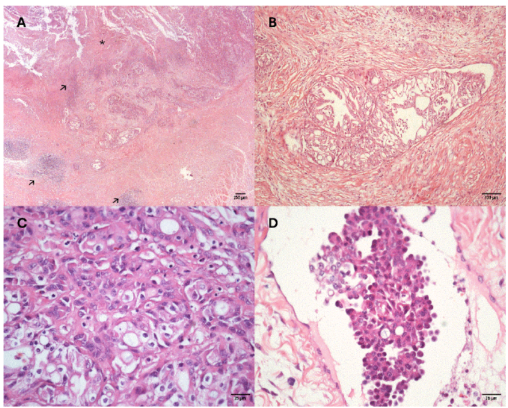
Histopathological examination revealed a multilobulated, non-encapsulated, irregular epithelial neoplasm infiltrating the vulvar submucosa (Figure 1a), with moderate cellularity and a mixed arrangement, comprising tubular structures (Figure 1b) and solid areas (nests), supported by a moderate desmoplastic reaction. Neoplastic cells were polygonal to round, moderately sized, with eosinophilic cytoplasm, poorly defined borders, central to paracentral nuclei with coarse chromatin, and 0-1 small, conspicuous, basophilic nucleoli. Occasionally, neoplastic cells exhibited marked cytoplasmic vacuolization with peripheral nuclei, giving a signet ring appearance (Figure 1c). Twenty-two mitotic figures were identified in 2.37 mm², along with marked anisocytosis, anisokaryosis, and pleomorphism, rare binucleations, and multiple foci of lymphovascular invasion characterized by emboli of solid nests of neoplastic cells (Figure 1d).
Adjacent to the neoplasm, there were focally extensive areas of necrosis and ulceration of the vulvar mucosa, marked hemorrhage, and a dense mixed inflammatory infiltrate composed of abundant neutrophils and, to a lesser extent, lymphocytes, histiocytes, and plasma cells associated with numerous coccoid bacterial colonies. In the vulvar wall, the inflammatory response was of similar nature, nodular to diffuse, and moderate in intensity.
Histological sections on marked slides (4 µm) were subjected to immunohistochemical (IHC) processing to evaluate the expression of pan-cytokeratin, S-100, neuron-specific enolase (NSE), and uroplakin III (UPIII). For IHC reactions, primary antibodies were applied under conditions described in Table 1 and incubated overnight in combination with the Novolink Polymer Detection System (Leica). Immunoreactivity was visualized using 3,3'-diaminobenzidine, with slides counterstained in Harris hematoxylin. Positive and negative controls (omission of the primary antibody) were included in all experiments.

Bitch, canine vestibular carcinoma. A) Neoplastic epithelial cells associated with mucosal ulceration (*) and marked inflammation (arrows) in the submucosa and muscular layer of the vulvar region, 2x, H&E. B) Neoplastic epithelial cells organized in irregular tubules, 10x, H&E. C) Polygonal to round neoplastic cells in solid areas, exhibiting variable cytoplasmic vacuolization, occasionally with signet ring appearance, 40x, H&E. D) Solid neoplastic emboli, 40x, H&E.
Immunohistochemical results showed strong and diffuse membranous and cytoplasmic labeling for pan-cytokeratin in the neoplastic cells (Figure 2a); heterogeneous, moderate cytoplasmic expression of NSE (Figure 2b); negative expression of S-100 (Figure 2c); and membranous and cytoplasmic labeling for UPIII, ranging from mild to strong, heterogeneous, being more prominent at the plasma membrane of cells with greater cytoplasmic vacuolization (Figure 2d).
Based on the anatomical location, macroscopic and microscopic features, and immunohistochemical expression profile, a morphological diagnosis of CVC of urothelial origin was established.
Antibodies used for immunohistochemical characterization of canine vestibular carcinoma, organized according to clonality, dilution, and antigen retrieval methods

Bitch, canine vestibular carcinoma. Immunohistochemistry. A) Strong, diffuse cytoplasmic labeling for pan-cytokeratin in neoplastic cells, 10x. Inset: positive labeling in a neoplastic embolus, 40x. B) Heterogeneous, mild to moderate labeling for NSE in neoplastic tissue, 10x. Inset: mild positive labeling in a neoplastic embolus, 40x. C) Negative labeling for S-100, 10×. Inset: negative labeling in a neoplastic embolus, 40x. D) Heterogeneous labeling with isolated cells showing strong membranous immunoreactivity for uroplakin III, 40x. Inset: positive labeling in a neoplastic embolus, 40x. Chromogen: 3,3'-diaminobenzidine; counterstained with Harris hematoxylin.
DISCUSSION
Clinical signs and lesions of neoplasms affecting the vagina, vestibule, vulva, and clitoris in bitches can vary depending on tumor type, grade, and stage (Brodey and Roszel, 1967; Tatcher and Bradley, 1983; Magne et al., 1985; McEntee, 1990; Mathews, 2001; Agnew and MacLachlan, 2016). The combination of clinical and laboratory findings and the unfavorable progression of the vulvovaginal neoplasm precluded a clinical diagnosis and determination of the primary site of CVC, which can exhibit infiltrative and aggressive behavior (Brodey and Roszel, 1967; Tatcher and Bradley, 1983; Magne et al., 1985; McEntee, 1990; Agnew and MacLachlan, 2016), as observed here.
Although vulvovaginal cytology has diagnostic value for urothelial-origin CVC (Magne et al., 1985), marked inflammation and ulceration prevented lesion identification by this method. Urinary dysfunction can be explained by tumor location, while systemic anemia and neutrophilic leukocytosis likely resulted from severe hemorrhage and extensive suppurative inflammation and necrosis in the vulva, secondary to neoplastic proliferation (Brodey and Roszel, 1967; Tatcher and Bradley, 1983; Magne et al., 1985; McEntee, 1990; Agnew and MacLachlan, 2016).
Microscopic findings were consistent with other reports of urothelial-origin CVC in bitches (Magne et al., 1985; McEntee, 1990). Although no macroscopic metastases were observed, features such as disorganized growth, loss of polarity, marked pleomorphism, numerous mitoses, and multiple foci of lymphovascular invasion - frequently reported in high-grade urothelial tumors (Meuten and Meuten, 2016) - likely explain the rapid growth and aggressive behavior in the vulvar region.
Carcinomas of the vagina, vestibule, vulva, and clitoris may arise from different epithelia: stratified squamous mucosal epithelium; keratinized stratified squamous epithelium of the vulvar lips; vestibular submucosal glands (Bartholin’s glands); or apocrine glands at the clitoral base (Magne et al., 1985; McEntee, 1990; Agnew and MacLachlan, 2016; Verin et al., 2018). These regions may also be secondarily affected by neoplasms originating from the urethral lining, which in bitches consists of urothelium in the proximal third and non-keratinized squamous epithelium in the distal two-thirds (Tarvin et al., 1978; Cullen et al., 1981; Magne et al., 1985; McEntee, 1990; Bacha et al., 2012; Meuten and Meuten, 2016; Liebich, 2019; Agnew and MacLachlan, 2016). Therefore, in poorly differentiated CVCs lacking features of other epithelial tumors, urothelial origin should be considered, as in this case.
Histopathology revealed signet ring-like cells (Melamed-Wolinska bodies), strongly suggestive of urothelial differentiation (Tarvin et al., 1978; Magne et al., 1985; McEntee, 1990; Agnew and MacLachlan, 2016; Meuten and Meuten, 2016). Because of poor differentiation and pleomorphism, immunohistochemistry was performed to confirm tumor histogenesis.
Pan-cytokeratin labeling confirmed the epithelial nature of the tumor, consistent with other epithelial neoplasms, including urothelial tumors, adenocarcinoma, and CCC (Neihaus et al., 2010; Agnew and MacLachlan, 2016; Verin et al., 2018; Rout et al., 2016; Moss et al., 2023; Ramos-Vara and Borst, 2016). Therefore, pan-cytokeratin alone could not determine the specific epithelial origin.
Considering lesion location, involvement of external genital structures, and histopathological features of a poorly differentiated epithelial tumor, differential diagnoses included urothelial-origin CVC, adenocarcinoma, and CCC. Vestibular gland adenocarcinoma, a rare tumor described in cows, arises from cuboidal to columnar cells with microvilli and apical deposits arranged in tubular patterns (Tanimoto et al., 1994; Nascimento and Santos, 2021), features not observed here, ruling out adenocarcinoma.
Microscopically, this CVC shared some features with CCC, which is multilobulated, poorly defined, infiltrative, and potentially metastatic, composed of round to oval cells with occasional cytoplasmic vacuolization arranged in solid, tubular, or rosette patterns (Verin et al., 2018). CCC likely arises from apocrine glands at the clitoral base and may express neuroendocrine markers, similar to canine anal sac apocrine carcinomas (Neihaus et al., 2010; Suzuki et al., 2013; Verin et al., 2018; Rout et al., 2016; McAloney et al., 2021; Moss et al., 2023).
Moderate NSE expression in this case resembled CCC, where NSE labeling ranges from mild to moderate, in 10-90% of cells (Verin et al., 2018). Although NSE is typically found in neural and neuroendocrine cells, its expression is not exclusive to these cell types (Ramos-Vara and Borst, 2016). In this case, NSE expression may reflect malignant transformation of urothelial cells, as reported in canine and human urothelial carcinomas (Soh et al., 2012; Fernández-Aceñero et al., 2011).
No other neuroendocrine morphological or immunophenotypic features were observed. The distribution of diffuse neuroendocrine cells in dogs remains poorly understood (Champion et al., 2025). NSE expression has been reported in CCC with neuroendocrine features, ureter and bladder neuroendocrine tumors in dogs, and human vulvar Merkel cell carcinoma (Hierro et al., 2000). Negative S-100 labeling, as in CCC (Verin et al., 2018), helped exclude a neuroectodermal origin. Additional studies using neuroendocrine markers (NSE, S-100, chromogranin, synaptophysin) could clarify diffuse neuroendocrine cell distribution in the canine genitourinary tract and improve understanding of rare genitourinary tumors.
UPIII is naturally expressed on the apical membrane of urothelial cells (Ramos-Vara et al., 2003; Sledge et al., 2015), with low sensitivity but high specificity for canine urothelial cells (Meuten and Meuten, 2016; Ramos-Vara and Borst, 2016). UPIII is detected in over 90% of canine urothelial carcinomas and is used to confirm urothelial origin in primary or unknown-site tumors, even when only 5-10% of neoplastic cells express it (Meuten and Meuten, 2016; Ramos-Vara and Borst, 2016).
In high-grade urothelial carcinomas, UPIII labeling is often patchy, with partial or absent expression due to loss of differentiation and aggressive tumor behavior, as seen here (Sledge et al., 2015; Meuten and Meuten, 2016; Ramos-Vara and Borst, 2016). Therefore, UPIII was the key marker confirming the urothelial origin of CVC.
CONCLUSION
Definitive diagnosis of urothelial-origin CVC was achieved through correlation of histomorphological findings with immunohistochemistry results, which proved essential for excluding differential diagnoses and confirming tumor histogenesis via UPIII expression. These results highlight the importance of thorough pathological investigation using complementary markers, particularly in poorly differentiated or anaplastic neoplasms of the canine urogenital tract.
REFERENCES
- AGNEW, D.W.; MACLACHLAN, N.J. Tumors of the genital systems. In: MEUTEN, D.J. (Ed.). Tumors in domestic animals 5.ed. Ames: Wiley-Blackwell, 2017. p.689-722.
- BACHA JR, W.J.; BACHA, L.M. Color atlas of veterinary histology Hoboken: John Wiley & Sons, 2012.
- BRODEY, R.S.; ROSZEl, J.F. Neoplasms of the canine uterus, vagina, and vulva: a clinicopathologic survey of 90 cases. J. Am. Vet. Med. Assoc., v.151, p.1294-1307, 1967.
- CHAMPION, C.P.; MILLER, A.D.; PARRY, S.; DEMETER, E.A. Immunohistochemical and histomorphologic characterization of canine neoplasms of the disseminated neuroendocrine system. Vet. Pathol., v.62, p.490-501, 2025.
- CULLEN, W.C.; FLETCHER, T.F.; BRADLEY, W.E. Histology of the canine urethra. 1. Morphometry of the female urethra. Anat. Rec., v.199, p.177-186, 1981.
- FERNÁNDEZ-ACEÑERO, M.J.; CÓRDOVA, S.; MANZARBEITIA, F.; MEDINA, C. Immunohistochemical profile of urothelial and small cell carcinomas of the bladder. Pathol. Oncol. Res., v.17, p.519-523, 2011.
- FOSTER, R.A.; PREMANANDAN, C. Female reproductive system and mammae. In: ZACHARY, J.F. (Ed.). Pathologic basis of veterinary disease 7.ed. San Diego: Elsevier, 2022. p.1263-1307.
- HIERRO, I.; BLANES, A.; MATILLA, A. et al Merkel cell (neuroendocrine) carcinoma of the vulva: a case report with immunohistochemical and ultrastructural findings and review of the literature. Pathol. Res. Pract., v.196, p.503-509, 2000.
- KYDD, D.M.; BURNIE, A.G. Vaginal neoplasia in the bitch: a review of forty clinical cases. J. Small Anim. Pract., v.27, p.255-263, 1986.
- LIEBICH, H.G. (Ed.). Veterinary histology of domestic mammals and birds: textbook and colour atlas 5.ed. Sheffield: 5m Books, 2019.
- MAGNE, M.L.; HOOPES, P.J.; KAINER, R.A. et al Urinary tract carcinomas involving the canine vagina and vestibule. J. Am. Anim. Hosp. Assoc v.21, p.767-772, 1985.
- MATHEWS, K.A. Surgical treatment of vaginal tumors in the dog. Compend. Contin. Educ. Pract. Vet., v.23, p.330-341, 2001.
- McALONEY, C.A.; TAIKOWSKI, K.L.; CRONIN, J.P. et al Pathology in practice. J. Am. Vet. Med. Assoc., v.259, p.1-3, 2021.
- McENTEE, K. Cervix, vagina, and vulva. In: ______. (Ed.). Reproductive pathology of domestic animals San Diego: Academic Press, 1990. p.191-223.
- MEUTEN, D.J.; MEUTEN, T.L.K. Tumors of the urinary system. In: MEUTEN, D.J. (Ed.). Tumors in domestic animals 5.ed. Ames: Wiley-Blackwell, 2017. p.632-688.
- MITCHELL, K.E.; BURGESS, DM.M.; CARRIGAN, M.J. Clitoral adenocarcinoma and hypercalcaemia in a dog. Aust. Vet. Pract., v.42, p.279-282, 2012.
- MOSS, C.; SUGAI, N.; PERSONS, R. et al Case report: clitoral adenocarcinoma in a mixed-breed female dog. Front. Vet. Sci., v.10, p.1264538, 2023.
- NASCIMENTO, E.F.; SANTOS, R.L. Patologia da cérvix, da vagina e da vulva. In: _____. Eds.). Patologia da reprodução dos animais domésticos 4.ed. Rio de Janeiro: Guanabara Koogan, 2021. p.77-81.
- NEIHAUS, S.A.; WINTER, J.E.; GORING, R.L. et al Primary clitoral adenocarcinoma with secondary hypercalcemia of malignancy in a dog. J. Am. Anim. Hosp. Assoc., v.46, p.193-196, 2010.
- PIECH, T.L.; CHU, S.; BOZYNSKI, C.C.; ROYAL, A.B. Pathology in practice. J. Am. Vet. Med. Assoc., v.254, p.1167-1170, 2019.
- RAMOS-VARA, J.A.; BORST, L.B. Immunohistochemistry: fundamentals and Applications in Oncology In: MEUTEN, D.J. (Ed.). Tumors in domestic animals 5.ed. Ames: Wiley-Blackwell, 2017. p.44-87.
- RAMOS-VARA, J.A.; MILLER, M.A.; BOUCHER, M. et al. Immunohistochemical detection of uroplakin 3, cytokeratin 7, and cytokeratin 20 in canine urothelial tumors. Vet. Pathol, v.40, p.55-62, 2003.
- ROUT, E.D.; HOON-HANKS, L.L.; GUSTAFSON, T.L. et al What is your diagnosis? Clitoral mass in a dog. Vet. Clin. Pathol., v.45, p.197-198, 2016.
- SCHLAFER, D.H.; FOSTER, R.A. Female genital system. In: MAXIE, M.G. (Ed.). Jubb, Kennedy, and Palmer's pathology of domestic animals 6.ed. St. Louis: Elsevier, 2016. p.358-464.
- SLEDGE, D.G.; PATRICK, D.J.; FITZGERALD, S.D. et al Differences in expression of uroplakin 3, cytokeratin 7, and cyclooxygenase-2 in canine proliferative urothelial lesions of the urinary bladder. Vet. Pathol., v.52, p.74-82, 2015.
- SOH, M.; DUNLEVY, J.R.; GARRETT, S.H. et al Increased neuron specific enolase expression by urothelial cells exposed to or malignantly transformed by exposure to Cd2+ or As3+. Toxicol. Lett., v.212, p.66-74, 2012.
- SUZUKI, K.; MORITA, R.; HOJO, Y. et al Immunohistochemical characterization of neuroendocrine differentiation of canine anal sac glandular tumours. J. Comp. Pathol., v.149, p.199-207, 2013.
- TANIMOTO, T.; FUKUNAGA, K.; OHTSUKI, Y. Adenocarcinoma of the major vestibular gland in a cow. Vet. Pathol, v.31, p.246-247, 1994.
- TARVIN, G.; PATNAIK, A.; GREENE, R. Primary urethral tumors in dogs. J. Am. Vet. Med. Assoc., v.172, p.931-933, 1978.
- TATCHER, C.D.; BRADLEY, R.L. Vaginal and vulvar tumors in the dog: a retrospective study. J. Am. Vet. Med. Assoc., v.183, p.690-692, 1983.
- VERIN, R.; CIAN, F.; STEWART, J. et al Canine clitoral carcinoma: a clinical, cytologic, histopathologic, immunohistochemical, and ultrastructural study. Vet. Pathol., v.55, p.501-509, 2018.
- ZAMBELLI, D.; VALENTINI, S.; BALLOTTA, G.; CUNTO, M. Partial vaginectomy, complete vaginectomy, partial vestibule-vaginectomy, vulvo-vestibule-vaginectomy and vulvo-vestibulectomy: different surgical procedure in order to better approach vaginal diseases. Animals, v.12, p.196, 2022.
The research data are available within the article itself.