 Clinical, haematological, and microbiological profiles of respiratory diseases in the Brejo region of Paraíba
ABSTRACT
The objective was performed to analyze clinical, hematological, and microbiological profiles through culture of secretions obtained by tracheal lavage in horses with respiratory infections. The study involved 19 male and female horses aged between 3 months and 10 years presenting signs of respiratory disease. They underwent anamnesis, clinical examination, and laboratory tests. For tracheal lavage, sedation with 10% xylazine (1.1mg/kg intravenously) was performed, followed by trichotomy on the ventral edge of the mid-distal region of the neck, anesthetic button using 2% lidocaine without a vasoconstrictor, and surgical antisepsis. The trachea was accessed using 14G catheter, through which n.4 urethral probe was introduced and attached to 20mL syringe containing lactated Ringer’s solution. Immediately the solution was aspirated along with the tracheal secretion. The samples were refrigerated and transported to the laboratory in isothermal boxes for microbiological culture. The microbiological culture the following pathogens were identified: Rhodococcus in 8/19 (42.1%), Aspergillus in 6/19 (31.6%), and Klebsiella, Streptococcus, and Escherichia coli/Proteus in 1/19 (5.3%) each; 2/19 (10.5%) samples were negative. The tracheal lavage is an effective method for collecting tracheal secretions and the association of clinical and laboratory tests provide rapid, early, and conclusive diagnosis in animals with suspected pulmonary disease.
Keywords:
respiratory diseases; tracheal wash; fungi; pneumonia; bacteria
RESUMO
O objetivo deste estudo foi analisar os perfis clínico, hematológico e microbiológico por meio da cultura de secreções obtidas por lavagem traqueal em equinos com infecções respiratórias. Foram avaliados 19 equinos, machos e fêmeas, com idade entre três meses e 10 anos, que apresentavam sinais de doença respiratória. Eles foram submetidos a exames, clínicos, laboratoriais e de anamnese. Para a lavagem traqueal, foi realizada sedação com xilazina 10% (1,1mg/kg por via intravenosa), seguida de tricotomia na borda ventral da região médio-distal do pescoço, botão anestésico com lidocaína 2% sem vasoconstritor e antissepsia cirúrgica. A traqueia foi acessada por cateter 14G, por meio do qual foi introduzida uma sonda uretral nº4, acoplada a uma seringa de 20mL contendo solução de Ringer com lactato. Imediatamente a solução foi aspirada juntamente com a secreção traqueal. As amostras foram refrigeradas e transportadas ao laboratório em caixas isotérmicas para a realização de cultura microbiológica, identificando-se os seguintes patógenos: Rhodococcus em 8/19 (42,1%), Aspergillus em 6/19 (31,6%) e Klebsiella, Streptococcus e Escherichia coli/Proteus em 1/19 (5,3%) cada; 2/19 (10,5%) amostras foram negativas. O lavado traqueal é um método eficaz para coleta de secreções traqueais, e a associação de exames clínicos e laboratoriais proporciona diagnóstico rápido, precoce e conclusivo em animais com suspeita de doença pulmonar.
Palavras-chave:
doenças respiratórias; lavado traqueal; fungos; pneumonia; bactérias
INTRODUCTION
Respiratory pathologies continue to be a major problem in equine medicine. They are common causes of mortality in foals up to 12 months of age and an important cause of death in the equine population in general (Rahman et al., 2022). The most common diseases involving the lower respiratory tract are inflammatory respiratory disease, recurrent airway obstruction, exercise-induced pulmonary hemorrhage, and pneumonia (Niedźwiedź et al., 2016; Crispe et al., 2018; Woodrow et al., 2023). The development of respiratory diseases is complex and multifactorial, and it most often occurs with non-maturation or decreased immune function (Bishop et al., 2023). This immunodeficiency can lead to the development of infectious agents such as viruses, bacteria, parasites, protozoa, and fungi (Wilkins and Lascola, 2015), all of which are important for the initiation or progression of clinical disease.
Viral respiratory diseases in horses are important causes of death. Some viruses cause acute response, resulting in clinical signs like fever and systemic illness, leading to impaired pulmonary function (Frippiati et al., 2025). On the other side, other viruses may cause subclinical respiratory infections (El-Hage et al., 2021). Fungal infections in horses are uncommon but potentially fatal. Depending on the geographic area, the prevalence can be highly variable, and such infections have been reported to occur at all levels of the equine respiratory tract (Stewart and Cuming, 2015). Bacterial agents have been frequently isolated from horses with respiratory disease. Commensal bacteria of the nasopharynx are the most common etiological agents, followed by enteric microorganisms (Hallowell et al., 2024). Several authors have reported significant differences in bacterial populations and antimicrobial susceptibility patterns between regions, and even between outpatient practices and referral hospitals in similar geographic locations (Potier and Durham, 2020). When not accurately determined, this can lead to the development of resistance mechanisms in infectious agents, which is a growing problem in both veterinary and human medicine.
Thus, this work was carried out to analyze clinical, hematological, and microbiological profiles through the culture of secretions obtained by tracheal lavage of horses with respiratory infections, with the goal of achieving accurate and timely diagnosis.
ETHICAL ASPECTS
The research was submitted to the Ethics Committee on Animal Use of the federal University of Paraíba, and approved under the number 8233280420.
MATERIALS AND METHODS
This study was conducted at a private clinic specializing in equine care, located in the city of Lagoa Seca, Paraíba, Brazil. Nineteen animals, ranging in age from two months to 10 years and of various sexes and breeds, were selected for analysis. All animals exhibited clinical signs of respiratory disease, including coughing, nasal discharge, abnormal pulmonary and tracheal auscultation, hyperthermia, apathy, reluctance to exercise, and/or reduced appetite. Each animal underwent anamnesis and a clinical examination, which included the measurement of heart rate (HR), respiratory rate (RR), rectal temperature, capillary refill time, mucous membrane coloration, and intestinal motility.
The tracheal fluid samples were collected according to the methodology used by Helson and Arroyo (2015). During tracheal lavage, the animals were sedated with 10% xylazine (1.1 mg/kg intravenously), followed by a wide trichotomy on the ventral edge of the mid-distal region of the neck, application of a local anesthetic button using 2% lidocaine without a vasoconstrictor, and surgical antisepsis. After donning appropriate surgical attire, the surgeon accessed the trachea using a 14G catheter. Once access was established, the catheter mandrel was removed, leaving only the Jelco in place, through which a No. 4 urethral catheter was introduced. A 20mL syringe containing lactated Ringer’s solution was attached to the urethral catheter. When the solution was injected into the trachea, tracheal lavage was performed. Immediately after infusion, the fluid was quickly aspirated along with the secretions present in the trachea.
Following collection, the catheter and probe were removed from the trachea, and a repellent spray was applied to the puncture site. The samples were kept refrigerated in the same syringes in which they were collected and transported in isothermal boxes for culture.
For bacterial culture, the material was seeded on 5% sheep blood agar (Merck), and the Petri dish was inoculated using the streak-stripping technique to allow colonies to grow in isolation, facilitating pathogen identification. The plates were incubated at 37°C for 24-48 hours. After the growth period, the colonies were subjected to phenotypic and biochemical tests, including catalase, oxidase, nitrate reduction, urease production, ortho-nitrophenyl-β-D-galactopyranoside (ONPG), and CAMP tests to investigate the ‘equi factor’, considered one of the differential tests for the identification of Rhodococcus equi.
Fungal culture was performed by fragmenting the samples with a sterile scalpel. The fragments were then seeded in duplicate on Petri dishes containing Sabouraud-dextrose agar supplemented with chloramphenicol (30mg/100mL). The plates were incubated in microbiological incubators at 25°C and 37°C for 7 days, with fungal growth assessed from the second day onward. Identification was based on both macro- and micro-morphological characteristics. The macroscopic evaluation included colony size, edge characteristics, texture, relief, and pigmentation, while the microscopic evaluation was carried out through direct examination of an aliquot of the colony stained with lactophenol cotton blue. The colony was subsequently subcultured in duplicate.
Analysis of variance was performed for each variable using the F test. This was followed by Tukey’s test (a mean comparison test) to identify differences between them at the 5% level. To enable the tests, the analysis was first divided into a general descriptive statistical assessment of all qualitative variables-namely, culture, sex, age, ocular mucosa, and oral mucosa-generating their absolute frequencies, relative frequencies, and percentages, along with their respective graphs. A descriptive statistical analysis was then carried out for all quantitative variables, including fibrinogen, leucocytes, red blood cells, hemoglobin, hematocrit, platelets, weight, RR, HR, and temperature. This analysis provided information on the mean, standard deviation, minimum and maximum values, median, interquartile range, Pearson’s correlation coefficient, and coefficient of variation.
Graphs were plotted based on correlations between the variables, using p= 0.05 as a reference to assess significance. All p values of <0.05 were considered significant, while p values of >0.05 were considered non-significant.
RESULTS
The results obtained in relation to sex showed that 11/19 (57.9%) animals were female and 8/19 (42.1%) were male. Additionally, 17/19 (89.4%) were foals and 2/19 (10.6%) were adults. The age of the animals ranged from 2 months to 10 years.
In the evaluation of the oral mucosa, 2/19 (10.5%) animals had congested mucosa, while 17/19 (89.5%) had pink mucosa. Regarding the ocular mucosa, 16/19 (84.2%) animals had pink mucosa, 1/19 (5.3%) had pink mucosa with petechiae, and 2/19 (10.5%) had congested mucosa.
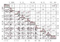
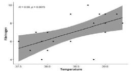
Figure 1 shows the correlation between all clinical and hematological variables. A positive relationship was observed between fibrinogen and leucocytes, although there was no significant difference, and between fibrinogen and temperature, where a significant difference was observed.
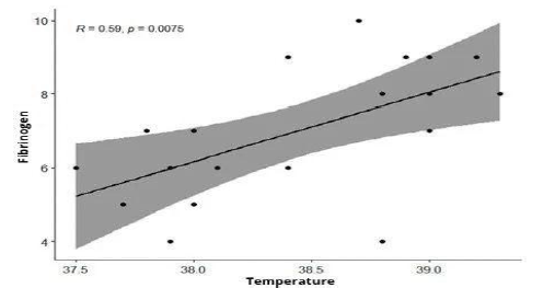
Specifically, as temperature increased, the fibrinogen level also increased, showing a correlation of 59% with a p-value of 0.0075 (Fig. 2).
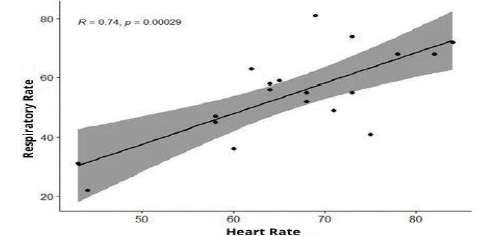
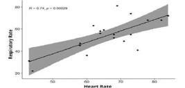
The clinical parameters HR and RR showed a correlation of 74%, and the p-value of 0.00029 indicated a significant correlation (Fig. 3).
Regarding fibrinogen levels (Table 1), 2/19 (10.5%) animals had the maximum value within the reference range (2-4g/L).
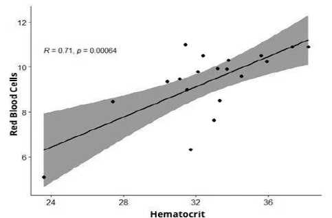
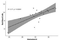
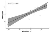
A total of 2/19 (10.5%) had a level of 5 g/L, 4/19 (21.1%) had a level of 6 g/L, 3/19 (15.8%) had a level of 7 g/L, 3/19 (15.8%) had a level of 8 g/L, 4/19 (21.1%) had a level of 9g/L, and 1/19(5.3%) had a level of 1 g/L. In the red series (red blood cells, hemoglobin, and hematocrit), positive correlations with significant differences were observed across all three variables (Figs. 4 and 5).
In the microbiological culture of secretions obtained via tracheal lavage, the following results were obtained (Table 2): R. equi in 8/19 (42.1%) animals, Aspergillus spp. in 6/19 (31.6%), Klebsiella pneumoniae in 1/19 (5.3%), Streptococcus equi in 1/19 (5.3%), and Escherichia coli/ Proteus in 1/19 (5.3%). The culture was negative in 2/19 (10.5%) animals.
Microorganisms found in microbiological cultures of secretions obtained through tracheal lavage
DISCUSSION
In respiratory infections in horses, foals and young horses are more susceptible to certain microbiological agents such as fungi and bacteria. This is most often due to the fact that these animals still have an immune system in the process of consolidation, which is not yet fully prepared to respond to opportunistic agents. Among young and adult animals, the greatest susceptibility is observed in young, athletic, and older horses. In a study on the risk factors associated with the survival of horses with septic pleuropneumonia, Arroyo et al. (2017) demonstrated that the average age was 2.5 years. Depending on the age group, animals may be more susceptible to specific diseases such as rhodococcosis, which is a common cause of pneumonia in foals between three weeks and six months of age on endemic properties (Reuss and Cohen, 2015).
The results observed in most of the animals during the evaluation of the mucous membranes indicated that the animals did not present systemic impairment related to the diseases. The evaluation of mucous membrane color is crucial during the clinical examination and is one of the parameters monitored in the assessment and follow-up of disease, alongside complementary tests such as the blood count. This is one of the most auxiliary tests requested because the results can provide information about changes in the animal’s condition, bone marrow functions, and clues of yields or even diagnosis as to the presence of underlying disease (Satué et al., 2023). Furthermore, the blood count is effective for evaluating development and performance of horses that can demonstrate the effectiveness of the conduct performed.
The correlation between fibrinogen and temperature is important in indicating certain diseases because fibrinogen is an acute-phase protein involved in inflammation in most species. It activates the immune system, enhances phagocytosis, and aids in the clearance of inflammatory by-products. Although there are other more sensitive markers, in horses, fibrinogen has remained the mainstay of blood analysis for inflammation in horses and other large animal species, largely because of the ease and minimal expense of testing (Long and Nolen-Walston, 2020). In a study conducted in Filand with newly weaned horses, Junkkari et al. (2017) observed higher plasm fibrinogen concentration in all animals that demonstrated clinical respiratory signs (17/60), include higher temperature. The present study demonstrated a positive correlation with a significant difference between fibrinogen and temperature, highlighting the importance of measuring this parameter in respiratory diseases.
Correlating fibrinogen with the red blood cell series (erythrocytes, hemoglobin, and hematocrit), the results were significant, underscoring its importance in clinical diagnosis. In the present study, the values obtained for red blood cells were within the reference range for the erythrocyte series in horses, showing no alterations. However, hemoglobin levels were below the reference range in five animals, and hematocrit values were also below the reference in four animals. Nevertheless, these indices in the animals of the present study-most of which were young-did not exceed the reference values; by contrast, their values remained well below the maximum acceptable range.
Although the correlation analysis did not demonstrate a significant result between fibrinogen and leucocytes, monitoring this relationship is highly relevant in clinical practice because it serves as a good indicator of acute-phase inflammation, showing an increase in the initial stage of disease Passamonti et al. (2015) noted that measuring plasma fibrinogen provides an early response during the initial phase of disease, or even in subclinical infections, because fibrinogen levels typically rise within 24 to 72 hours and return to baseline concentrations within 1 to 2 weeks. Infectious and inflammatory processes-even when subtle-tend to alter the leucocyte count, leading to leukocytosis, which aids in diagnosis and may corroborate the findings related to fibrinogen elevation. Other than that, leukocytosis may occur in some stressful situations as transportation, overcrowding, and introduction of new animals (Salco et al., 2020). This results in an increased neutrophil:lymphocyte ratio due to neutrophilia and lymphopenia, and his phenomenon can occur even when the overall degree of leukocytosis remains unchanged (Popescu and Diugan, 2017). The lack of statistical significance in this study may be attributed to the differing timing of increase and decrease between the two variables.
The results obtained in this study demonstrated a positive response in relation to the diagnosis of respiratory diseases caused by microbiological infections (fungi and bacteria). Given the importance of early and accurate diagnosis, tracheal lavage and culture were employed to identify the etiological agent involved in each respiratory disorder. Considering the importance of implementing an early diagnosis and appropriate therapeutic plan, the tracheal lavage and culture were used to determine the etiological agent involved in each respiratory disorder. This technique allows for a qualitative evaluation of the tracheal microbiota in domestic animals. As an accessible and minimally invasive method-typically requiring only restraint of the snout for physical control and allowing for repetition without compromising the animal’s health-it facilitates a more concise and targeted diagnosis. Another important point is that its use has been shown to significantly improve the diagnosis of certain diseases, especially when combined with other complementary examinations, such as endoscopy (Cascardo et al., 2022).
Horses that test positive for bacteria in tracheal lavage cultures are at high risk of also having a viral or fungal infection, which may have led to secondary bacterial infections (Martineau et al., 2023). Thus, in the present study, a variety of microbiological agents were identified in the culture results; however, most animals were diagnosed with R. equi and Aspergillus spp.
Rhodococcus equi infection has a significant detrimental impact on the equine breeding industry, with a high incidence and affecting foals in all parts of the world. A study conducted by Hagist (2016) on a farm in northern Germany demonstrated the presence of R. equi in 54% (54/100) of cases, cultured from samples obtained via tracheal lavage of foals that showed ultrasonographic signs of pneumonia. In the same study, the bacterium was detected in all 24 foals that died with ultrasonographic lesions.
Regarding the results observed in the cultivation of Aspergillus, Dobiáš et al. (2023) reported that clinicians should exercise caution when attributing significance to the presence of fungal elements-whether free or in large quantities-in tracheal aspirates because they are commonly identified in healthy horses. A case report on pulmonary aspergillosis in horses suggested that infection may have occurred following the inhalation of spores present in moldy bedding and hay, with the hypothesis that the development of the clinical condition was facilitated by a pre-existing issue such as asthma (Melo et al., 2024). In the database from the study conducted by Dobiáš et al. (2023), it was shown that the trachea of horses can be heavily colonized by Aspergillus spp., particularly in individuals with severe asthma. In such cases, these fungi can be detected through microbiological cultures obtained via the tracheal lavage technique, corroborating the findings of the present study.
The results obtained for Pseudomonas spp., E. coli, S. equi, and K. pneumoniae corroborate the findings of other authors who used the same technique employed in the present study, such as Rahman et al. (2022), in a retrospective study of horses with pneumonia in California, who observed these same microorganisms. In studies conducted by different authors, the microorganisms identified may vary depending on the risk factors and epidemiological conditions of the locations where the animals are kept. However, it is important to highlight that those identified in the present study should be considered as differential diagnoses in horses with respiratory diseases because they have the potential to be multidrug-resistant. Care should always be taken to prevent environmental contamination and hospital-acquired infections.
CONCLUSION
Tracheal lavage has proven to be effective in the diagnosis of respiratory diseases because it is a minimally invasive, simple, fast, and low-cost technique. It can be considered the gold standard for such cases and is increasingly used in hospitals, veterinary clinics, and field care. When combined with microbiological culture and blood count, this technique has proven essential for achieving an accurate diagnosis. The present study revealed that the microorganisms identified in horses showing respiratory signs were R. equi, Aspergillus spp., K. pneumoniae, E. coli, and S. equi.
ACKNOWLEDGMENTS
This study was supported by the Equestre: clinic, surgery, and reproduction; and University Veterinary Hospital of the Federal University of Paraíba (UFPB), Areia/PB.
REFERENCES
- ARROYO M.G.; SLOVIS, N.M.; MOORE, G.E.; TAYLOR, S.D. Factors associated with survival in 97 horses with septic pleuropneumonia. J. Vet. Intern. Med, v.31, p.894-900, 2017.
- BISHOP R.C.; MIGLIORISI, A.; HOLMES, J.R. et al Microbial populations vary between the upper and lower respiratory tract, but not within biogeographic regions of the lung of healthy horses. J. Equine Vet. Sci, v.140, p.105141, 2023.
- CASCARDO B.; BERNARDES, C.; SOUZA, G.N. et al Diagnostic value of tracheal wash cytology for monitoring exercise-induced pulmonary haemorrhage in thoroughbred racehorses. J. Equine Vet. Sci, v.118, p.104-128, 2022.
- CRISPE, EJ.; SECOMBE, C.J.; PERERA, D.I. et al Exercise-induced pulmonary haemorrhage in Thoroughbred racehorses: a longitudinal study. Equine Vet. J, v.51, p.45-51, 2018.
- DOBIÁŠ R. et al Diagnóstico de aspergilose em cavalos. J. Fungi. 9(2):161, 2023.
- EL-HAGE, C.; MEKURIA, Z.; DYNON, K. et al Association of equine herpesvirus 5 with mild respiratory disease in a survey of EHV1, -2, -4 and -5 in 407 Australian horses. Animals, v.11, n.12, p.3418, 2021.
- FRIPPIATI T.; VAN DEN WOLLENBERG, L.; VAN ERCK-WESTERGREN, E. et al Respiratory viruses affecting health and performance in equine athletes. Virology, v.603, p.1-16, 2025.
- HAGIST, C. Genotypisierung von rhodococcus equi stämmen aus Deutschland, isoliert bei fohlen und anderen tierarten 2016. 134f. Thesis (Postgraduate course in Veterinary Medicine) - Veterinary School of Hanover.
- HALLOWELL, K.L.; HEPWORTH-WARREN, K.L.; DEMBEK, K. An updated description of bacterial pneumonia in adult horses and factors associated with death. J. Vet. Intern. Med, v.38, p.2766-2775, 2024.
- HELSON, J.D.V.M.; ARROYO, L.G. Respiratory disease in the horse diagnostic approaches in the horses. Vet. Clin. North Am. Equine Pract, v.31, p.307-336, 2015.
- JUNKKARI, R.; SIMOJOKI, H.; HEISKANEN, M.L. et al A comparison of unheated loose housing with stables on the respiratory health of weaned-foals in cold winter conditions: an observational field-study. Acta Vet. Scand, v.59, p.73, 2017.
- LONG, A.; NOLEN-WALSTON, R. Equine inflammatory markers in the twenty-first century: a focus on serum amyloid A. Vet. Clin. North Am. Equine Pract, v.36, p.147-160, 2020.
- MARTINEAU, M.; CASTAGNET, S.; KOKABI, E. et al Detection of Mycoplasma spp. in horses with respiratory disorders. Equine Vet. J, v.55, p.747-754, 2023.
- MELO, U.P.; FERREIRA, C.; BARRETO, S.W.M. Pulmonary aspergillosis in a horse: a case report. Braz. J. Vet. Med, v.46, p.1-8, 2024.
- NIEDŹWIEDŹ, A.; BOROWICZ, H.; KUBIAK, K. et al Evaluation of serum cytokine levels in recurrent airway obstruction. Pol. J. Vet. Sci, v.14, p.785-791, 2016.
- PASSAMONTI, F.; VARDI, D.M.; STEFANETTI, V. et al Rhodococcus equi pneumonia in foals: an assessment of the early diagnostic value of serum amyloid A and plasma fibrinogen concentrations in equine clinical practice. Vet. J, v.203, p.211-218. 2015.
- POPESCU, S.; DIUGAN, E.A. The relationship between the welfare quality and stress index in working and breeding horses. Res. Vet. Sci, v.115, p.442-450, 2017.
- POTIER, J.F.N.; DURHAM, A.E. Antimicrobial susceptibility of bacterial isolates from ambulatory practice and from a referral hospital. J. Vet. Intern. Med, v.34, p.300-306, 2020.
- RAHMAN, A.; UZAL, F.A.; HASSEBROEK, A.M.; CARVALLO, F.R. Retrospective study of pneumonia in non-racing horses in California. J. Vet. Diagn. Invest., v.34, p.587-593, 2022.
- REUSS, S.M.; COHEN, N.D. Update on bacterial pneumonia in the foal and weanling. Vet. Clin. North Am. Equine Pract, v.31, p.121-135, 2015.
- SALCO, R.; BOWERS, J.; HERNANDEZ, V. et al Prevention of respiratory infections with alpha- and gamma-herpesviruses in weanling foals by using a modified live intra-nasal equine influenza vaccine. Can. Vet. J, v.61, p.517-520, 2020.
- SATUÉ, K.; FAZIO, E.; GARDÓN, J.C.; MEDICA, P. Contribution of hemogram plan in the horse´s clinical evaluation. J. Equine Vet. Sci, v.12, p.104292, 2023.
- STEWART, A.J.; CUMING, R.S. Update on fungal respiratory disease in horses. Vet. Clin. North Am. Equine Pract, v.31, p.43-62, 2015.
- WILKINS, P.A.; LASCOLA, K.M. Update on interstitial pneumonia. Vet. Clin. North Am. Equine Pract, v.31, p.137-157, 2015.
- WOODROW, J.S.; HINES, M.; SOMMARDAHL, C. et al Initial investigation of molecular phenotypes of airway mast cells and cytokine profiles in equine asthma. Front. Vet. Sci., v.9, p.1-14, 2023.
The research data are available within the article itself.