 Use of the Brain4care® BcMM/2000 monitor in the assessment of non-invasive intracranial pressure waves in dogs with traumatic head injury
ABSTRACT
The objectives of the current study were to employ a non-invasive intracranial pressure waveform monitoring device (ICP-Ni) in dogs with traumatic brain injury (TBI) to assess its effectiveness in detecting intracranial pressure waveforms consistent with intracranial hypertension (ICH), determine if the waveform normalized following treatment, and investigate whether waveform alterations indicative of ICH correlate with clinical outcomes. Eleven dogs presented with TBI between May 2019 and December 2020 were evaluated. The low number of animals was due to COVID-19 pandemic phase. The Modified Glasgow Coma Scale (MGCS), heart rate (HR), respiratory rate (RR), blood pressure (BP), temperature, clinical outcomes, laboratory parameters, non-invasive intracranial waveform, and P2/P1 ratio before and after treatment were evaluated. The MGCS ranged from 8 to 18 (median=13), at initial assessment, and from 6 to 18 (median=17) after treatment. In six cases with an increased P2/P1 ratio and MGCS<8, there was an unfavorable clinical outcome. The use of an ICP-Ni monitor associated with the assessment by the coma scale in dogs with cranial trauma helped in the evaluation of intracranial pressure waveforms and decisions regarding therapeutic choices, including the use of hyperosmolar agents, with survival rates of 63,6%.
Keywords:
intracranial pressure; intracranial hypertension; dogs; critical care; nervous system
RESUMO
Os objetivos do presente estudo foram utilizar um dispositivo não invasivo de monitoramento das ondas de pressão intracraniana (ICP-Ni), em cães com trauma cranioencefálico (TCE), para avaliar sua eficácia na detecção de formas de onda de pressão intracraniana consistentes com hipertensão intracraniana (HIC), determinar se a forma de onda normalizou após o tratamento e investigar se as alterações clínicas e as alterações na forma de onda indicativas de HIC se correlacionaram com os resultados clínicos. Foram avaliados 11 cães que apresentaram TCE entre maio de 2019 e dezembro de 2020. A baixa quantidade de animais deve-se à fase de pandemia de COVID-19. Foram analisados a Escala de Coma de Glasgow Modificada (ECGM), a frequência cardíaca (FC), a frequência respiratória (FR), a pressão arterial (PA), a temperatura, a evolução, os parâmetros laboratoriais, as ondas não invasivas da PIC e a relação P2/P1 antes e após o tratamento. A ECGM variou de 8 a 18 (mediana=13), na avaliação inicial, e de 6 a 18 (mediana=17) após o tratamento. Em seis casos com relação P2/P1 aumentada e ECGM < 8, houve desfecho clínico desfavorável. O uso de um monitor ICP-Ni em cães com trauma craniano auxiliou na avaliação das formas de onda da pressão intracraniana e nas decisões quanto às escolhas terapêuticas, incluindo o uso de agentes hiperosmolares, com taxa de sobrevivência de 63,6%.
Palavras-chave:
pressão intracraniana; hipertensão intracraniana; cães; cuidados intensivos; sistema nervoso
INTRODUCTION
Traumatic brain injury (TBI) is caused by external trauma that leads to anatomical alterations of the skull, as well as functional compromise of the meninges, brain, or its vasculature, resulting in momentary or permanent cerebral changes, alterations in consciousness levels, epileptic seizures, coma, and even death (Bell et al., 2022). In the initial approach to human patients with TBI, imaging techniques such as computed tomography (CT) and/or magnetic resonance imaging (MRI) are conventionally advocated, in addition to invasive intracranial pressure (ICP) monitoring (Carney et al., 2017). The latter is considered the basis of critical patient management; however, in veterinary medicine, several factors limit this procedure, such as the risk of infection and hemorrhage, and the requirement for intensive care unit management (Giannasi et al., 2020).
Due to the limited access to invasive ICP monitoring in dogs, alternatives have been investigated, such as the application of the Modified Glasgow Coma Scale (MGCS) (Platt et al., 2016), use of MRI to assess cerebral parenchyma and imaging changes indicative of elevated ICP (Beltran et al., 2014; Bittermann et al., 2014; Noh et al., 2019; Rapoport et al., 2020; Vali et al., 2021; Wyatt et al., 2021), evaluation of clinical and laboratory parameters (Hall et al., 2014), and measurement of biomarkers, such as specific neuronal enolase serum concentration (Chai et al., 2020), with the aim of establishing prognosis. However, all these approaches exhibit availability limitations and/or low specificity, culminating in significant challenges in diagnosing elevated ICP in dogs with TBI and effectively managing these patients.
Among the non-invasive ICP monitoring techniques available, one consists of evaluating the components of the ICP wave, which has three peaks that reflect the propagation of the arterial pulse pressure inside the skull. When ICP was normal, P1 was higher than P2 and P3. Peaks P1, P2, and P3 correspond to the systolic, tidal, and dicrotic waves, respectively. Morphological changes in wave tracing, such as increased amplitude and P2 elevation, may indicate decreased intracranial compliance and probable intracranial hypertension (Kawoos et al., 2015).
A Brazilian company has developed a device for the non-invasive monitoring of ICP waves (ICP-Ni), which uses a strain gauge type sensor, that detects skull bone deformations caused by changes in the ICP. This type of sensor is used to measure the deformations of certain material, particularly in engineering. For the monitor, the sensor is placed on the skin on the surface of the skull and records changes in waveforms. This technique does not provide the ICP value in mmHg; however, the data obtained are analyzed using a software that provides a report on the waves and the relationships between them. The device has been tested in rats (Cabella et al., 2016), dogs (Bahr Arias et al., 2022; Bahr Arias and Weizenmann, 2024) and humans (Bollela et al., 2017) with promising results. Recently, this equipment has been used in dogs with and without neurological diseases, and the method detected abnormal pulse waveforms that suggested increased (ICP) (Bahr Arias et al., 2022). In six dogs with myelopathies, ICP-Ni was monitored before, during, and after contrast medium injection into the subarachnoid space for myelography, and was effective in detecting changes in ICP dynamics induced by contrast injection (Rocha et al., 2023). The methodology applied was recently described (Bahr Arias and Weizenmann, 2024).
Thus, this study aimed to use this monitor in dogs with head trauma, to verify its effectiveness in detecting alterations in the tracing of ICP waves compatible with ICH, whether there was normalization of the tracing after treatment, and whether the clinical and waveform alterations indicative of ICH correlated with clinical outcomes.
ETHICAL ASPECTS
This study was approved by the Animal Ethics Committee under protocol number 8266.2015.10, and the procedures were conducted in accordance with the ethical guidelines established for experimental animals.
MATERIAL AND METHODS
This study was a prospective case series, which included 11 dogs treated at a Veterinary Teaching Hospital (HV/UEL), between May 2019 and December 2020, all with a history of traumatic brain or head injury.
All patients were hospitalized and underwent clinical evaluation and systolic blood pressure measurement used the Doppler method. Clinical assessment and initial stabilization were performed, with a focus on the respiratory and circulatory systems. This involved venous access, fluid therapy with maintenance rate of 5ml/kg/hour and administration of oxygen therapy through a face mask, to preserve or restore blood pressure to guarantee appropriate cerebral perfusion and substrates delivery to the brain, minimizing secondary brain damage. Treatment was adjusted according to the severity of brain and systemic lesions, age and weight of the patient. The patients were positioned to keep the head and torso elevated relative to the pelvic limbs as a maneuver to reduce intracranial pressure. After the patient was stabilized, a neurological evaluation was performed, and the modified Glasgow coma scale was used to assess injury severity and guiding therapeutic decisions. Analgesics were administered based on severity of trauma and pain intensity, with the preferred agents being tramadol hydrochloride (3-5mg/kg, subcutaneous or intramuscular/thrice a day), methadone (0.1-0.2mg/kg, subcutaneous or intramuscular), or continuous fentanyl infusion (2-5μg/kg/h). The protocol used varied according to the patient’s clinical presentation with individual treatment, as the study was clinical and not experimental.
Complementary examinations were performed to exclude and/or diagnose alterations in other systems, such as thoracic and appendicular skeleton radiographs, skull radiograph, and abdominal ultrasound. Blood samples were collected for blood count, serum biochemical profile (blood urea nitrogen, creatinine, alanine aminotransferase, alkaline phosphatase, albumin, glucose and total protein levels), blood gas analysis, and lactate dosage. Subsequently, the patients underwent non-invasive monitoring of ICP waves. The time interval between initial care and monitoring ranged from 1 to 4 hours.
Non-invasive ICP monitoring was performed using the Brain4care® BcMM2000 monitor following the previously described methodology (Bahr Arias et al., 2022; Rocha et al., 2023; Bahr Arias and Weizenmann, 2024). Briefly, monitoring was conducted in a quiet room, with limited lighting, and the room temperature was maintained at 25°C with the assistance of an air conditioning device. Two veterinarians managed the dogs, placing them in lateral rebumbency on a table with the stereotactic apparatus base. Subsequently, the environment was maintained in silence, with cotton placed inside the dogs’ auditory canals to help control noise, and/or a compress over their eyes to reduce visual stimuli as needed for each case. Once relaxed, the sensor was positioned on the parietal region without shaving, following previously described protocol (Bahr Arias and Weizenmann, 2024). If agitation prevented monitoring, sedatives were administered. ICP-Ni was recorded for two to five minutes. After the non-invasive monitoring was completed, the collected data were transmitted over the internet to the analytical platform of the Brain4care® company to be assessed using LabView® software. The software then provide a blind analysis, comprising waveform recording, mean ICP pulses, and P2/P1 ratio. A P2/P1 ratio greater than or equal to 0.8, defined as the ratio between the height of P2 and P1 waves (Figure. 1), is likely associated with alterations in cerebral compliance and increased ICP in dogs (Rabelo, 2012; Bahr Arias et al., 2022; Rocha et al., 2023) and humans (Fan et al., 2008). ICP-Ni monitoring was conducted once in patients with waves suggestive of normal brain compliance and, sequentially, if monitoring suggested ICH.
After monitoring, patients were hospitalized and continued to receive fluid and oxygen therapy, analgesics, and care such as elevation of the head, and individualized nutritional management. The MGCS was performed daily from the day of admission until discharge, death or euthanasia. To evaluate the MGCS, scores at the time of the initial evaluation and clinical outcomes were used.
When ICP-Ni monitoring indicated ICH or the coma scale was equal to or less than eight points, in addition to the standard clinical treatment for TBI patients, specific treatment for ICH control was administered. This consisted of mannitol administration at a dose of 1 g/kg/bolus/IV, or a hypertonic saline solution (3% NaCl) at a dose of 3-5mL/kg/h in cases refractory to mannitol or when mannitol administration was contraindicated as in cases of hypovolemia, hypotension how about for acute renal failure and rebound elevation of ICP (Diringer, 2016). ICP-Ni wave monitoring was performed again at 30 min and 2h after medication administration.
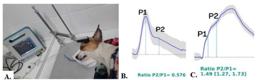
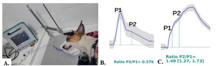
A. Dog with traumatic brain injury (TBI) submitted to non-invasive monitoring of ICP waves (ICP-Ni). Monitoring performed with the patient in lateral recumbency and the sensor placed on the parietal region using a stereotactic device. B. Tracing of non-invasive intracranial pressure waves (ICP-Ni). Normal wave ratio: P1>P2 and P2/P1=0.576. C. Abnormal wave relationship, with P2>P1 and P2/P1=1.49.
The descriptive statistical analysis consisted of estimating the mean, median, standard deviation and interquartile range of the quantitative variables [weight, trauma time, MGCS, MGCS evolution, evolution time (MGCS days), heart rate (HR), respiratory rate (RR), systemic blood pressure (SBP), rectal temperature (TR), blood glucose, lactate, time to outcome (days), mean P2/P1 before treatment and mean P2/P1 after treatment] and in the evaluation of simple and relative frequencies categorical variables (sex, age group, etiology, ratio of pre- and post-treatment ICP waves, concomitant conditions, prognosis, and clinical outcome).
The Shapiro-Wilk normality test was performed to determine the statistical approach to quantitative variables. The variables were analyzed in relation to patient's clinical outcomes. To assess the differences between the groups, the Mann Whitney tests were used for the non-parametric approach to MGCS, MGCS evolution, time of MGCS evolution and lactatemia, and the t-Student test for the parametric approach to heart rate variables (beats per minute), respiratory rate (movements per minute), blood pressure (mmHg), body temperature (T⁰C) and blood glucose. The chi-square test was used to assess the association between the qualitative variables and outcomes. Statistical significance was set at 5% (p=0.05), and all analyses were conducted using R 4.0.4 software (Team, 2021).
RESULTS
The epidemiology, MGCS during initial care and at discharge, observed neurological alterations, location of the brain lesion, concomitant alterations, additional treatment, ICP-Ni results, and outcomes are shown in Table 1. Five mixed-breed dogs, two Shi Tzu dogs, and one dog of each of the following breeds were included: Chow Chow, Lhasa Apso, Pinscher, and Pit Bull. The mean age was 5.6 years (3 months to 13 years), with an average weight of 9.53 kg (2.4-23kg), with 54.5% (6) males; the time elapsed between the trauma and clinical care ranged from 1-5 days (median=5.2h). The main cause of TBI was car accidents, in 81.8% of cases.
Eight dogs were discharged between one and 14 days (mean=4.8 days) after the initial treatment, whereas three dogs died between four and eight days after hospitalization (mean=5.6 days). Two dogs were euthanized, and one died despite treatment. The MGCS of the 11 dogs in initial care ranged from 8 to 18 points (median=13 points), and the MGCS of the dogs that survived ranged from 13 to 18 points (median=18 points), after an average of 4.87 days post-trauma.
Regarding ICP-Ni monitoring and MGCS scores, three patients (dogs 1, 2, and 3) had a P1>P2 wave ratio, with a mean ratio of 0.70 and did not receive hyperosmolar agents. In these patients, the median MGCS was 13 points, and one patient (dog 3) achieved 18 points in seven days. One patient (dog 2) had impaired assessment due to blindness due to ocular disease. All three of these dogs were discharged.
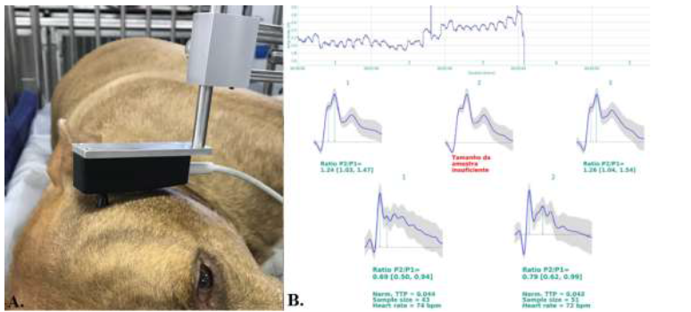
Eight patients exhibited a P2>P1 and received either mannitol or saline. In four dogs (dogs 4, 5, 6 and 8) normalization of P2/P1 was observed (Fig. 2). The average P2/P1 ratio was 1.2 before treatment and 0.62 after treatment. Dog 4 had ocular proptosis, with a normal coma scale; however, the P2/P1 ratio indicated a probable change in brain compliance. After application of mannitol, the relationship between the waves normalized. Dogs 4, 5 and 6 were discharged; dog 8 remained hospitalized and had an epileptic seizure on the eighth day of hospitalization, with a score of 8 on the MGCS; in the new monitoring of the ICP-Ni, a P2>P1 ratio was observed, with euthanasia being performed due to the unfavorable prognosis and the owner’s decision.
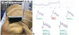
Dog number 5 submitted to non-invasive monitoring of intracranial pressure (ICP-Ni). A. Patient in right lateral recumbency position and sensor positioned in the parietal region using a stereotactic device. The second graph highlights that the sample size was insufficient, that is, the monitoring time was not adequate, making the sample unviable and requiring new register. B. Pressure wave tracing, P2>P1 and P2/P1>0.8 (1.24 and 1.26) in the upper curves, normalization of waves 26 min after the administration of mannitol, with a P2/P1 ratio <0.8(0.69 and 0.79).
Dog 9 had a score of 14 points on the MGCS, pulmonary contusion, and mild dehydration and received 3% NaCl as initial treatment. Follow-up monitoring was not possible, as there was evident clinical improvement after the administration of the therapeutic agent, with an evolution in the MGCS to 17 points. Additionally, other injuries, such as pubic and humeral fractures, were identified, requiring orthopedic surgical procedures, that extended the hospitalization period by eight days. In dog 7, after the fifth day of trauma, P2>P1 (0.82) and an MGCS score of 14 points were observed; however, the owner did not authorize further ICP-Ni monitoring.
Patients who were discharged from the hospital had, at the time initial clinical evaluation, a mean lactate level of 1.29 mmol/L (0.98-1.58), while those who progressed to death or euthanasia had an average of 6.8 mmol/L (6.42-7.02). Analysis of blood glucose levels revealed a tendency towards hyperglycemia in patients who died or were euthanized, but the data evaluated did not demonstrate statistically significant results.
The hospitalization period ranged from one to 14 days, with a median of four days, which was extended owing to the occurrence of a mandibular fracture in dog 6 and the necessity of esophageal tube feeding. Dogs 1, 2, 3, 4, 5, 6, 7, and 9 had favorable prognoses and were discharged from the hospital. The primary concomitant lesions included mandibular fractures, ocular protrusions, corneal ulcers, humeral and femoral fractures and lacerative facial wounds.
There was no statistical difference between the P2/P1 ratio before and after treatment in dogs that survived or died (Table. 2). In this analysis, dogs 7 and 9, which were not monitored after treatment, were excluded.
Statistical analysis of variables related to the evaluation of MGCS, time elapsed until clinical outcome (days), and the P2/P1 wave ratios before and after a specific therapeutic intervention for intracranial hypertension (ICH) in dogs with traumatic brain injury (TBI) evaluated between May 2019 and December 2020, who underwent non-invasive intracranial pressure monitoring (ICP-Ni) using the Brain4care® monitor
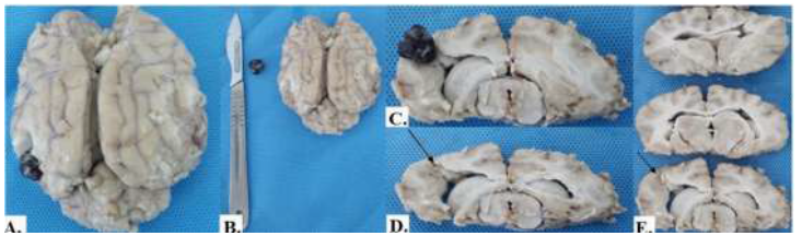
The mean P2/P1 ratio in patients whose P2 remained greater than P1 (dogs 10 and 11) was 1.36 (±0.35) after treatment, with a median of eight points on the MGCS, and a duration of 5.5 days. (Table. 2). In these patients (dogs 10 and 11) with P2>P1, the administration of 3% NaCl did not reduce this ratio even after mannitol treatment. In dog 10, the coma scale remained at eight points throughout the period, and due to the worsening condition, euthanasia was performed on the fifth day after admission. In dog 11, the MGCS score remained low and unchanged (8), and death occurred on the fourth day of hospitalization. Necropsy revealed a clot it in the left occipital lobe of the cerebral cortex. (Figure. 3). In 4 cases with na increased P2/P1 ratio and MGCS < 8, there was an unfavorable clinical outcome, that is, the mortality rate was 36.36%.

Post-mortem macroscopic image of the brain of dog 11. A and B. A clot observed in the cortical region of the left occipital lobe, measuring 1 cm in diameter. C, D, and E. Transverse sections of the brain demonstrating the deep cortical lesion (arrow).
DISCUSSION
Owing to the existing limitations in veterinary medicine for the evaluation of patients with TBI, alternative methods of obtaining prognosis or improving therapy have been researched, such as the use of MGCS, laboratory parameters, CT, and MRI (Bell et al. al., 2022; Cameron et al., 2021; Her et al., 2022). In this study, the Brain4care® BcMM2000 monitor was used to verify whether changes in the dynamics of the ICP-Ni waves, together with clinical and laboratory parameters, could assist in patient evaluation, therapeutic decision, and analysis of disease progression.
Although the number of cases evaluated was small, it was observed that non-invasive ICP monitoring was useful in detecting ICP-Ni waves suggestive of ICH. Even though there was no statistically significant difference in the P2/P1 ratio between dogs that survived and those that died, in five dogs with a MGCS greater than 8, a level at which, according to Freeman and Platt (2012), hyperosmolar agents would not typically be indicated, a P2/P1 ratio > 0.8 was observed in the ICP-Ni. Consequently, these agents were administered, and four of these patients were discharged. Mortality was lower than in a previous study carried out at the same place as the present study, in which hyperosmolar agents were only used in cases of severe neurological impairment (Vianna and Bahr Arias, 2013).
The most frequent cause of traumatic brain injury (TBI) was car accidents, as observed in other studies (Vianna and Bahr Arias, 2013; Sharma and Holowaychuk, 2015; Cameron et al., 2021). This type of trauma can lead to injury to various organ systems, as observed in the present study. Therefore, the management of dogs with TBI is complex and requires a detailed evaluation that allows for the identification of clinical, neurological, and laboratory changes (Sharma and Holowaychuk, 2015; Cameron et al., 2021).
Given the significant heterogeneity in the assessment and treatment of canine TBI within the veterinary community and as there is still no consensus on the best form of treatment (Evans and Fernandez, 2019), the management of the dogs in the present study was adjusted based on established protocols and the individual needs of each patient, as performed in other clinical studies with dogs with TBI (Sharma and Holowaycuk, 2015; Chai et al., 2020). Thus, in general, the main objectives of treatment of TBI are mantaining brain perfusion, associated with analgesics, antiepileptics and osmolar agents according to the clinical and neurological evaluation.
A three-level approach is proposed for the management of dogs with TBI. At level 1, the animal must receive oxygen, the patient’s head and torso should be elevated to 30 º, and its blood volume must be reestablished. At level 2, treatment is based on the use of osmotic diuretics if the patient is normovolemic and has an MGCS < 8. At level 3, in patients who do not respond to treatment at levels 1 and 2, it is recommended to maintain the patient in an induced coma in intensive care units and even consider surgical intervention to reduce ICP (Freeman and Platt, 2012; Platt et al., 2016). In the present study, with the assistance of ICP-Ni monitoring, alterations suggestive of ICH were observed even in patients with MGCS scores considered reserved (9-14 points) to good (15-18 points), which led to the administration of hyperosmotic agents, with a decrease in the P2/P1 ratio shortly after drug administration. Mannitol is the gold standard drug for the control of ICH; its main effects are plasma expansion and reduction of blood viscosity, with a consequent increase in cerebral blood flow and improvement in tissue oxygenation; in addition to the osmotic effect, that reduces interstitial fluid and brain edema (Diringer, 2016). In the present study, mannitol was considered effective in most cases and was replaced with hypertonic saline solution as an additional therapy in cases that did not respond to the initial mannitol treatment.
The administration of hypertonic saline solution is recommended in patients with cerebral edema and high ICP in refractory cases (Ballocco et al., 2019), and it seems to be more effective than mannitol in reducing ICP in humans (Eskandari et al., 2013), but there is insufficient evidence to consider this hyperosmolar solution as the first choice for TBI, especially in dogs. The effect of mannitol and hypertonic saline solution on the treatment of ICH was evaluated in three animals with TBI: two cats and one dog. Both solutions promoted a decrease in ICP and an improvement in cerebral perfusion pressure, with differences in the duration of these effects (Ballocco et al., 2019).
It was observed that patients with P1>P2 or P2/P1 ratio < 0.8 and higher MGCS scores did not require specific treatment for ICH and progressed to hospital discharge. There are controversies regarding the normal P2/P1 ratio in dogs, due to the existence of few studies that evaluate the ICP waves obtained by invasive or non-invasive monitoring. The same doubt was raised in a series of five human cases of TBI, in which P2/P1 ratios >1.0 were considered indicative of ICH (Link et al., 2022). In another study, of 41 human patients with various intracranial conditions, in which non-invasive and invasive monitoring was compared, it was concluded that the P2/P1 ratio >1.2 predicted ICH (Brasil et al., 2021). In the present study, P2/P1>1.25 and MGCS scores of eight points without good evolution over a prolonged period of hospitalization were associated with increased mortality.
Low MGCS scores are related to worse neurological status and a lower probability of survival (Platt et al., 2016) in both dogs and cats (Cameron et al., 2021). In a study involving 72 dogs with TBI, a low MGCS score was the strongest predictor of death, with a score of 11 being 84% sensitive and 73% specific for predicting this outcome (Sharma and Holowaychuk, 2015). However, in the present study, even some patients with high MGCS scores, presented of P2/P1>0.8, and one possibility is that there may be changes in brain compliance without serious neurological manifestations, or other factors interfering with monitor reading.
The occurrence of epileptic seizures is an important complication of TBI, that contributes to an increase in brain injury and consequent morbidity and mortality (Steinmetz et al., 2013; Platt et al., 2016). In the present study, one patient had epileptic seizures on the eighth day after TBI, and euthanasia was required. Current guidelines from the Brain Trauma Foundation advocate the administration of anti-epileptic drugs to prevent epileptic seizures after TBI; however, the prophylactic use of these drugs in dogs remain debatable (Platt et al., 2016).
The lack of response to the instituted treatment, associated with the low MGCS score, may indicate intracranial mass effects, which were observed in one of the patients, whose necropsy confirmed the presence of a clot and significant tissue damage. CT or MRI can be useful in identifying structural lesions associated with increased ICP (Bittermann et al., 2014; Vali et al., 2021), but these exams were not available at the hospital where this study was carried out.
The limitations of this study were the small number of patients and the lack of monitoring of the patients’ ICP-Ni waves after treatment. The reduced number of animals was due to the impact of reduced attendance during the COVID-19 pandemic phase. COVID-19 pandemic has affected all levels of the education system in countries worldwide (Nicola et al., 2020). In addition, the impossibility of continuous monitoring in patients who maintained a P2>P1 ratio, made it impossible to diagnose possible ICP oscillations during hospitalization. Unfortunately, unlike humans, in whom the monitor can be used on awake patients (Moraes et al., 2023), the device can only be used on dogs if they are still (Bahr Arias et al., 2022; Rocha et al., 2023; Bahr Arias and Weizenmann 2024) wich would require sedation or even anesthesia in patients who have shown clinical improvement.
There was a higher incidence of death in dogs with low MGCS scores and changes indicative of ICH detected by the monitor, especially with P2/P1 ratio > 1.2. However, owing to the existence of few studies using this monitor in dogs, and the fact that invasive ICP monitoring was not performed for comparative purposes, it is not known exactly which value of this ratio would be the threshold to be considered for inferring ICH. Based on the results obtained, the non-invasive ICP-Ni monitoring system in dogs with TBI made it possible to compare the ICP-Ni waves during the initial treatment and after treatment and allowed the observation of an improvement in the P2/P1 ratio after the application of hyperosmolar agents, which provided additional information for the management of dogs with TBI.
ACKNOWLEDGEMENTS
To the “Coordination for the Improvement of Higher Education Personnel” (CAPES) for the PhD scholarship of T.C. Weizenmann.
REFERENCES
- BAHR ARIAS, M.V.; CONCEIÇÃO, R.T.; GUIMARÃES, F.C. et al. Preliminary evaluation of a non-invasive device for monitoring intracranial pressure waveforms in dogs. J. Small. Anim. Pract, v.63, p.624-631, 2022.
- BAHR ARIAS, M.V.; WEIZENMANN, T.C. Methodology for non-invasive monitoring of intracranial pressure waves in dogs with traumatic brain injury using the Brain4care® BCMM/2000 monitor. Vet. Zootec. UNESP, v.31, 2024.
- BALLOCCO, I.; EVANGELISTI, M.A.; DEIANA, R. et al. A pilot study evaluating the effect of mannitol and hypertonic saline solution in the treatment of increased intracranial pressure in 2 cats and 1 dog naturally affected by traumatic brain injury. J. Vet. Emerg. Crit. Care, v.29, p.578-584, 2019.
- BELL, A.L.; ROZANSKI, D.V.M.; BABYAK, J. A multicenter retrospective comparison of trauma in toy breeds versus giant breeds: a Veterinary Committee on Trauma registry study. J. Vet. Emerg. Crit. Care, v.32, p.26-33, 2022.
- BELTRAN, E.; PLATT, S.R.; McCONNELL, J.F. et al. Prognostic value of early magnetic resonance imaging in dogs after traumatic brain injury: 50 cases. J. Vet. Intern. Med, v.28, p.1256-1262, 2014.
- BITTERMANN, S.; LANG, J.; HOWARD, J. et al. Magnetic resonance imaging signs of presumed elevated intracranial pressure in dogs. Vet. J, v.201, p.101-108, 2014.
- BOLLELA, V.R.; FRIGIERI, G.; VILAR, F.C. et al. Noninvasive intracranial pressure monitoring for HIV-associated cryptococcal meningitis. Braz. J. Med. Biol. Res, v.50, p.6392, 2017.
- BRASIL, S.; SOLLA, D.J.F.; NOGUEIRA, R.C. et al. A novel noninvasive technique for intracranial pressure waveform monitoring in critical care. J. Pers. Med, v.11, p.1302, 2021.
- CABELLA, B.; VILELA, G.H.; MASCARENHAS, S. et al. Validation of a new noninvasive intracranial pressure monitoring method by direct comparison with an invasive technique. Acta Neurochr. Suppl., v.122, p.93-96, 2016.
- CAMERON, S.; WELTMAN, J.G.; FLETCHER, D.J. The prognostic value of admission point-of-care testing and modified Glasgow Coma Scale score in dogs and cats with traumatic brain injuries (2007-2010): 212 cases. J. Vet. Emerg. Crit. Care, v.32, p.75-82, 2021.
- CARNEY, N.; TOTTEN, A.M.; O'REILLY, C. et al. Guidelines for the management of severe traumatic brain injury. Oper. Neurosurg, v.80, p.6-15, 2017.
- CHAI, O.; MAZAKI-TOVI, M.; KLAINBART, S. et al. Serum concentrations of neuron-specific enolase in dogs following traumatic brain injury. J. Comp. Pathol, v.179, p.45-51, 2020.
- DIRINGER, M.N. The evolution of the clinical use of osmotic therapy in the treatment of cerebral edema. Acta Neurochir. Suppl., v.121, p.3-6, 2016.
- ESKANDARI, R.; FILTZ, M.R.; DAVIS, G.E. et al. Effective treatment of refractory intracranial hypertension after traumatic brain injury with repeated boluses of 14.6% hypertonic saline. J. Neurosurg, v.119, p.338-346, 2013.
- EVANS, E.K; FERNANDEZ, A.L. Current trends in the management of canine traumatic brain injury: an internet-based survey. Can. Vet. J., v. 60, p.73-79, 2019.
- FAN, J.Y.; KIRKNESS, C.; VICINI, P.; BURR, R.; MITCHELL, P. Intracranial pressure waveform morphology and intracranial adaptive capacity. Am. J. Crit. Care, v.17, p.545-554, 2008.
- FREEMAN, C.; PLATT, S.R. Head trauma. In: PLATT, S.; GAROSI, L. Small animal neurological emergencies. [s.l.]: Manson Publishing, 2012. 672p.
- GIANNASI, S.; KANI, Y., HSU, F.C. et al. Comparison of direct measurement of intracranial pressures and presumptive clinical and magnetic resonance imaging indicators of intracranial hypertension in dogs with brain tumors. J. Vet. Intern. Med., v.34, p.1514-1523, 2020.
- HALL, K.E.; HOLOWAYCHUCK, M.K.; SHARP, C.R. et al. Multicenter prospective evaluation of dogs with trauma. J. Am. Vet. Med. Assoc, v.244, p.300-308, 2014.
- HER, J.; YANKE, A.B.; GERKEN, K. et al. Retrospective evaluation of the relationship between admission variables and brain herniation in dogs (2010-2019): 54 cases. J. Vet. Emerg. Crit. Care, v.32, p.50-57, 2022.
- KAWOOS, U.; McCARRON, R.M.; AUKER, C.R. et al. Advances in Intracranial Pressure Monitoring and Its Significance in Managing Traumatic Brain Injury. Int. J. Mol. Sci., v.16, p.28979-28997, 2015.
- LINK, C.; BOTELHO, A.F.; DHAESE, T.M. et al. Noninvasive intracranial pressure in patients with traumatic brain injury. Res. Soc. Dev., v.11, p.1-11, 2022.
- MORAES, F.M.M.; ADISSY, E.N.B.; ROCHA, E. Multimodal monitoring intracranial pressure by invasive and noninvasive means. Nat. Sci. Rep., 18404, 2023.
- NICOLA, M.; ALSAFI, Z.; SOHRABI, C.; KERWIN, A. et al. The socio-economic implications of the coronavirus pandemic (COVID-19): a review. Int. J. Surg., v.78, p.185-193, 2020.
- NOH, D.; CHOI, S.; CHOI, H. et al. Evaluating traumatic brain injury using conventional magnetic resonance imaging and susceptibility weighted imaging in dog. J. Vet. Sci., v.20, p.1-7, 2019.
- PLATT, S.; FREEMAN, C.; BELTRAN, E. Canine head trauma: an update. In Pract., v.38, n.1, 2016.
- RABELO, R.C. Small animal emergencies: clinical and surgical management in critically ill patients. [Philadelphia]: Elsevier, 2012. 210p.
- RAPOPORT, K.; MATEO, I.; PEERY, D. et al. The prognostic value of the Koret CT score in dogs following traumatic brain injury. Vet. J, v.266, 2020.
- ROCHA, N.L.F.C.; CARDOSO G.S.; NOGUEIRA, J.F. et al. Non-invasive monitoring of intracranial pressure waveforms using Braincare® BCMM 2000 monitor in dogs with myelopathies undergoing myelography. Pesqui. Vet. Bras,. v.43, p.e07132, 2023.
- SHARMA, D.; HOLOWAYCHUK, M.K. Retrospective evaluation of prognostic indicators in dogs with head trauma: 72 cases (January-March 2011). J. Vet. Emerg. Crit. Care, v.25, p.631-639, 2015.
- STEINMETZ, S.; TIPOLD, A.; LOSCHER, W. Epilepsy after head injury in dogs: a natural model of posttraumatic epilepsy. Epilepsia, v.54, p.580-588, 2013.
-
TEAM, R.C.R. A language and environment for statistical computing. Vienna, Austria: R Foundation for Statistical Computing, 2021. Available in: https://www.R-project.org/. Acessed: 27 Apr. 2023.
» https://www.R-project.org - VALI, Y.; GIELEN, I.; SOROORI, S. et al. The diagnostic value of intravenous contrast computed tomography in addition to plain computed tomography in dogs with head trauma. BMC Vet. Res., v.17, p.46, 2021.
- VIANNA, C.G.; BAHR ARIAS, M.V. Prospective study of 32 dogs with traumatic brain injury. Rev. Bras. Ciênc. Vet., v.35, p.93-99, 2013.
- WYATT, S.; LLABRES-DIAS, F.; BELTRAN, E. Early CT in dogs following traumatic brain injury has limited value in predicting short-term prognosis. Vet. Radiol. Ultrasound, v.62, p.181-189, 2021.
Data-in-article.