 Comparative hemodynamic effects between AVM and HFO: implications in veterinary clinical practice
ABSTRACT
High-frequency oscillatory ventilation (HFO) is a mechanical ventilation technique used in veterinary medicine to treat various respiratory diseases, particularly in small animal patients, but its potential application in critical care settings and its effects on cardiopulmonary function remain insufficiently explored. This study compared HFO and conventional mechanical ventilation (CMV) in neonatal piglets, assessing systemic and regional oxygenation and ventilatory parameters. HFO resulted in significantly higher maximal inspiratory pressure (MIP) (23.9±4.4 vs 18.6±0.9, p<0.001) and mean airway pressure (MAP) (8.8±1.6 vs 6.1±0.2) than CMV. Mean arterial pressure was significantly lower in the HFO group. After 60 minutes, regional renal fossa tissue oxygenation (rNIRS) was significantly higher in the CMV group (114.23±13.03 vs. 99.31±10.7, p<0.05), whereas no significant difference was observed in cerebral NIRS (cNIRS). Blood gas analysis showed no significant differences between groups. While HFO demonstrated different ventilatory mechanics in this piglet model, the comparable gas exchange suggests it warrants further investigation as a potential ventilation strategy in veterinary patients.
Keywords:
high-frequency oscillatory ventilation (HFO); veterinary medicine; SDRA ventilatoy support; ventilation; hemodynamics
RESUMO
A ventilação oscilatória de alta frequência (HFO) é uma técnica de ventilação mecânica utilizada na medicina veterinária para tratar diversas doenças respiratórias, particularmente na clínica de pequenos animais, mas sua aplicação potencial em ambientes de cuidados críticos e seus efeitos na função cardiopulmonar permanecem insuficientemente explorados. Este estudo comparou HFO e ventilação mecânica convencional (CMV) em leitões neonatos, avaliando a oxigenação sistêmica e regional e os parâmetros ventilatórios. A HFO resultou em pressão inspiratória máxima (MIP) (23,91±4,35 vs. 18,56±0,97, P<0,0001) e pressão média da via aérea (MAP) (8,82±1,61 vs. 6,14±0,18) significativamente maior do que a CMV. A pressão arterial média foi substancialmente menor no grupo HFO, e, após 60 minutos de ventilação, a oxigenação regional do tecido da fossa renal (rNIRS) foi consideravelmente maior no grupo CMV (114,23±13,03 vs. 99,31±10,7, P<0,05), enquanto não foi observada diferença expressiva nos valores do NIRS cerebral (cNIRS). Por sua parte, a análise dos valores obtidos das gasometrias seriadas tampouco apresentou diferenças significativas entre os grupos. Embora a HFO tenha demonstrado mecânica ventilatória distinta neste modelo, a troca gasosa comparável sugere que ela merece investigação adicional como estratégia de ventilação potencial em pacientes veterinários.
Palavras-chave:
ventilação oscilatória de alta frequência (VOAF); medicina veterinária; suporte ventilatório em SDRA; hemodinâmica
INTRODUCTION
Veterinary medicine has advanced significantly in the management of critical patients, especially in respiratory diseases such as acute respiratory distress syndrome (ARDS), which is well-documented in humans. According to Wilkins et al. (2007), ARDS in veterinary patients (vet-ARDS) is characterized by an acute onset (<72 hours) of respiratory distress, concurrent disease or predisposing factor (severe pulmonary disease, trauma, sepsis, visceral torsion, heatstroke, systemic inflammatory response syndrome), pulmonary fluid accumulation without increased non-cardiogenic capillary pressure, and refractory hypoxemia.
Mechanical ventilation in veterinary medicine has become a fundamental strategy for the treatment of respiratory pathologies and critical patient support (Boiron et al., 2019) and has experienced exponential growth in recent years both in the intensive care of companion animals and in its application for ventilatory support during anesthesia (Fantoni et al., 2022; Rodrigues et al., 2022; O’Keefe and Donaldson., 2023). However, it is necessary to explore the use of ventilatory modes that have been reported as beneficial in ARDS therapy, such as the use of high-frequency oscillatory ventilation (HFO), to enhance their utilization and applicability in veterinary patients (Balakrishnana, 2021).
HFO is characterized by the delivery of small tidal volumes (1-2 ml/kg) at high frequency (above 1 Hz) and it is used to support patients with ARDS and other severe pulmonary pathologies. Its use decreases the possibility of ventilator-induced lung injury (VILI) by reducing the risk of volutrauma and maintains alveolar insufflation with constant airway pressure, using sinusoidal flow oscillation. This results in the prevention of atelectrauma, improved oxygenation, and effective ventilation even with tidal volumes lower than the alveolar dead space (Meyers et al., 2019).
Although advantageous in the management of diverse pathological conditions, positive pressure mechanical ventilation is fundamentally anti-physiological, as it alters the physiological oscillations of pleural pressure (normally negative during inspiration) and transpulmonary pressure (De Monte et al., 2018). Furthermore, it affects ventricular filling and the functionality of large vessels, which can have hemodynamic implications for ventilated patients (Vieillard-Baron et al., 2016). Positive airway pressure impedes venous return and increases right ventricular afterload, reducing right ventricular output and subsequent left ventricular filling, leading to hemodynamic effects (Pinsky et al., 1985; Vieillard-Baron et al., 2001).
The development of protective ventilatory strategies is crucial for the proper management of patients requiring invasive ventilatory therapy. Research utilizing animal models, spanning both veterinary and human medicine, provides a conduit for translating mechanical ventilation therapies to veterinary applications (Jiang et al., 2006; Wang et al., 2008; Otáhal et al., 2016; Weia et al., 2019). Evaluating the comparative effects of conventional mechanical ventilation (CMV) and HFO on gas exchange, hemodynamic parameters, and tissue perfusion is a tool to select the ventilation strategy selection in clinical practice.
The aim of this study was to determine the systemic repercussions, assessed by monitoring heart rate, oxygen saturation, invasive systemic arterial pressure, and values obtained from the monitoring of cerebral and regional oxygenation (NIRS, Near InfraRed Spectroscopy), as indicators of flow distribution and oxygen utilization balance. Complementarily, the arterial partial pressure of carbon dioxide and oxygen, along with pH and lactate, measured by serial arterial blood gas analysis, were assessed for ventilatory-respiratory evaluation during HFO and CMV in neonatal piglets under sedo-analgesia.
ETHICAL ASPECTS
The research was submitted to the Ethics Committee on Animal Use (Honorary Commission for Animal Experimentation, CHEA) of the School of Medicine (University of the Republic) and approved under the number 070151-500087-21.
MATERIALS AND METHODS
Eleven newborn males of the species Sus scrofa domestica (domestic pig), between 12 to 48 hours old, weighing between 1300 to 1800 g, acquired from a local farm were used during these experiments. They were randomly assigned to one of two groups: HFO (n=5) or CMV (n=6).
After intramuscular premedication with ketamine (25mg/kg) and midazolam (0.2mg/kg), pulse oximetry monitoring was initiated on the second toe of the right hind limb. Umbilical artery and vein catheterization then allowed for invasive arterial pressure monitoring, blood gas sampling, and administration of maintenance fluids (5% glucose solution mixed with saline solution 50/50) and sedo-analgesia. This was performed with a constant infusion of Midazolam, Ketamine, and Fentanyl at 3mL/Kg per hour (prepared at a concentration of 1ug/mL of Fentanyl, 1mg/mL of Ketamine, and 0.06mg/mL of Midazolam). Orotracheal intubation was then performed with a 3.5mm endotracheal tube for subsequent connection to the ventilator (Accutronic®, Fabian model) and initiation of ventilatory mode.
Ventilation parameters were adjusted according to standardized values, establishing for CMV a Peak Inspiratory Pressure (PIP) of 20cmH2O, Positive end-expiratory pressure (PEEP) of 5cmH2O, Inspiratory time (Ti) 0.33s, frequency of 20 breaths per minute, and Inspired oxygen fraction (FiO2) 0.21; while for individuals ventilated in HFO, values of PIP 20cmH2O, PEEP 5cmH2O, respiratory frequency 5Hz, Mean Airway Pressure (MPAW) 8cmH2O, amplitude of 35mbar and FiO2 0.21 were used.
Once ventilatory support was ensured, connection to monitors was completed (electrocardiography electrodes, esophageal thermometer placement, and NIRS monitoring of somatic and cerebral regional oximetry, for determination of oxygen availability in the vascular bed in relation to infrared light absorbance (Ward et al., 2006; Marin and Moore, 2011). To monitor comfort, a NIPE™ (Newborn Infant Parasympathetic Evaluation) monitor (MDoloris Medical Systems. Loos, France) was used to continuously assess heart rate variability via electrocardiography obtained from a multiparameter monitor (Ohmeda, enGuard CM4, Massimo SET). Analgesic intervention was indicated when NIPE™ values fell below 50 (Sakthivel et al., 2024).
Following verification of normal monitoring parameters and blood gas analysis, a 5µg/kg bolus of Fentanyl was administered, with the option to repeat after 3 minutes if the pedal withdrawal reflex persisted. Intercostal nerve block and incision line infiltration were performed in the 4th and 5th intercostal space with 2% Lidocaine, total dose 4mg/Kg for thoracotomy. Once thoracotomy and pericardiectomy were performed, an 18G catheter was placed in the right ventricle to reach the pulmonary artery to obtain invasive pulmonary artery pressure wave recording.
Hemodynamic measurements from multiparametric monitoring (Ohmeda, enGuard CM4, Massimo SET) included continuous assessment of Heart Rate, HR; Oxygen Saturation, SpO2; Systolic Systemic Arterial Pressure, sSAP; Mean Arterial Pressure, MAP; Diastolic Arterial Pressure, PAD; and Systolic Pulmonary Arterial Pressure, sPAP.
Monitoring data was recorded every 15 minutes during ventilation once the piglets were stabilized, and blood gas analyses were performed every 30 minutes to determine the repercussions on the internal environment of the established ventilatory modes. Ventilation under each modality (CMV or HFO) was maintained for 2.5 hours. Following trial completion, the animals were euthanized with a barbiturate overdose. After verifying deep unconsciousness and complete absence of response to stimuli, cardiac arrest was induced with potassium chloride.
These records were analyzed using GraphPad Prism 8.0.2(263). Data from multiparameter monitoring, regional NIRS oxygenation, and serial blood gas analyses were processed. Descriptive statistics were used to examine variable behavior. Subsequently, a two-way ANOVA with Bonferroni post-hoc testing was performed to determine the effect of ventilatory modes over time on the variables of interest. The mean absolute baseline value and its standard deviation are provided for each case.
RESULTS
In all subjects of the trial, the application of either proposed ventilatory strategy was successfully achieved. According to the data obtained, PIP was significantly higher (p<0.0001) in the HFO ventilation group, as well as MPAW. Conversely, the FiO2 required for normoxia maintenance differed between the two groups, with the CMV group requiring significantly higher values (p<0.0005). The evolution of ventilatory parameters for each group is presented in (Table 1).
Hemodynamic variables showed no significant differences between ventilatory modes during the evaluation period (Table 2).
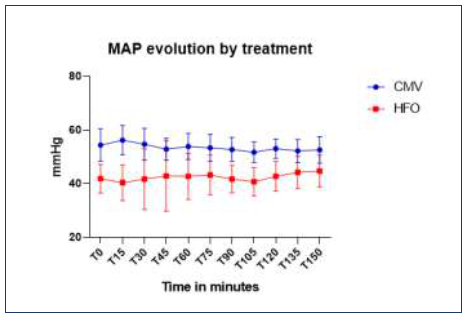
Below, Fig. 1 illustrates MAP evolution over time for each group, showing no variation during the ventilatory period. Both groups maintained stable values within clinically normal ranges throughout the experiment.

Mean Arterial Pressure (MAP) evolution by treatment along time in newborn piglets during HFO or CMV ventilation strategy.
To further evaluate tissue perfusion and gas exchange quality, cerebral (cNIRS) and renal (sNIRS), and serial arterial blood gas analysis, were done as complementary assessments.
Figure 2 (“a” and “b”) shows the evolution of regional oxyhemoglobin saturation obtained by NIRS placed at the renal fossa (sNIRS) and encephalic sensor (cNIRS).

a and b; evolution of regional oxyhemoglobin saturation by somatic (sNIRS), (a), and encephalic (cNIRS), (b), detected with Near InfraRed Spectroscopy Sensors (NIRS) during HFO or CMV ventilation in newborn piglets.
In Fig. 2, (a) the normalized regional oxygen saturation measured by sNIRS is shown. Baseline values, expressed as a percentage relative to an absolute value of 100 within each group, were 43.2±5.1 for the CMV group and 55.6±10.1 for the HFO group. Inter-group comparison revealed no statistically significant difference (p>0.05) for these initial values. Figure 2, (b), shows normalized regional oxygen saturation measured by cNIRS. The baseline values were 42,6±3,4 for the CMV group and 55,2±8,2 for the HFO group.
Analysis of sNIRS values revealed a significant difference (p<0.05) starting at 60 minutes of ventilation, which persisted until the end of the trial. Regional oxygenation values obtained from the renal fossa sensor, were higher in individuals ventilated with CMV compared to those ventilated with HFO (114±13 for AVM; vs 99±10 for HFO). However, no significant differences were detected in cNIRS values between groups during ventilation with CMV versus HFO
To evaluate changes in the homeostatic environment of the subjects, serial arterial blood gas analyses were performed. The results are presented in (Table 3) No significant differences were observed in any of the monitored variables of interest between the two ventilation modes throughout the trial.
DISCUSSION
Mechanical ventilation therapy is essential in the management of patients with ARDS, and its application has been refined in recent decades. Nevertheless, the nature of this technique involves the use of positive pressure within the thoracic cavity, which is non-physiological and can be associated with pulmonary damage (De Monte et al., 2018). Excessive volume (volutrauma) or pressure (barotrauma) applied to the lung will have negative consequences if ventilation is not performed within specific parameters and monitored. This damage is defined as ventilator-associated lung injury (VALI) or ventilator-induced lung injury (VILI) (Guzel et al., 2013).
In patients with acute lung injury and ARDS, it has been demonstrated that mechanical ventilation with lower tidal volumes compared to those traditionally used can decrease disease-associated mortality (Goutorbe et al., 2008). In this regard, the use of HFO in the ventilatory therapy of patients with ARDS has been supported by physical characteristics inherent to this modality, which promote protective ventilation compared to CMV. These characteristics include a lower incidence of hyperinflation and injury from alveolar recruitment and derecruitment, continuous airway pressure, and tidal volumes below dead space (Pillow, 2005).
In this study, both ventilatory strategies were successfully utilized for the support of patients in intensive care settings, demonstrating no differential impact on internal environment homeostasis. Normal and sustained PaO2 values were observed for both ventilation modes, alongside adequate CO2 elimination levels. Although HFO has been reported to increase CO2 elimination in pulmonary ARDS (Friesecke et al., 2015), no significant difference in CO2 elimination was observed between groups during this trial. This may be explained by the fact that these were animals without prior lung injury, where this reported benefit with the use of HFO in affected lungs may not have been evident. Furthermore, the implementation of conservative baseline ventilation parameters in both modes, thereby minimizing this difference, may have contributed (Bancalari, 2003; Chakkarapani et al., 2020; Lattari, 2023).
HFO used in the ventilatory therapy of ARDS has been associated with a decrease in cardiac output. This is attributed to the fact that higher airway pressures maintained in patients ventilated with HFO negatively impacted left ventricular stroke volume. This, combined with a decrease in diastolic diameter (i.e., reduced filling) and shortening of ejection time, can have hemodynamic implications (Simma et al., 2000). In the subjects of this trial, higher values of PIP and MPAW were evidenced in those ventilated with HFO, and these factors may be implicated in the observed differences in MAP between the two groups.
Blood pressure (BP) is a vital sign reflecting cardiovascular status. The results indicated lower mean arterial pressure (MAP) values for individuals ventilated with High-Frequency Oscillation (HFO); however, these findings were not mirrored by the systolic arterial pressure (SAP) values. Although a statistically significant difference was observed in MAP values between the two groups (52,43±1,27 CVM vs 43,50±3,29HFO), this finding is not clinically significant. MAP remained within normal clinical ranges and stable throughout the entire experiment for both treatments. This observed statistical difference is likely attributable to the limited number of animals per group and the inherent variability associated with this physiological parameter (Guilherme et al., 1999).
To further analyze these data, a review of the oxygen saturation values obtained via pulse oximetry is necessary, as well as the evolution of the oxyhemoglobin proportion measured with NIRS in the renal fossa and cerebral vascular bed. This technology has demonstrated its value in providing critical hemodynamic monitoring information, particularly regarding tissue oxygenation and microcirculation, data which cannot be obtained with standard monitoring (arterial pressure and pulse oximetry) (Green et al., 2016).
Considering that tissue oxygenation depends on perfusion, it is noteworthy that patients ventilated with HFO exhibited lower regional oxygenation values measured by NIRS in the renal fossa (sNIRS). This finding could be considered consistent with the lower MAP values recorded in these individuals compared to those ventilated with CMV. Both parameters may reflect some degree of hemodynamic impact in this group, without affecting cerebral perfusion, possibly due to the presence of autoregulatory mechanisms (Peterson et al., 2011; Siwicka-Gieroba et al., 2022), as no significant difference was observed in cerebral NIRS values (cNIRS) between the two groups. Furthermore, cerebral oxygen delivery is conditioned by blood oxygen content, allowing the tissue to increase oxygen extraction in response to decreased cerebral blood flow, tolerating flow reductions of up to 50-60% (Bain et al., 2014), potentially maintaining stable cNIRS values even if there had been some impact on oxygen supply (Meng, 2021).
Cerebral tissue oxygen extraction rate is not only modified by perfusion variations. Changes in homeostasis, such as increased PaCO2 and decreased pH, can heighten cerebral oxygen delivery due to a decrease in oxygen affinity for hemoglobin, a rightward shift in its dissociation curve, and alterations in cerebral blood flow (Brown et al., 2018; Silvera et al., 2022; Hoiland et al., 2019). Analysis of the arterial blood gas values of interest in the experimental groups revealed that both ventilation modes maintained PaCO2 levels within normocapnic ranges for neonate patients (Gannon et al,. 1998; Ambalavanan and Carlo, 2001), and no significant difference in arterial pH values was found between the two groups. Therefore, both modalities preserved acid-base balance.
Multiple findings supporting the development of HFO as a ventilatory strategy in intensive care were obtained from experimental studies conducted in animals. Thus, as early as 1980, Bohn et al. reported that when using HFO in anesthetized beagles with PIP of 4 to 8 cmH2O and frequencies of 15Hz reaching low tidal volumes (approx. 1,9mL/Kg), optimal CO2 elimination rates were reached. Furthermore, they demonstrated that HFO within protective pressure and volume ranges was effective in maintaining normocapnia for several hours.
Likewise, Hamilton et al. (1983) compared the use of CMV versus HFO on the quality of gas exchange and oxygenation in experimentally induced acute lung injury in rabbits, observing that the use of HFO reduced VALI while maintaining efficient gas exchange. That same year, Rehder et al. (1983) published the use of HFO compared with CMV for ventilatory support in dogs under experimental conditions maintained for 36 hours and found no cardiovascular instability in the individuals ventilated with HFO. Similarly, Gillespie and Hyatt (1985) reported that the use of HFOV in ventilatory therapy maintained for 5 to 7 hours in dogs was able to preserve ventilatory mechanics and adequate gas exchange with tidal volumes of 2.5 mL/kg and frequencies of 15 to 30 Hz.
To our knowledge, there are no published reports on the clinical use of HFO in veterinary patients. This holds true for both healthy lungs and those with lung disease, and HFO has not been mentioned in recent reviews on ARDS prevalence and management or ventilatory therapy in companion animals (Balakrishnan, 2021; Loewen and Bach, 2022). Despite limitations inherent to the small number of animals per group and the use of piglets with healthy lungs, our study contributes to the understanding and application of HFO in veterinary patients. This study facilitated care team training and offered crucial hemodynamic and ventilatory-respiratory insights, leading to a deeper understanding of each modality's effects
Based on our findings in newborn piglets and the established efficacy of HFO in human and canine models, we suggest HFO as a potential strategy to mitigate ventilation-associated lung injury and preserve ventilatory-respiratory function. The incorporation of this ventilatory modality in veterinary medicine is feasible with the availability of safe technology and a trained working group. With the required monitoring elements and control of ventilatory variables, the use of HFO in veterinary patients could be viable as a therapeutic strategy, even potentially as an alternative to be explored in lung injured animals.
REFERENCES
- AMBALAVANAN, N.; CARLO, W.A. Hypocapnoea and hypercapnoea in respiratory management of newborn infants. Clin. Perinatol., v.28, p.517-531, 2001.
- BAIN, A.R.; MORRISON, S.; AINSLIE, P.N. (2014) Cerebral oxygenation and hyperthermia. Front. Physiol., v.5, p.92, 2014.
- BALAKRISHNAN, A. Current standards and practices in small animal mechanical ventilation. Adv. Anim. Care, v.2, p.69-83, 2021.
- BANCALARI, M.A. Ventilación de alta frecuencia en el recién nacido: un soporte respiratorio necesario. Rev. Chil.Pediatr., v.74, p.475-486, 2003.
- BOHN, D.J.; MIYASAKA, K.; MARCHAK, B.E. et al. Ventilation by high-frequency oscillation. J. Appl. Physiol., v.48, p.710-716, 1980.
- BOIRON, L.; HOPPER, K.; BORCHERS, A. Risk factors, characteristics, and outcomes of acute respiratory distress syndrome in dogs and cats: 54 cases. J. Vet. Emerg. Crit. Care, v.29, p.1-7, 2019.
- BROWN, M.K.; POELTLER, D.M.; HASSEN, K.O. et al. Incidence of hypocapnia, hypercapnia, and acidosis and the associated risk of adverse events in preterm neonates. Respir. Care, v.63, p.943-949, 2018.
- CHAKKARAPANI, A.A.; ADAPPA, R.; MOHAMMAD ALI, S.K. et al. "Current concepts of mechanical ventilation in neonates" - Part 1: Basics. Int. J. Pediatr. Adolesc. Med., v.7, p.13-18, 2020.
- DE MONTE, V.; BUFALARI, A.; GRASSO, S.; FERRULLI, F. et al. Respiratory effects of low versus high tidal volume with or without positive end-expiratory pressure in anesthetized dos with healthy lungs. Am. J. Vet. Res., v.79, p.496-504, 2018.
- FANTONI, D.T.; IDA, K.K.; SOARES, J.H.N.; AMBROSIO, A.M. Editorial: mechanical ventilation in anesthesia and critical care small animal patients. Front. Vet. Sci., v.9, p.942731, 2022.
- FRIESECKE, S.; STECHER, S.S.; ABEL, P. High-frequency oscillation ventilation for hypercapnic failure of conventional ventilation in pulmonary acute respiratory distress syndrome. Crit Care, v.19, p.201, 2015.
- GANNON, C.M.; WISWELL, T.E.; SPITZER, A.R. Volutrauma, PaCO2 levels, and neurodevlopmental sequelae following assisted ventilation. Clin. Perinatol., v.25, p.159-175, 1998.
- GILLESPIE, D.J.; HYATT, R.E. Effect of ventilation by high-frequency oscillation on lung and chest wall mechanics in the dog. Lung, v.163, p.317-325, 1985.
- GOUTORBE, P.; ASENCIO Y.; BORDES J. et al. Protective ventilation in ARDS: as soon as possible. An immediate use of HFOV. Cases J., v.1, p.124, 2008.
- GREEN, M.S.; SEHGAL, S.; TARIQ, R. Near-infrared spectroscopy. Semin. Cardiothorac. Vasc. Anesth., v.20, p.213-224, 2016.
- GUILHERME, B.A.J.; NIVALDO, A.V.J.; NATAL, F.J. Fetal influences on neonatal blood pressure. J. Perinatol., v.19, p.593-595, 1999.
- GUZEL, O.; YILDAR, E.; KARABAGLI, G. et al. Comparison of the effects of spontaneous and mechanical ventilation on blood gases during general anaesthesia in dogs. Kafkas Univ. Vet. Fak. Derg., v.19, p.19-25, 2013.
- HAMILTON, P.P.; ONAYEMI, A.; SMYTH, J.A. et al. Comparison of conventional and high-frequency ventilation: oxygenation and lung pathology. J. Appl. Physiol., v.55, p.131-138, 1983.
- HOILAND, R.L.; FISHER, J.A.; AINSLIE, P.N. Regulation of the cerebral circulation by arterial carbon dioxide. Compr. Physiol., v.9, p.1101-1154, 2019.
- JIANG, L.; WANG, Q.; LIU, Y. et al. Total liquid ventilation reduces lung injury in piglets after cardiopulmonary bypass. Ann. Thorac. Surg., v.82, p.124-130, 2006.
-
LATTARI, A. Respiratory support in neonates and infants. MSD Manual Professional Edition. 2023. Available in: https://www.msdmanuals.com/ professional/pediatrics/respiratory-problems-in-neonates/respiratory-support-in-neonates-and-infants Accessed in: 18/02/2025.
» https://www.msdmanuals.com/ professional/pediatrics/respiratory-problems-in-neonates/respiratory-support-in-neonates-and-infants - LOEWEN, J.M.; BACH, J.F. Respiratory distress in small animals: pathophysiology and clinical approach. J. Vet. Emerg. Crit. Care, v.32(S1), p.3-15, 2022.
- MARIN, T.; MOORE, J. Understanding near-infrared spectroscopy. Adv Neonatal Care, v.11, p.382-388, 2011.
- MENG, L. Heterogeneous impact of hypotension on organ perfusion and outcomes: a narrative review. Br. J. Anaesth., v.127, p.845-861, 2021.
- MEYERS, M.; RODRIGUES, N.; ARI, A. High-frequency oscillatory ventilation: a narrative review. Can. J. Respir. Ther., v.55, p.40-44, 2019.
- O’KEEFE, T.; DONALDSON, R. Mechanical ventilation in dogs and cats with tick paralysis. Front. Vet. Sci., v.10, p.1071191, 2023.
- OTÁHAL, M..; MLČEK, M..; VÍTKOVÁ, I..; KITTNAR, O. A novel experimental model of acute respiratory distress syndrome in pig. Physiol. Res., v.65, p.643-651, 2016.
- PETERSON, E.C.; WANG, Z.; BRITZ, Regulation of cerebral blood flow. Int. J. Vasc. Med., v.2011, p.823525, 2011.
- PILLOW, J.J. High-frequency oscillatory ventilation: mechanisms of gas exchange and lung mechanics. Crit. Care Med., v.33, p.S135-S141, 2005.
- PINSKY, M.R.; MATUSCHAK, G.M.; KLAIN, M. Determinants of cardiac augmentation by increases in intrathoracic pressure. J. Appl. Physiol., v.58, p.1189-1198, 1985.
- REHDER, K.; SCHMID, E.R.; KNOPP, T.J. Long-term high frequency ventilation in dogs. Am. Rev. Respir. Dis., v.128, p.476-480, 1983.
- RODRIGUES, R.R.; AMBRÓSIO, A.M.; ENGBRUCH, A.M. et al. Intraoperative protective mechanical ventilation in dogs: a randomized clinical trial. Front. Vet. Sci., v.9, p.842613, 2022.
- SAKTHIVEL, M.; SU, V.; NATARAJA, R.M.; PACILLI, M. Newborn and Infant Parasympathetic Evaluation (NIPE™) monitor for assessing pain during surgery and interventional procedures: a systematic review. J. Pediatr. Surg., v.59, p.672-677, 2024.
- SILVERA, F.; GAGLIARDI, T.; VOLLONO, P.F. et al. Study of the relationship between regional cerebral saturation and pCO2 changes during mechanical ventilation to evaluate modifications in cerebral perfusion in a newborn piglet model. Braz. J. Med. Biol. Res., v.55, p.e11543, 2022.
- SIMMA, B.; FRITZ, M.; FINK, C.; HAMMERER, I. Conventional ventilation versus high-frequency oscillation: hemodynamic effects in newborn babies. Crit. Care Med., v.28, p.227-231, 2000.
- SIWICKA-GIEROBA, D.; ROBBA, C.; GOŁACKI, J.; BADENES, R.; DABROWSKI, W. Cerebral oxygen delivery and consumption in brain-injured patients. J. Personal. Med., v.12, p.1763, 2022.
- VIEILLARD-BARON, A.; AUGARDE, R.; PRIN, S. et al. Influence of superior vena caval zone condition on cyclic changes in right ventricular outflow during respiratory support. Anesthesiology, v.95, p.1083-1088, 2001.
- VIEILLARD-BARON, A.; MATTAHAY, M.; TEBOUL, J.L. et al. Experts’ opinion on management of hemodynamics in ARDS patients: focus on the effects of mechanical ventilation. Intensiv. Care Med., v.42, p.739-749, 2016.
- WANG, H.M.; BODENSTEIN, M.; MARKSTALLER, K. Overview of the pathology of three widely used animal models of acute lung injury. Eur. Surg. Res., v.40, p.305-316, 2008.
- WARD, K.R.; IVATURY, R.R.; BARBEE, R.W. Near infrared spectroscopy for evaluation of the trauma patient: a technology review. Resuscitation, v.68, p.27-44, 2006.
- WEIA, F.; WENA, S.; WUA, H. et al. Partial liquid ventilation-induced mild hypothermia improves the lung function and alleviates the inflammatory response during acute respiratory distress syndrome in canines. Biomed. Pharmacother., v.118, p.109344, 2019.
- WILKINS, P.A.; OTTO, C.M.; BAUMGARDNER, J.E. et al. Acute lung injury and acute respiratory distress syndromes in veterinary medicine: consensus definitions: the dorothy russell havemeyer working group on ALI and ARDS in veterinary medicine. J. Vet. Emerg. Crit. Care, v.17, p.333-339, 2007.
The research data are available within the article itself.