 Potential of ozone therapy as an adjuvant in the treatment of equine purpura hemorrhagica - case report
ABSTRACT
Equine Purpura Hemorrhagica (EPH) is an acute disease secondary to an immune-mediated hypersensitivity reaction characterized by generalized vasculitis, often caused by complications from infection by Streptococcus equi equi. The disease is characterized by generalized edema in the subcutaneous tissue, hemorrhages in the mucous membranes, and fistulas in the extremities. Diagnosis is based on clinical signs, history, and complementary tests such as blood count, biochemical tests, skin biopsy, and pathogen isolation. This paper aims to report on ozone therapy as an adjuvant therapy in the treatment of purpura hemorrhagica in horses. A 6-year-old Quarter Horse, female, with a history of equine adenitis previously treated with a underdose of antibiotics, the animal presented apathy, dyspnea, limb edema, petechial hemorrhages in the ocular and vaginal mucosa and fistulas in the limbs for 15 days. Allopathic therapy was implemented alongside ozone therapy via rectal insufflation and minor autohemotherapy at VG1 points and immunity points. After 30 days of treatment, the animal showed regression of clinical signs and complete recovery. The combination of therapies was effective in the treatment of purpura hemorrhagica in horses; however, further studies are needed to standardize ozone therapy and minor autohemotherapy protocols in the treatment of equine conditions.
Keywords:
equine adenitis; minor autohemotherapy; integrative medicine; immune response
RESUMO
A púrpura hemorrágica equina (PHE) é uma doença aguda secundária a uma reação de hipersensibilidade imunomediada, caracterizada por vasculite generalizada, frequentemente causada por complicações da infecção por Streptococcus equi equi. Caracteriza-se por edema generalizado no tecido subcutâneo, hemorragias nas mucosas e fístulas nas extremidades. O diagnóstico é baseado nos sinais clínicos, no histórico e em exames complementares, como hemograma, exames bioquímicos, biópsia de pele e isolamento do patógeno. Este trabalho tem como objetivo relatar a ozonioterapia como terapia adjuvante no tratamento da púrpura hemorrágica em equinos. Uma égua da raça Quarto de Milha, fêmea, de seis anos de idade, com histórico de adenite equina previamente tratada com subdosagem de antibióticos, apresentou apatia, dispneia, edema de membros, hemorragias petequiais na mucosa ocular e vaginal e fístulas nos membros há 15 dias. A terapia alopática foi implementada juntamente com a ozonioterapia por meio de insuflação retal e auto-hemoterapia menor nos pontos VG1 e pontos de imunidade. Após 30 dias de tratamento, o animal apresentou regressão dos sinais clínicos e recuperação completa. A combinação das terapias foi eficaz no tratamento da púrpura hemorrágica em equinos; no entanto, mais estudos são necessários para padronizar os protocolos de ozonioterapia e auto-hemoterapia menor no tratamento de condições equinas.
Palavras-chave:
adenite equina; auto-hemoterapia menor; medicina integrativa; resposta imune
INTRODUCTION
Equine Purpura Hemorrhagica (EPH) is a disease with an as-yet unknown etiology, where most cases involve a history of streptococcal infections in the upper respiratory tract. It is characterized by generalized edema in the subcutaneous tissue and the presence of hemorrhages in the mucous membranes, being described as an acute and non-contagious disease. One of the key features of this pathology is cutaneous vasculitis due to the deposition of antigen-antibody immune complexes, which trigger an inflammatory process. As a result of this process, there is an increase in endothelial permeability in the capillaries, macroscopically characterized by petechial hemorrhages in the mucous membranes and edema due to the extravasation of higher molecular weight proteins, erythrocytes, and fluids. Also, Immune complex deposition in capillaries leads to fistula formation, primarily in the animal's extremities (Jacobsen, 2019; Tomassian, 2005).
Among the predisposing factors for the development of purpura hemorrhagica, the sensitization triggered by the bacterial protein leads to an exacerbated reaction, as seen in cases of reinfection by Streptococcus equi subsp. equi, the etiological agent of equine strangles. The large antigen load encountered by a sensitized animal overwhelms the horse's defense cells, which employ a compensatory defense mechanism by forming antigen-antibody complexes. These circulate through the vessels, potentially adhering to them and causing vasculitis. Clinical signs typically appear two to four weeks after contact with the agent, and the onset of respiratory disease (Nicholson, 2025; Tomassian, 2005).
Due to capillary fragility and fluid leakage into the interstitial space, an increase in volume is noticeable in certain areas of the body, with the face, muzzle, and limbs being frequently affected. Thus, the diagnosis of purpura hemorrhagica is based on clinical signs and anamnesis, including identification of a previous upper respiratory tract infection and a history of prior sensitization, as vaccinated animals for strangles may develop EPH. A biopsy can be performed to confirm the suspected purpura, with the recommended sampling material being the skin and subcutaneous tissue from areas where significant edema is observed (Campos et al., 2008).
Since this condition involves a biological agent, treatment focuses on eliminating the causative agent and administering drugs that reduce the exaggerated immune response and vasculitis, thereby reducing edema and minimizing the chances of the animal developing further complications. Common therapies include the administration of antibiotics, but prior inappropriate use of these drugs in sub doses or at inadequate intervals leads to antimicrobial resistance. In this context, ozone therapy emerges as a powerful adjuvant due to its immunomodulatory function, helping control autoimmune processes and facilitating the animal's recovery (Costa et al., 2014; Espada, 2020).
Ozone therapy emerges as a potential therapeutic approach that utilizes a combination of oxygen and medicinal ozone (95%-99.95% oxygen and 0.05%-5% ozone) and can be used as an adjuvant treatment for a variety of diseases (Madrid…, 2015). It is highly effective against microorganisms, acting directly on their nucleic acid or liposomal envelope. Once the membrane is damaged, its permeability increases, facilitating the entry of ozone molecules into the cells, producing molecular-level reactions, releasing O2 free radicals, and destroying the microorganism (Zeng and Lu, 2018). The objective of this work is to report a case using ozone therapy as a complementary treatment for equine purpura hemorrhagica.
ETHICAL ASPECTS
This research was not submitted to the Ethics Committee on Animal Use.
CASE REPORT
A 6-year-old female Quarter Horse, weighing 487 kilograms, received an on-site examination by Equine and Integrative Health Research and Extension Group - GRUPEQUI - UFAL. During anamnesis, the caretaker reported that the animal had recently been relocated from another state and had shown signs of apathy, fatigue, and limb edema for several days. The caretaker also mentioned that the horse had previously been under the care of another veterinarian, who suspected of equine strangles and treated the horse 15 days earlier with antibiotic therapy based on Penicillin G Benzathine (12,000UI/Kg/IM/SID/5d). With the previous treatment, the animal showed improvement, but after five days, the symptoms worsened. The caretaker noted that there was no recent history of vaccination against strangles.
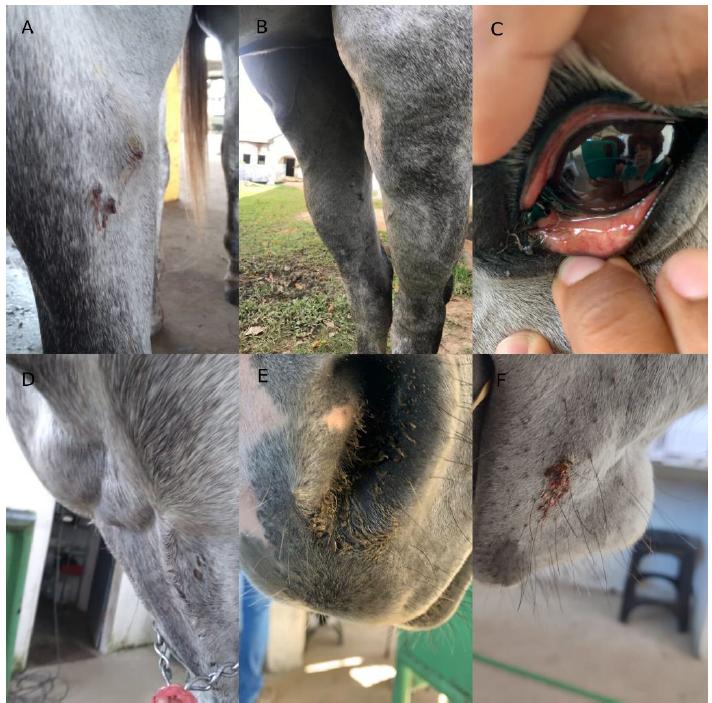
During the clinical examination (Fig. 1), the horse showed apathy, the presence of fistulas and edema in all four limbs, more pronounced in the hind limbs; mucopurulent nasal discharge; petechial hemorrhages on the ocular and vaginal mucosa; exercise intolerance; easy fatigue; and enlarged submandibular lymph nodes. Physiological parameters, including heart rate, respiratory rate, capillary refill time, rectal temperature, and intestinal motility, were within normal limits. Given the patient’s history, anamnesis, and clinical examination, purpura hemorrhagica was considered. Treatment began on the property, and hospitalization was later opted for due to the worsening of the clinical condition.
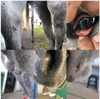
Horse presenting signs of equine purpura hemorrhagica (EPH). A. The right forelimb has the presence of fistulas. B. Generalized edema in the hind limbs. C. Petechial hemorrhages on the ocular mucosa. D. Enlargement of the submandibular lymph node. E. Presence of mucopurulent nasal secretion. F. Presence of fistulas in the face region.
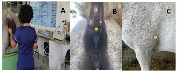
Treatment was initiated on the property with systemic antibiotic therapy, including Gentamicin (6.6mg/kg/IV/SID) for 5 days and Penicillin G Benzathine (20,000 UI/Kg/IM/24h/SID) for 3 days, along with the use of ozone therapy via rectal insufflation (10mL/kg) at a concentration of 15µg/mL. This consisted of 3 cycles, with five consecutive days of treatment followed by a two-day break between cycles. Ozonated minor autohemotherapy (10mL/IM) was administered every 72 hours, totaling 3 applications at acupuncture points VG1 and immunity points (Fig. 2). However, after seven days of treatment, hospitalization was deemed necessary as the patient began exhibiting high fever spikes and dehydration.
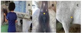
therapies in a horse with purpura hemorrhagica. A. Animal submitted to ozone therapy via rectal insufflation. B and C. Acupuncture points where ozonated minor autohemotherapy was administered. B. VG1 acupuncture point, located above the anus. C. Immunity acupuncture point, located on the olecranon tuberosity, applied bilaterally.
Upon admission to the GRUPEQUI - UFAL outpatient clinic, the horse underwent complementary tests (blood count, serum biochemistry, skin biopsy for histopathology, culture, isolation, and antibiogram of the fistula secretion) to confirm the clinical suspicion of purpura hemorrhagica. The blood count and biochemical tests revealed normocytic normochromic anemia, dehydration, leukocytosis due to lymphocytosis, hyperfibrinogenemia, and serum creatine kinase levels at their upper limits.
The skin and subcutaneous tissue from the radio-ulnar region of the right forelimb, on the medial side, were selected for the biopsy due to the pronounced edema and more recent fistulas in this area. The material was collected using a punch biopsy, removing a circular sample measuring 1.1x0.8x0.4cm, which was preserved in 10% buffered formalin and sent for histopathological examination. The analysis revealed the expansion of the dermis and focally ulcerated epidermis, with an intense, focally extensive inflammatory infiltrate composed of granulomas and areas of hemorrhage characterized by dense aggregates of both intact and degenerated neutrophils, surrounded by foamy macrophages with abundant cytoplasm, epithelioid macrophages forming multinucleated giant cells, lymphocytes, and plasma cells. This indicated a marked, focally extensive pyogranulomatous dermatitis with ulceration and hemorrhage.
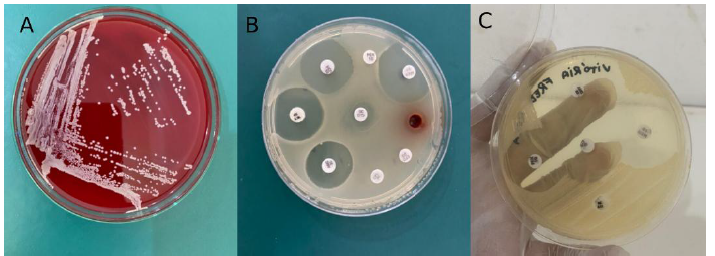
For culture and antibiogram, secretions from the fistulas were collected using sterile swabs, stored in a culture medium, and sent under refrigeration to the Microbiology Laboratory of the Federal University of Alagoas. For microbiological examination, the material was inoculated on a sheep blood agar base and MacConkey agar using the streak-plate technique. The inoculated plates were incubated in a bacteriological incubator for up to 48 hours at 37°C to isolate bacterial agents. The agent was identified through Gram staining and the examination of morphological, staining, and phenotypic characteristics, adhering to the methodology outlined by Koneman. E.W., (2007). In the bacterial culture (Figure 3), based on phenotypic findings, Streptococcus sp. was determined to be the causative agent.
For the antibiogram (Figure 3), the isolated and identified bacterial specimen was inoculated into Brain Heart Infusion (BHI) broth, incubated at 37°C for 24 hours, and used for sensitivity testing. Antimicrobial activity was evaluated using the disk diffusion method on agar. The samples were inoculated on Petri dishes containing Mueller-Hinton agar, with antibiotic disks placed on the agar surface, and the plates were incubated at 37°C for 24 hours. After this period, the inhibition halos were measured. The results indicated resistance to bacitracin and tetracycline, intermediate susceptibility to penicillin, and sensitivity to amoxicillin, amikacin, gentamicin, sulfazotrim, rifampicin, amoxicillin, and meropenem.
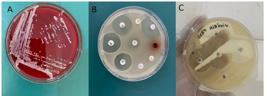
of exudate from a horse affected by Purpura Hemorrhagica on blood agar. Phenotypically showing Streptococcus equi colonies. B and C. Antibiogram test showing the sensitivity of the agent to the following antibiotics: amikacin, sulfazotrim, rifampicin, gentamicin, meropenem, amoxicillin, penicillin, bacitracin, and tetracycline.
Based on the clinical findings and complementary tests, a diagnosis of purpura hemorrhagica was confirmed.
After the laboratory results, with high fever spikes reaching 40.5°C and elevated leukocyte levels, a new treatment plan was established. The treatment included systemic antibiotic therapy with Ceftiofur (4mg/kg/IM/SID) for 10 days, Dexamethasone (0.05mg/kg/IV/SID) for 8 days, and continued ozone therapy via rectal insufflation, following the same protocol mentioned earlier. Dipyrone (25mg/kg/IV/SID) was prescribed for 5 days to be administered during fever episodes. To reduce swelling and stimulate lymphatic circulation, hydrotherapy in the form of showers was applied to both the forelimbs and hindlimbs, along with daily walks, limited to a maximum of ten minutes. Additionally, a multivitamin supplement (Hemolitam® Vetnil/20ml/VO/SID) was administered.
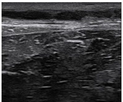
Due to the limb edema, it was observed that the saphenous veins in both hindlimbs were visibly enlarged upon inspection. An ultrasound revealed vascular wall thickening and anechoic content, consistent with thrombophlebitis (Fig. 4). Local therapy was initiated with alternating warm and cold compresses, followed by topical application of heparin sodium ointment (Trombofob®), resulting in gradual improvement over the following days.
After observing additional fever spikes, another blood test was conducted, revealing leukocytosis (total leukocytes 22,200/mm³; segmented neutrophils 18,870/mm³). This indicated that the previously prescribed antibiotic was no longer effective, necessitating the use of a different active ingredient. As a result, a new systemic antibiotic protocol was implemented with Enrofloxacin (6.6mg/kg/IV/SID) for 7 days, along with anti-inflammatory therapy using Dexamethasone (0.05mg/kg/IV/SID) for 5 days. The final cycle of ozone therapy via transrectal insufflation was also administered. As the animal’s clinical signs improved, the Dexamethasone was tapered off using a combination of Dexamethasone (phenylpropionate) and Dexamethasone (sodium phosphate), which provided prolonged action. This combination was administered every 7 days in gradually decreasing doses.
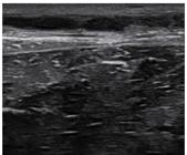
in the saphenous vein in a horse with purpura hemorrhagica. Vascular wall thickening and anechoic content are observed, indicating a condition compatible with thrombophlebitis
The animal responded satisfactorily to the proposed therapy and, after 20 days of hospitalization, was discharged, returning to its normal athletic and reproductive functions. The horse was monitored for up to one year after discharge, with no recurrence of the condition.
DISCUSSION
Purpura Hemorrhagica is a condition that affects equines in various regions, but studies explaining its dynamics are still not well elucidated. Little is known about the factors that trigger the disease, although Streptococcus equi subsp. equi infection and the animal's immune response are mentioned in the limited number of studies available.
The clinical signs observed, such as limb edema, fistulas, petechial hemorrhages in the ocular and vaginal mucosa, apathy, and exercise intolerance, along with the history of infection by Streptococcus equi subsp. equi, are consistent with the findings described by Tomassian (2005) and Nicholson (2025). The results of the laboratory tests, showing normocytic normochromic anemia, leukocytosis with a predominance of segmented neutrophils, hyperfibrinogenemia, and increased creatine kinase, also corroborate the study by Pursterla et al. (2003).
The microscopic findings from the skin biopsy indicated a marked, focally extensive pyogranulomatous dermatitis with ulceration and hemorrhage. This is in line with Reed et al. (2018), who noted that inflammatory infiltrates rich in neutrophils are important histological characteristics for confirming the diagnosis of purpura hemorrhagica.
Ozone therapy can be used to promote controlled oxidation in the patient, stimulating the production of endogenous antioxidants and modulating epigenetics, thereby activating cytoprotective mechanisms. Additionally, ozone therapy has significant antimicrobial capacity, where the oxidation of bacterial cell walls in direct contact with ozone gas causes a fatal hydro-electrolytic imbalance in microorganisms. Ozone can also modulate mitochondrial biogenesis and cellular lifespan, directly impacting their functionality. Regulating low levels of reactive oxygen species induced by ozone helps restore proper mitochondrial function, reducing cell death and necrotic processes. This makes ozone therapy essential for cellular modulation, particularly in autoimmune processes such as purpura hemorrhagica (Basile and Baccarin, 2022; Valdenassi et al., 2022).
Pyrexia is reported by some authors as one of the first signs, occurring between 3 and 14 days after exposure. It is noted that the fever is persistent and can exceed 42°C. This aligns with the findings in the reported case, where the animal experienced several daily episodes of fever, reaching 40.5°C (Boyle et al., 2018).
Regarding vaccination, there is a disparity of opinions among authors. While some oppose vaccination against strangles due to potential side effects, such as the development of local abscesses and purpura, others recommend preventive vaccination in endemic areas to control and prevent the disease, reducing the severity of the disease in vaccinated animals and facilitating eradication. The animal in this case had no prior contact with the vaccine, but specific predisposing factors for immunosuppression, such as a change in environment and transport stress, can be noted (Boyle et al., 2018; Harrington et al., 2002; Pusterla et al., 2003).
It is seen that serological testing may be necessary to indicate the need for vaccination and to identify animals that may be prone to developing purpura hemorrhagica, particularly animals with specific SeM antibodies > 1:1600. Vaccination is only recommended for animals with low titers to avoid complications such as purpura hemorrhagica (Reed et al., 2018).
Although some authors point to susceptibility in older horses with residual immunity, foals experiencing a decline in maternal antibody protection, and vaccinated animals, these groups tend to have limited susceptibility and may develop a mild form of adenitis, known as "catarrhal or atypical strangles." These animals shed virulent Streptococcus equi subsp. equi, which in susceptible horses typically young ones can cause more severe disease (Boyle et al., 2018).
In the reported case, visible enlargement of the saphenous veins was observed upon inspection, leading to an ultrasound examination that revealed vascular wall thickening and anechoic content, which is consistent with thrombophlebitis. This condition had not been previously reported in cases of purpura hemorrhagica, except for vasculitis in smaller caliber vessels (Kaese et al., 2005).
It is evident that early diagnosis and assertive treatment directly influence the prognosis of the animal. Ozone therapy, as an adjuvant treatment, contributed to an improved prognosis, likely due to the immunomodulatory action promoted by ozone. This contrasts with what has been previously reported in the literature, where conventional treatment alone often leads to high mortality rates. For example, Vale (2021, 2022) reported that, out of five animals treated, only one survived.
The indiscriminate and improper use of antibiotics causes significant harm to animals, as incorrect dosing and duration can lead to antibiotic resistance. The practice of administering antibiotics without a veterinarian's prescription is commonly used by caretakers and laypersons, which compromises subsequent treatments, rendering them ineffective (Vale, 2021). The sub-dosage of Penicillin G Benzathine previously used on this animal likely contributed to the intermediate resistance to penicillin, rendering the treatment ineffective and worsening the animal's clinical condition.
After resolving the case, the animal was kept under periodic observation for any signs of relapse. One year later, the animal showed no issues and returned to its normal athletic and reproductive functions.
CONCLUSION
With the improvement of the animal, the role of integrative therapies as a complement to the allopathic treatment of Purpura Hemorrhagica in horses became evident, particularly considering the immunomodulatory and antimicrobial effects of ozone therapy. Further studies on equine purpura hemorrhagic are needed to improve the diagnosis and treatment of the disease, as it is often underdiagnosed and diagnosed late, which compromises the prognosis.
REFERENCES
- BASILE, R.C.; BACCARIN, R.Y.A. Ozonioterapia em animais domésticos: conceitos básicos e diretrizes. Ars Vet., v.38, p.199, 2022.
- BOYLE, A.G.; TIMONEY, J.F.; NEWTON, J.R. et al Streptococcus equi infections in horses: guidelines for treatment, control, and prevention of strangles-revised consensus statement. J. Vet. Intern. Med., v.32, p.633-647, 2018.
- CAMPOS, S.B.S.; SILVA, T.V.; BRAZIL, D.S. et al Purpura Hemorrágica em equino: relato de caso. In: CONGRESSO BRASILEIRO DE MEDICINA VETERINÁRIA, 35., 2008, Gramado. Anais… Gramado: SOVERGS, 2008.
- COSTA, R.A.; POZZOBON, R.; LUBECK, I. et al Surto de púrpura hemorrágica em equinos. In: ENCONTRO INTERNACIONAL DE SANIDADE DE ANIMAIS DE PRODUÇÃO, 2., 2014/ENCONTRO NACIONAL DE DIAGNÓSTICO VETERINÁRIO, 8., 2014, Uruguaiana. Anais… Uruguaiana: Universidade Federal do Pampa, 2014. p.1-2.
- ESPADA, M.A. Ozonioterapia: uma antiga e revolucionária terapia medicinal. Rev. InterCiênc. IMES Catanduva, v.1, p.57, 2020
- HARRINGTON, D.J.; SUTCLIFFE, I.C.; CHANTER, N. The molecular basis of Streptococcus equi infection and disease. Microbes Infect., v.4, p.501-510, 2002.
- JACOBSEN, T.K. Relatório de estágio curricular supervisionado na área de clínica, cirurgia e reprodução de equinos 2019. 65f. Trabalho de Conclusão de Curso (Graduação em Medicina Veterinária) - Universidade Federal de Santa Catarina, Centro de Ciências Rurais, Campus Curitibanos, Curitiba, SC.
- KAESE, H.J.; VALBERG, S.J.; HAYDEN, D.W. et al Infarctive Purpura Hemorrhagica in five horses. J. Am. Vet. Med. Assoc., v.226, n.11, 2005.
- KONEMAN, E.W. Koneman diagnóstico microbiológico 6.ed. [Madrid]: Médica Panamericana, 2007.
- MADRID declaration on ozone therapy. 2.ed. Madrid, Spain: ISCO3, 2015. Available in: www.isco3.org. Accessed in: 09 de Agosto de 2025.
- NICHOLSON, H. Cardiovascular | equine guidelines. In: Australian veterinary prescribing guidelines Melbourne: University of Melbourne, 2025.
- PUSTERLA, N.; WATSON, J.L.; AFFOLTER, V.K. et al Purpura haemorrhagica in 53 horses. Vet. Rec., v.153, p.118-121, 2003.
- REED, S.M.; BAYLY, W.M.; SELLON, D.C. Equine internal medicine 4.ed. [Oxford]: Elsevier, 2018.
- TOMASSIAN, Armen. Enfermidade dos cavalos 4.ed. Botucatu: Varela, 2005.
- VALDENASSI, L.; ROSSI, E.; CORSETT, M.T. et al Sjogren Syndrome successfully treated with oxygen-ozone auto-hemotherapy (o2-o3-AHT): a case report. Eur. Rev. Med. Pharmacol. Sci., v.26, p.5911-5917, 2022.
- VALE, V.R. Resistencia aos antimicrobianos na medicina veterinária 2021. 31f. Trabalho de conclusão de (Bacharelado em Medicina Veterinária) - Centro Universitário do Planalto Central Apparecido dos Santos, Gama-DF.
- ZENG, J.; LU, J. Mechanisms of action involved in ozone-therapy in skin diseases. Int. Immunopharmacol., v.56, p.235-241, 2018.
The research data are available within the article itself.