 Medial patellar desmotomy in cattle and horses in the field: surgical and clinical-pathological aspects - case report
ABSTRACT
Dorsal fixation of patella (FDP) is one of the main causes of lameness in cattle and horses, resulting in locomotor instability. The present study describes surgical and clinical-pathological aspects of medial patellar desmotomy performed in the field in four cows and three horses with intermittent FDP, mild to severe degree. Animals were submitted to general clinical exam and locomotor evaluation in stationary position and in movement, being referred to surgical intervention. The technique was well succeeded in all cases, demonstrating its efficiency and maintaining the animals alive and functional. Furthermore, the economic viability of the procedure in the field was considered favorable, representing an accessible alternative to FDP management in field situations.
Key-words:
surgery; ruminants; patella; orthopedics
RESUMO
A fixação dorsal da patela (FDP) é uma das principais causas de claudicação em bovinos e equinos, resultando em instabilidade locomotora. Este estudo descreve os aspectos cirúrgicos e clínico-patológicos da desmotomia patelar medial realizada a campo em quatro vacas e três cavalos com FDP intermitente, de grau leve a severo. Os animais passaram por exame clínico geral e avaliação locomotora em estação e em movimento, sendo encaminhados para a intervenção cirúrgica. A técnica foi bem-sucedida em todos os casos, demonstrando-se eficaz na manutenção da sobrevida e funcionalidade dos animais. Além disso, a viabilidade econômica do procedimento a campo foi considerada favorável, representando uma alternativa acessível para o manejo da FDP em situações de campo.
Palavras-chave:
cirurgia; ruminantes; patela; ortopedia
INTRODUCTION
Among the main locomotor diseases in cattle and horses one is the dorsal fixation of patella, a condition that affects patellofemoral joint, uni or bilateral, causing locomotor disorders, joint instability and animal disposal when not treated (Dobler et al., 2023).
The disease has a multifactorial nature, and its predisposition is not entirely known, and may be associated with nutritional management, high workload, trauma to the joint region, limbs angular deviations and genetic nature (Peitzmeier et al., 2015).
FDP condition can be treated with counterirritants, physiotherapy (Watanabe et al., 2013) and surgically, standing out the desmotomy of the medial patellar ligament. This technique has a high level of success but must be well elected as there is the possibility of post-surgical complications (Tnibar, 2003).
This work aims to describe the surgical and clinical-pathological aspects of medial patellar desmotomy performed in the field for the treatment of FDP in four cows and three horses, all the procedures being successfully executed and efficient to maintain the affected animals alive, in addition to be economically viable.
CASUISTRY
Seven cases are reported in four cows and three horses, raised in an extensive pasture system, presenting clinical signs of joint instability and locomotor difficulty, attended at a private property and presenting a good general clinical state.
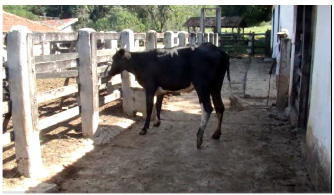
The cattle reported were all females, with the following characteristics: Cow 1, Girolando breed, six years old, weighed 430kg, history of locking the left hind limb with a five-month evolution, as reported by the producer. During locomotion the limb locked, and the animal dragged the claw on the ground (Fig. 1); Cow 2, also Girolando breed, four years old, weighed 420kg, with difficulty to move associated with crackle sounds in the patellofemoral joint. The owner informed that these symptoms began a few weeks before giving birth and have been present since then, at least for six months; Cow 3, Brahman breed, five years old, weighed 518kg, similar history to Cow 2; Cow 4, mixed-breed, two years old, weighed 300kg, with joint instability in right pelvic limb since birth.

Cow presenting history of "locking" the left hind limb, dragging the hoof claw on the ground (Font: Personal file).
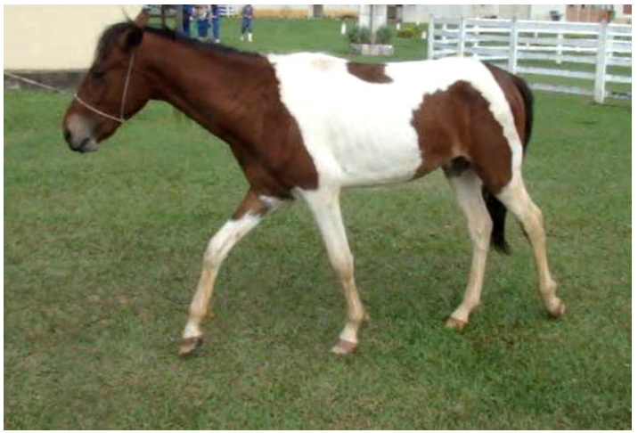
The evaluated horses were males, with the following characteristics: Horse 1, Mangalarga Marchador breed, three years old, weighed 325kg; Horse 2, mixed breed, four years old, weighed 355kg; Horse 3, Tordilho breed, four years old, weighed 400kg. All animals presented a similar history of difficulty to move, characterized by episodes of locking, dragging the hoof claw on the ground (Fig. 2), with crackle sounds in the patellofemoral joint, registered for three, two and six months respectively.
All animals underwent general and specific clinical examination, being the evaluation performed with animals in stationary position, walking in line and in circles where all presented intermittent dorsal fixation of patella, mild to severe degree. The other clinical parameters did not show alterations.

Magalarga Marchador horse presenting "locking" in left hind limb and dragging the hoof claw on the ground (Font: Personal file).
For all the affected animals the surgical approach was the medial patellar desmotomy. The surgery was performed in the field, with Cow 1 and Cow 3 in standing position, while Cow 2 and Cow 4 were positioned in lateral recumbency. The horses were all operated in standing position. All the animals were subjected to 24-hour food fasting and 8-hour water fasting. The pre-surgical preparation included hair trichotomy of the entire pelvic limb region followed by its antisepsis with degerming and topical povidone-iodine to perform the surgery.
In the cattle the pre anesthetic medication (MPA) was made with Xylazine Hydrochloride 2% (0,1mg/kg) intramuscular (IM) and in the horses with Detomidine 1% dose of 20mcg/kg or 0,2mL/100kg intravenously (IV). The anesthetic protocol consisted of local blockage with Lidocaine Hydrochloride 2% using 2mL of local anesthetic over the medial margin of the middle patellar ligament, and more 5 to 10mL around the distal portion of the medial patellar ligament.
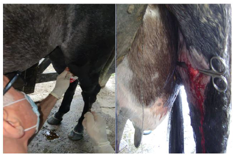
To perform the surgical procedure, a skin incision was made with scalpel blade directly over the medial patellar ligament taking the trochlea crest as an anatomical guide. After location of the medial patellar ligament its section was performed with a desmotome in all the animals, with exception of Cow 1 which was performed with scissors and scalpel support (Fig. 3). Subcutaneous and skin dermorrhaphy were made with Nylon sutures nº 2-0 in Sultan pattern ("x" shaped stitches).
For the cattle post-operative period was administered antibiotic therapy based on Oxytetracycline 20% (20mg/kg) and Flunixin Meglumine (1,1mg/kg), both IM, once a day (SID). The horses postoperative period consisted of antibiotic therapy administration based on Penicillin, 8.333UI of benzylpenicillin, 3,33mg of streptomycin-based sulfate per kg of body weight and Flunixin Meglumine (1,1mg/kg), both IM, SID, for five days. In all animals, the surgical wound was cleaned with Topical Povidone-iodine 10% and Ganadol antibacterial ointment, daily, for 10 days, and after 15 days the stitches were removed. All the animals have completely recovered and were not observed any trans or post-surgical complications.

Bovine and equine operated in standing position for procedure of medial patellar desmotomy. A. Skin incision with scalpel blade, directly over the medial patellar ligament, taking the trochlea crest as an anatomical guide in the equine species. B. Medial patellar ligament location in bovine (Font: Personal file).
DISCUSSION
Medial patellar desmotomy is the most widespread surgical approach for the treatment of FDP, in comparison with other techniques, as surgical fenestration of the patellar ligament (splitting). It is described that in comparison with the splitting technique, the medial patellar desmotomy presents bigger complications in the post-operative, mainly related to lameness. However, complications associated with the two species addressed in this work have not been observed so far, which may be associated with the degree of joint impairment regarding surgical technique and professional experience. Possible observed complications included joint instability, which can predispose the distal patella fragmentation and osteochondrosis (McIlwraith, 1990).
Surgical positioning is reported both in lateral recumbency (Tnibar, 2003) and standing position (Reiners et al., 2005). Both are efficient to easily access the joint region, and the choice is made according to the surgeon's preference. In the present work, both lateral recumbency and standing position showed easy visualization and manipulation of the structures, besides comfort to the animal. Furthermore, the techniques, both surgical and anesthetic and its protocols, were performed in the field, without prejudice to the animals when compared to protocols and techniques applied in veterinary hospitals, resulting in long-term survival and low morbidity of the animals.
Another important related aspect is the cost of developing the technique. Based on the materials used and without considering the veterinary costs, the average value to perform the desmotomy procedure in cattle was about R$137.74 (value expressed in reais). That includes R$21.00 of (Xilazin® 2%, Xylazine hydrochloride 2%, Syntec do Brasil, Brazil), R$4.81 of (Lidovet® 2%, Lidocaine hydrochloride 2%, Bravet Laboratory Ltda, Brazil), R$2.80 of (scalpel blade, Labor Health Supply, Brazil), R$1.79 of (nylon sutures, Technofio Suture Company, Brazil), R$17.39 of (Oxitetraciclina LA®, Oxytetracycline 20%, Syntec do Brasil, Brazil), R$28.69 of (Flunixina®, Flunixin Meglumine, UCBVET Animal Health, Brazil), R$2.31 of (povidone-iodine/PVPI, Riodeine®, Rioquímica Pharmaceutical Industry, Brazil) and R$58.95 of (Ganadol®, healing ointment, Zoetis Brasil, Brazil).
For the horses the average value to perform the procedure was about R$215.85 (value expressed in reais). That includes R$74.70 of (Detomidin® 1%, Detomidine 1%, Syntec do Brasil, Brazil), R$4.81 of (Lidovet® 2%, Lidocaine hydrochloride 2%, Bravet Laboratory Ltda, Brazil), R$2.80 of (scalpel blade, Labor Health Supply, Brazil), R$1.79 of (nylon sutures, Technofio Suture Company, Brazil), R$41.78 of (Penicilin Agrovet®, 8.333UI of benzylpenicillin and 3.33mg of streptomycin sulfate base, Elanco, Brazil), R$28.69 of (Flunixina®, Flunixin Meglumine, UCBVET Animal Health, Brazil), R$2.31 of (povidone-iodine/PVPI, Riodeine®, Rioquímica Pharmaceutical Industry, Brazil) and R$58.95 do (Ganadol®, healing ointment, Zoetis Brasil, Brazil).
It was not observed in the literature works that count the costs allocated to the surgical procedure of desmotomy in the production animals reported. It is of great importance the use of surgical techniques, taking into account the low costs and the possibility to perform the procedure in the field, making possible productivity for the rural producer and offering health and well-being conditions to the animal, based on animal husbandry and welfare standards and guidelines, such as occurred in the animals reported in this work. Based on this, the performed technique shows efficiency, possibility to be performed in the field, regardless of species, and economic viability.
CONCLUSION
The medial patellar desmotomy surgical procedure was efficient and appropriate to treat both species, even in field conditions, and of fundamental importance to maintain life and well-being, in addition to being an economically viable procedure.
REFERENCES
- DOBLER, G.H.; RYBU, F.R.; OLIVEIRA, H.S. et al. Deslocamento dorsal de patela: relato de caso. Arq. Ciênc. Vet. Zool., v.26, p.99-113, 2023.
- McILWRAITH, C.W. Osteochondral fragmentatation of the distal aspecto of the patella in horses. Equine Vet. J., v.22, p.157-163, 1990.
- PEITZMEIER, M.D.; KOONTZ, Z. D.; LYNCH, T.M. et al. Outcomeof medial patellar ligamentdesmoplasty for treatment of intermittent upward fixation of the patella in 24 horses. Can. Vet. J., v.56, p.193-195, 2015.
- REINERS, S.R.; MAY, K.; DIGRASSIE, W. et al. How to perform a standing medial patellar ligament splitting. AAEP Proc., v.51, p.481-483, 2005.
- TNIBAR, A. Treatment of upward fixation of the patela in the horse: an update. Equine Vet. Educ., v.15, p.236-242, 2003.
- WATANABE, M.J.; PYLES, M.D.; ALONSO, J.M. et al. Utilização de contrairritante no tratamento da fixação dorsal de patela intermitente em equinos: relato de casos. Arq. Bras. Med. Vet. Zootec., v.65, p.317-321, 2013.