Eosinophilic annular erythema in a patient with hepatitis B-related cirrhosis
Dear Editor,
Eosinophilic Annular Erythema (EAE) is a rare, benign dermatosis characterized by annular or polycyclic erythe-matous plaques and a dermal eosinophil-rich infiltrate on histopathology. Although initially described in children, it has also been reported in adults, occasionally in association with chronic systemic disorders or malignancy.1,2
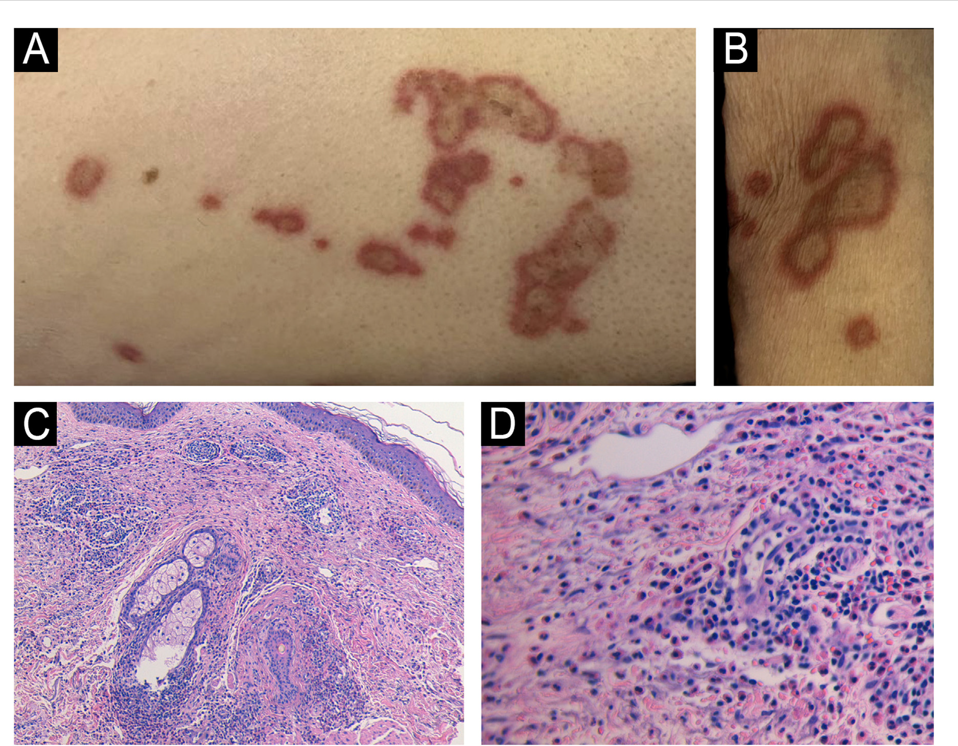
An 89-year-old man with decompensated cirrhosis sec-ondary to chronic hepatitis B infection developed multiple annular plaques on the neck, upper limbs, and proxi-mal thighs during hospital admission. The lesions were erythematous-violaceous, well-demarcated, with central clearing and mild pruritus (Fig. 1A-B). The patient recalled a similar self-limiting eruption approximately one year earlier.
(A) Annular erythematous-violaceous plaques with central clearing on the thigh. (B) Well-demarcated annular plaques on the upper limb. (C) Dense perivascular and interstitial eosinophil-rich dermal infiltrate with edema and erythrocyte extravasation (Hematoxylin & eosin, ×100). (D) Eosinophil-rich infiltrate without vasculitis or flame figures (Hematoxylin & eosin, ×400).
A 4-mm punch biopsy from the thigh revealed a dense perivascular and interstitial dermal infiltrate composed pre-dominantly of eosinophils, with associated dermal edema and erythrocyte extravasation (Fig. 1C-D). No vasculitis or flame figures were observed. Peripheral blood eosinophil count was within normal limits. The clinico-pathological correlation supported the diagnosis of eosinophilic annular erythema. The lesions gradually resolved over four weeks with only topical corticosteroids.
The differential diagnosis of annular plaques includes tinea corporis, annular urticaria, subacute cutaneous lupus erythematosus, erythema annulare centrifugum, and Wells syndrome.3 In EAE, diagnosis relies on clinical morphology together with the characteristic eosinophil-rich dermal infil-trate.
Although often idiopathic, adult-onset EAE has been linked to various systemic conditions, including hepatitis C infection, autoimmune thyroiditis, chronic renal fail-ure, autoimmune pancreatitis, and malignancy.4,5 Recent evidence suggests that EAE represents part of a broader spectrum of eosinophilic dermatoses driven by type 2 inflam-mation, in which IL-5-mediated eosinophil activation and dermal recruitment play a central pathogenic role. This paradigm is supported by reports of therapeutic response to IL-5 blockade in refractory cases.6
The association between EAE and liver disease is increasingly recognized. Hepatic dysfunction (whether viral, autoimmune or cholestatic) creates a systemic inflammatory environment characterized by altered cytokine metabolism, impaired antigen clearance, and enhanced Th2-skewed immunity, all of which may facilitate eosinophil activation and dermal migration. Cases linking EAE to autoimmune hepatitis, including situations in which cutaneous lesions preceded the diagnosis of hepatic autoimmunity, reinforce the possibility of EAE acting as a cutaneous marker of evolv-ing liver disease.7 Similarly, EAE has also been described in association with primary biliary cholangitis.8 In our patient, the onset of EAE during hepatic decompensation suggests that fluctuations in systemic inflammation related to chronic liver disease may act as a triggering factor.
Management of EAE remains challenging due to its relaps-ing course. Topical corticosteroids are frequently used, but systemic therapy is often required. Antimalarials such as chloroquine and hydroxychloroquine have demonstrated efficacy, although prolonged treatment may be necessary to achieve sustained remission.9,10 Other therapeutic options include dapsone, doxycycline, systemic corticosteroids, ciclosporin and methotrexate, with variable responses. In refractory disease, emerging therapies such as JAK inhibitors and biologics targeting Th2 cytokine pathways have shown benefit in isolated cases.6,8 This case expands the clini-cal spectrum of EAE and strengthens the hypothesis that advanced chronic liver disease may contribute to disease expression.
Research data availability
Does not apply.
References
- 1 Sempau L, Larralde M, Luna PC, Casas J, Staiger H. Eosinophilic annular erythema. Dermatol Online J. 2012;18:8.
- 2 Rongioletti F, Fausti V, Kempf W, Rebora A, Parodi A. Eosinophilic annular erythema: an expression of the clinical and patholog-ical polymorphism of Wells syndrome. J Am Acad Dermatol. 2011;65:e135-7.
- 3 Gray T, Lee J, Segars K, Knopp E, Miller R. Eosinophilic annular erythema: a striking clinical presentation with potential sys-temic implications. JAAD Case Rep. 2021;16:33-6.
- 4 Heras MO, Mun˜oz NP, Sancho MI, Millet PU. Eosinophilic annu-lar erythema in adults: report of two cases and review of the literature. An Bras Dermatol. 2017;92:65-8.
- 5 González-López MA, López-Escobar M, Fernández-Llaca H, González-Vela MC, López-Brea M. Eosinophilic annular erythema in a patient with metastatic prostate adenocarcinoma. Int J Dermatol. 2015;54:e80-2.
- 6 Zychowska M, Tutka K, Reich A. Mepolizumab therapy for recal-citrant eosinophilic annular erythema in an adult: a case report and review of treatment options. Dermatol Ther (Heidelb). 2020;10:893-9.
- 7 Awosika O, Totoraitis K, Eleryan M, Rengifo-Pardo M, Ehrlich A. A case of eosinophilic annular erythema as a presenting sign for autoimmune hepatitis. JAAD Case Rep. 2018;4:84-6.
- 8 Niu Y-L, He H-Y, Fang S. Successful treatment of refractory eosinophilic annular erythema with tofacitinib. An Bras Derma-tol. 2025;100:501194.
- 9 Ljubojevi´c Hadˇzavdi´c S, Bartoli´c L, Bradamante M. Prolonged treatment of eosinophilic erythema annulare with chloroquine. Acta Dermatovenerol Croat. 2018;26:262-3.
- 10 Chastagner M, Shourik J, Jachiet M, Battistella M, Lefevre G, Gibier JB, et al. Treatment of eosinophilic annular erythema: retrospective multicenter study and literature review. Ann Der-matol Venereol. 2022;149:123-7.
Edited by
-
Editor
Hiram Larangeira de Almeida Jr.