Durvalumab-induced lichenoid eruption: expanding a rarely recognized adverse event and review of the literature
Dear Editor,
Immune Checkpoint Inhibitors (ICIs) have markedly improved the prognosis of several malignancies but are associated with a variety of immune-related Adverse Events (irAEs), many of which affect the skin.1 The underlying pathophysiology involves loss of peripheral immune tolerance due to PD-L1 blockade, leading to overactivation of cytotoxic CD8+ T-cells and, in some cases, B-cell-mediated autoimmunity. This immune dysregulation results in T-cell infiltration and inflammation of the skin, manifesting as dermatologic irAEs.2 A wide spectrum of skin irAEs has been described with anti-PD-L1 agents, including lichenoid dermatitis, bullous pemphigoid, psoriasiform or spongiotic dermatitis, vitiligo and even severe toxicities such as Stevens-Johnson syndrome and toxic epidermal necrolysis-like reactions.1,2 Durvalumab, a fully human monoclonal antibody targeting PD-L1, is currently approved for unresectable stage III non-small cell lung cancer and advanced urothelial carcinoma. While its safety profile is consistent with that of other ICIs, cutaneous toxicities such as lichenoid reactions remain rare and likely underreported.3,4
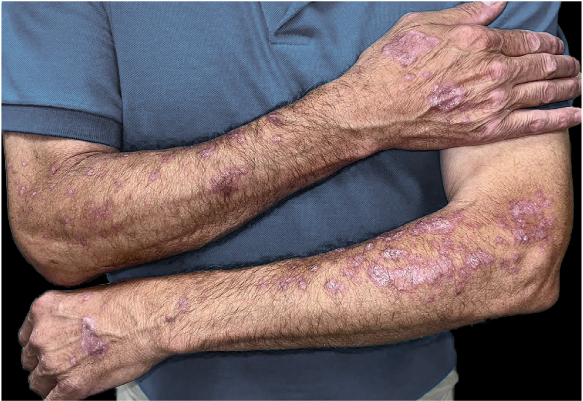
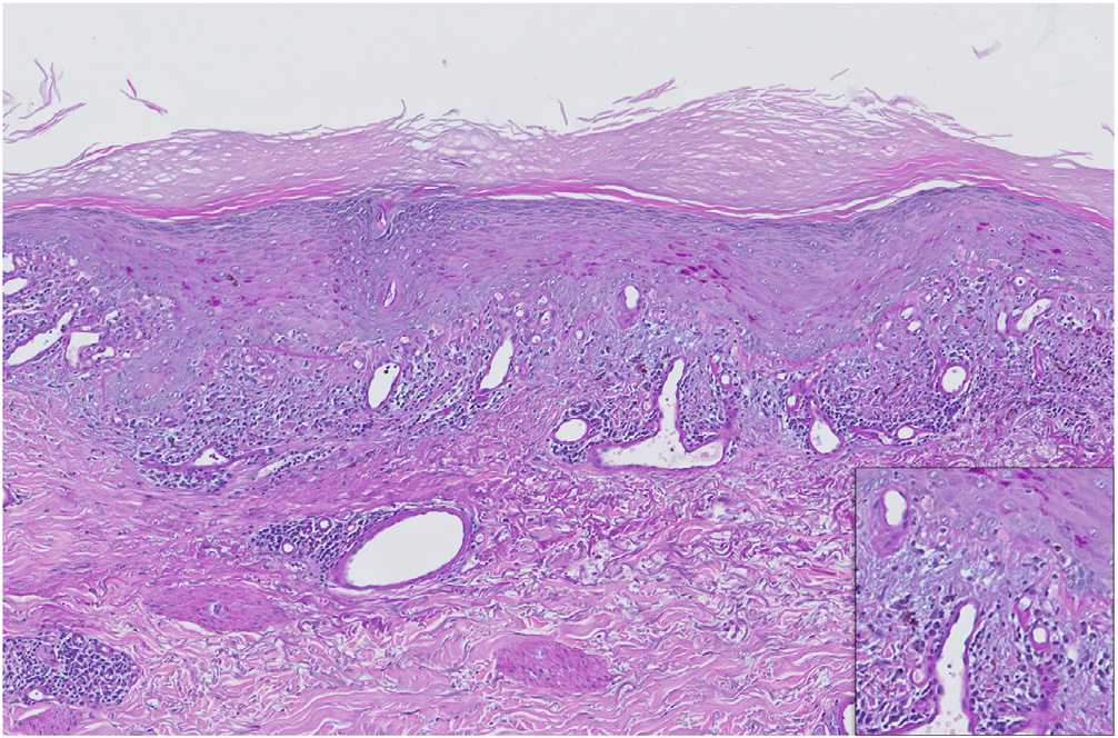
We present the case of a 64-year-old man with stage IIIB lung adenocarcinoma treated with chemoradiotherapy followed by one year of maintenance durvalumab. Three months after initiating immunotherapy, he developed symmetrical erythematous-violaceous plaques with Wickham’s striae on both forearms and dorsal hands (Fig. 1). The lesions were refractory to low-dose oral corticosteroids (10 mg/day) and potent topical corticosteroids. Histopathology revealed a lichenoid infiltrate with basal vacuolar degeneration, consistent with a drug-induced lichenoid reaction (Fig. 2). Treatment with calcipotriol/betamethasone and topical tacrolimus 0.1% led to partial improvement. As the lesions remained asymptomatic, durvalumab was continued. Complete resolution occurred following the completion of durvalumab therapy.

Clinical appearance of erythematous-violaceous plaques on the dorsal hands and forearms, three months after initiating durvalumab therapy and prior to starting dermatologic treatment.

PAS-stained section of an incisional biopsy from the patient, showing orthokeratotic hyperkeratosis with a thickened granular layer. At higher magnification, basal vacuolar degeneration with numerous apoptotic keratinocytes, blurred dermoepidermal junction, a band-like lymphoplasmacytic infiltrate, and abundant melanophages in the papillary dermis are evident. These findings are consistent with a lichenoid drug eruption.
Lichenoid dermatitis is a recognized irAE of ICIs, especially PD-1 inhibitors such as nivolumab and pembrolizumab.3,5,6 However, reports of this reaction under durvalumab are scarce. To our knowledge, only three previous cases of durvalumab-induced lichenoid eruptions have been documented.7-9 These cases display heterogeneous clinical presentations ranging from hypertrophic variants mimicking squamous cell carcinoma to lichenoid reactions progressing to bullous forms or involving mucosal surfaces, including the esophagus. Table 1 summarizes the four reported cases, including the present one. Notably, our patient’s presentation was purely cutaneous and non-bullous, managed entirely with topical therapy without the need for systemic immunosuppression or treatment interruption. The temporal relationship and complete resolution after durvalumab discontinuation further support causality.
Histopathologically, lichenoid eruptions induced by PD-1/PD-L1 inhibitors, such as durvalumab, may show greater spongiosis, epidermal necrosis, and a denser histiocytic infiltrate (CD163+) compared to idiopathic lichen planus. These features, although subtle, can support the diagnosis in the appropriate clinical context.10
This case broadens the phenotypic spectrum of durvalumab-induced lichenoid eruptions and highlights the importance of early recognition, as some cases may progress to bullous variants.7 These reactions typically appear early after treatment initiation and often respond well to symptom-guided conservative therapy. Prompt diagnosis and management can allow the continuation of potentially life-prolonging oncologic treatment without the need for systemic immunosuppression or treatment interruption.7-9 This case underscores the importance of conservative management, allowing continuation of potentially life-prolonging oncologic therapy when feasible.1,7
Research data availability
Does not apply.
References
- 1 Muntyanu A, Netchiporouk E, Gerstein W, Gniadecki R, Litvinov IV. Cutaneous immune-related adverse events to immune checkpoint inhibitors: a dermatology perspective on management. J Cutan Med Surg. 2021;25:59-76.
- 2 Ellis SR, Vierra AT, Millsop JW, Lacouture ME, Kiuru M. Dermatologic toxicities to immune checkpoint inhibitor therapy: a review of histopathologic features. J Am Acad Dermatol. 2020;83:1130-43.
- 3 Lindner AK, Schachtner G, Tulchiner G, Staudacher N, Steinkohl F, Nguyen VA, et al. Immune-related lichenoid mucocutaneous erosions during anti-PD-1 immunotherapy in metastatic renal cell carcinoma: a case report. Urol Case Rep. 2019;23:1-2.
- 4 Coleman E, Ko C, Dai F, Tomayko MM, Kluger H, Leventhal JS. Inflammatory eruptions associated with immune checkpoint inhibitor therapy: a single-institution retrospective analysis with stratification of reactions by toxicity and implications for management. J Am Acad Dermatol. 2019;80:990-7.
- 5 Obara K, Masuzawa M, Amoh Y. Oral lichenoid reaction showing multiple ulcers associated with anti-programmed death cell receptor-1 treatment: a report of two cases and published work review. J Dermatol. 2018;45:587-91.
- 6 Donaldson M, Owen JL, Chae YK, Choi JN. Management of persistent pruritus and lichenoid reaction secondary to nivolumab with narrowband ultraviolet B phototherapy. Front Oncol. 2018;8:405.
- 7 Manko S, Côté B, Provost N. A case of durvalumab-induced lichenoid eruption evolving to bullous eruption after phototherapy: a case report. SAGE Open Med Case Rep. 2021;9:2050313X21993279.
- 8 Myrdal CN, Sundararajan S, Curiel-Lewandrowski C. Widespread hypertrophic lichen planus following programmed cell death ligand 1 blockade. Case Rep Dermatol. 2020;12:119-23.
- 9 Mansilla-Polo M, Fayos-Gregori R, Alonso-Fernández G. Extensive lichen planus after initiation of durvalumab for cholangiocarcinoma. Med Clin (Barc). 2025;164:106926.
- 10 Schaberg KB, Novoa RA, Wakelee HA, Kim J, Cheung C, Srinivas S, et al. Immunohistochemical analysis of lichenoid reactions in patients treated with anti-PD-L1 and anti-PD-1 therapy. J Cutan Pathol. 2016;43:339-46.
Edited by
-
Editor:
Hiram Larangeira de Almeida Jr.