Histopathological evaluation of facial melasma treated with oral tranexamic acid alone and in combination with ketotifen
Abstract
Background: Tranexamic Acid (TA) has demonstrated effectiveness on melasma treatment, and Ketotifen (KET) may inhibit mast cell-mediated melanogenesis. The histologic basis of their depigmenting effects remains unclear.
Objectives: To evaluate histopathological changes from TA with KET over a 3-month treatment. Methods: In this randomized, double-blind trial, 50 women with facial melasma were assigned to KET 1 mg plus TA 250 mg (TA/KET group) or TA 250 mg (TA group), every 12 h for 3-months, with broad-spectrum sunscreen. Skin biopsies were performed at baseline and day-90. Pri-mary outcome was epidermal melanin density reduction; secondary outcomes included stratum corneum compaction, solar elastosis, basement membrane disruptions, and counts of mast cells, melanocytes (including pendulum melanocytes), and upper dermal VEGF density.
Results: Groups were comparable at baseline. Both showed reduced epidermal melanin without intergroup difference, with unchanged VEGF expression, mast cell and melanocyte count, and stratum corneum parameters. The TA/KET-group presented an increase in epidermal thickness, reduction in solar elastosis, pendulum melanocytes counting, and basal membrane disruptions. Study limitations: The short treatment and follow-up may have limited detection of histologic progression. Toluidine blue, while effective in detecting abundant mast cell populations, may lack sensitivity for precise quantification. PAS staining of the basement membrane may occa-sionally produce artifactual discontinuities, even when the membrane is structurally intact. Ketotifen was not tested alone.
Conclusion: Both interventions led to epidermal melanin reduction. The combination TA/KET induced greater dermal and epidermal remodeling changes that surpassed those with TA alone, showing features associated with photoaging reversal and skin homeostasis restoration.
KEYWORDS
Histology; Ketotifen; Melanosis; Tranexamic acid
Introduction
Melasma is a chronic, acquired pigmentary disorder charac-terized by symmetrical hyperpigmentation in sun-exposed areas. It predominantly affects women of childbearing age with intermediate and dark skin phototypes.1 In a Brazilian prevalence study, melasma was observed in 36.3% of adult women.2 Given its frequent facial distribution, melasma often has a significant impact on quality of life.3
Chronic exposure to ultraviolet (UV) and visible light is the primary extrinsic trigger of persistent epidermal hypermelanosis in melasma. This exposure promotes the upregulation of the melanocortin-1 receptor and a-MSH, and induces a senescent phenotype in dermal fibroblasts.4-6 Sex hormones also contribute to the pathogenesis, as pregnancy is a commonly associated risk factor, alongside a genetic predisposition.1 Melanocytes in melasma are hypertrophic and contain an increased number of melanosomes. These cells undergo paracrine stimulation via cytokines and medi-ators such as IL-17, iNOS, endothelin, prostaglandin E2, and growth factors (e.g., a-MSH, SCF, and í3-FGF), as well as by the overexpression of the estrogen receptor-í3.7
In addition to epidermal hyperpigmentation, structural alterations resembling photoaging, such as solar elastosis, basement membrane disruption, increased vascularization, and mast cell infiltration in the upper dermis, are also observed in melasma, partly due to impaired autophagy.8
According to current evidence-based clinical guidelines, the first-line treatment for melasma continues to rely on the use of broad-spectrum sunscreens in combination with topi-cal depigmenting agents.8,9 However, the limited response in many patients has stimulated interest in adjuvant therapies, including systemic agents.
Oral Tranexamic Acid (TA), a synthetic derivative of lysine, inhibits the conversion of plasminogen to plasmin, thereby disrupting the interaction between melanocytes and keratinocytes and reducing melanin synthesis.10 It also suppresses the production of prostaglandins and arachi-donic acid by keratinocytes, molecules known to stimulate melanogenesis. TA may further exert effects by indirectly reducing circulating a-MSH levels and acting as a compet-itive inhibitor of tyrosinase. Additionally, it is proposed to suppress UV-induced plasmin activity, reduce mast cell activation, and downregulate í3-FGF, leading to decreased dermal vascularization and mast cell infiltration.11
Among the various bioactive mediators released by mast cells, histamine has been shown to promote melanogenesis primarily through H2 receptor activation.12 Stem Cell Factor (SCF), which is overexpressed in melasma, supports mast cell survival, proliferation, migration, and activation via binding to the c-KIT receptor. Tryptase, another mast cell-derived protease, activates Matrix Metalloproteinases (MMP-1 and MMP-9), which degrade types I and IV colla-gen, contributing to extracellular matrix degradation and basement membrane disruption. Mast cells also stimulate angiogenesis by releasing VEGF, FGF-2, and TGF-í3.13,14
Ketotifen Fumarate (KET) is a mast cell stabilizer com-monly used in the treatment of asthma with allergic components. One proposed mechanism of action is the blockade of IgE-activated Ca2+ channels, which prevents the release of histamine and other mediators.15 In a clinical trial involving 74 women with melasma, a combination of KET and famotidine (an H2-receptor antagonist) resulted in a modest improvement in skin lightening.16
While the efficacy of tranexamic acid in melasma treat-ment is well established, the role of antihistamines remains less defined. As the histopathological basis of improvement from these agents remains poorly understood, investigat-ing the tissue-level changes associated with KET and TA is essential for supporting the development of new treatment strategies for melasma.
Methods
This was a prospective, randomized (1:1), comparative, parallel-group, double-blind, superiority clinical trial con-ducted from September to December 2022. Fifty women (aged 18-65 years) with untreated facial melasma, except for the use of sunscreen for at least one month, were enrolled. Exclusion criteria included the presence of other facial dermatoses, pregnancy or lactation, immunosuppres-sion, and risk factors for thromboembolism (e.g., use of hormonal contraceptives or hormone replacement therapy, smoking, obesity, sedentary lifestyle, or a personal or family history of thromboembolic events).
The study was conducted at the outpatient clinic of a public hospital in Brazil. The protocol was approved by the Institutional Research Ethics Committee (approval no 0333/2022), and all participants provided written informed consent. The study was registered in the Brazilian Registry of Clinical Trials (ReBEC: RBR-10jn7f39). Clinical, quality-of-life, and colorimetric efficacy data from this trial were published previously.17
Fifty eligible participants were randomized using a computer-generated sequence into two groups. The con-trol group (TA) received 250 mg of oral tranexamic acid plus a placebo every 12 h, while the intervention group (TA/KET) received 250 mg of oral tranexamic acid plus 1 mg of ketotifen every 12 h. All components were compounded into identical capsules, and both participants and evalu-ators were blinded to treatment allocation. The blinding code was maintained by the compounding pharmacy and was only broken after statistical analyses were completed. Sys-temic treatment lasted 90-days.18 All participants received a tinted broad-spectrum sunscreen (Photoaging Control SPF 50, EucerinTM) and were instructed to reapply it every 3-hs during the day throughout the study period.
Skin biopsies were obtained using a 3-mm punch at base-line (D0) and on day-90 (D90), from the same anatomical site, with a maximum variation of 1 cm. Samples were fixed in formalin, paraffin-embedded, and stained with Hematoxylin-Eosin (HE), Fontana-Masson, Periodic Acid-Schiff (PAS), and toluidine blue. Immunohistochemistry was performed using anti-Melan-A (Dako, undiluted, A103 clone, RTU) and anti-vascular endothelial growth factor (VEGF; Dako, 1:50 dilution, VG1 clone) antibodies.
Slides were photographed in triplicate at 40× magnifica-tion using a digital scanner (3DHistech, Budapest, Hungary), selecting representative interfollicular areas free of arti-facts. Images were saved in TIFF format. Quantitative analyses were conducted using ImageJ software (version 1.51e; NIH, USA) by a trained dermatologist who was blinded to both the biopsy time point (D0 or D90) and the treatment allocation. The only exception was the Toluidine Blue stain-ing, which was evaluated by two trained pathologists due to the complexity of its assessment.19
The primary outcome was the change in epidermal melanin content between D0 and D90, assessed by Fontana-Masson staining using split-channel analysis and image binarization. Staining intensity was quantified as the mean pixel intensity in the color histogram, ranging from 0 (black) to 255 (white). Secondary outcomes included: stra-tum corneum compaction and solar elastosis (evaluated on HE); basement membrane disruptions (PAS); mast cell density in the upper dermis (toluidine blue); melanocyte density and presence of pendulum melanocytes (Melan-A); and VEGF staining intensity (color deconvolution analysis in ImageJ). Due to the algorithm used for deconvolu-tion in ImageJ, intensity values are not restricted to the standard 8-bit scale (0-255), but reflect summed optical density, yielding higher raw pixel values proportional to stain deposition.20 Solar elastosis was qualitatively graded as: 0 = absent, 1 = mild/focal to moderate, and 2 = intense. Stratum corneum compaction was scored as: 0 = absent, 1 = partial, and 2 = diffuse.21
Sample size was calculated using G*Power software to detect a between-group difference greater than 20%, assum-ing a standard deviation of a similar magnitude. A 10% dropout rate was anticipated. With a one-tailed alpha of 0.05 and 80% statistical power, the required sample was 25 participants per group.
Normality was assessed using the Shapiro-Wilk test.22 Sta-tistical analyses were performed using IBM SPSS version 25. Between-group comparisons were conducted using a gen-eralized linear mixed-effects model with Sidak adjustment for multiple comparisons.23 Analyses followed the intention-to-treat principle, and missing data were handled via mixed model imputation. Statistical significance was set at p ≤ 0.05 (one-tailed).24
Results
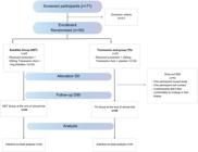
A total of 71 participants were initially screened, of whom 21 (29.5%) were excluded based on eligibility criteria. The trial ultimately enrolled 50 participants, equally allocated to the TA group (n = 25) and the TA/KET group (n = 25). Six participants discontinued the study (5 from the TA group and 1 from the TA/KET group) due to reasons unrelated to treatment-related adverse effects: two were lost to follow-up, and four declined to undergo the second biopsy (Fig. 1).
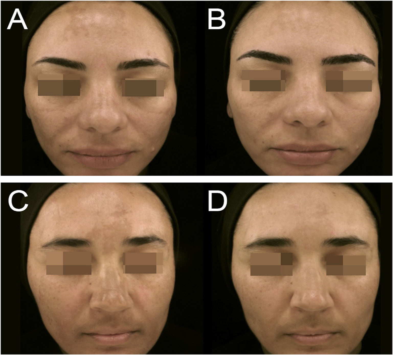
The groups were demographically comparable: the mean age (SD) was 48 (6) years in the TA/KET group and 45 (7) years in the TA group, and most participants in both groups had high phototypes (IV, V, or VI) (Table 1). Both groups showed clinical improvement in all evaluated parameters (mMASI, MelasQoL, and colorimetry) from baseline (D0) to post-treatment (D90), with no differences between them (Fig. 2). At day 90 (D90), for example, the mean reduction in mMASI was 47% (36-58%) in the TA group and 52% (44%-59%) in the KETO group (p > 0.25, 95% CI). During follow-up without treatment (D120), recurrence was observed in both groups, with no significant difference between them (p = 0.36).17
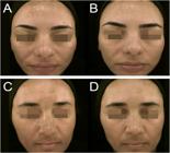
Photographic records are presented at baseline (DO), at the conclusion of systemic treatment (D90). TA group (A-B); KET group (C-D).
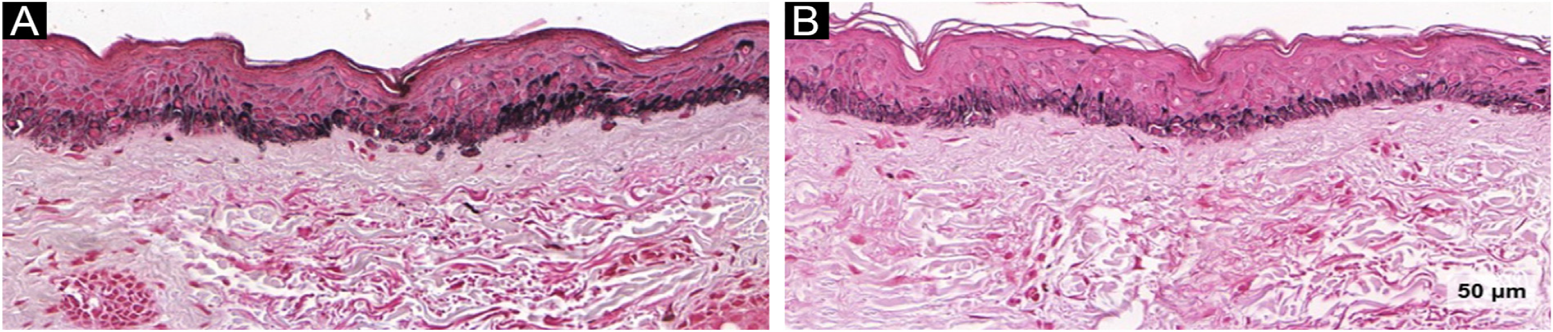
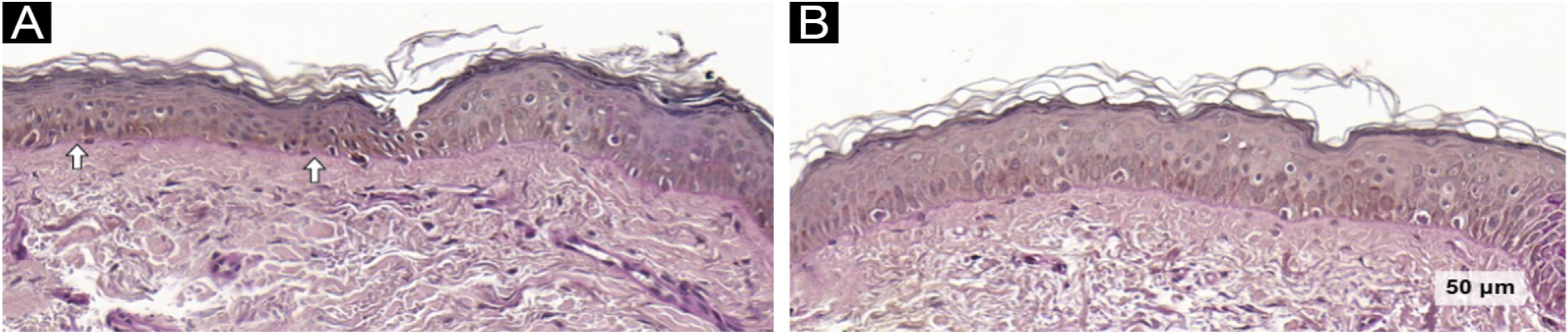
Histological and immunohistochemical findings are summarized in Table 2. An overall reduction in epidermal melanin content was observed in both groups after 90-days of treatment (-12.7; 95% CI: -18.0 to -7.4; p < 0.001), with no difference between the groups (Fig. 3).
Fontana-Masson images of TA group evidencing epidermal melanin reduction: (A) baseline and (B) post-treatment (Fontana-Masson, ×40).
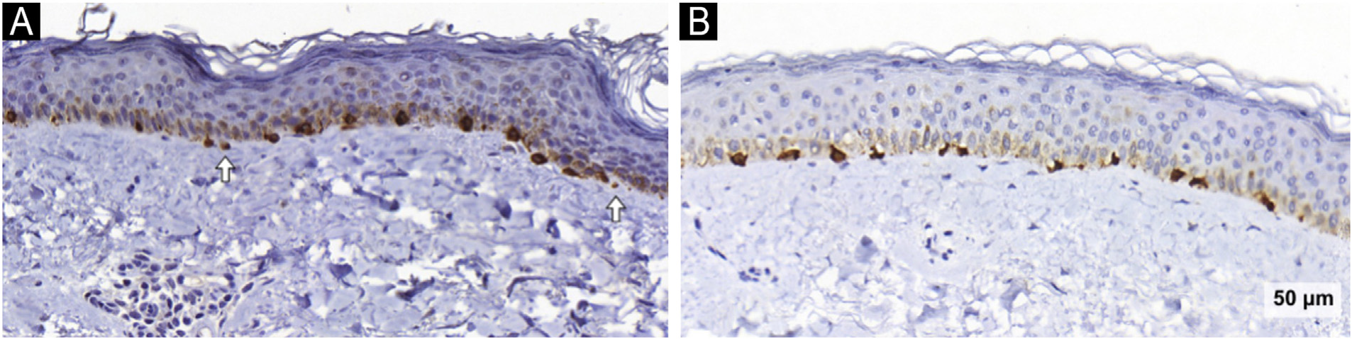
The melanocyte count, assessed via Melan-A staining, remained unchanged in both groups. However, a reduction in melanocyte area was observed across groups after treat-ment (-89; 95% CI: -167 to -11; p = 0.025), again with no between-group difference. Notably, melanocyte area decreased significantly in the TA/KET group (-132; 95% CI: -254 to -10; p = 0.034). In addition, the number of pendulum melanocytes was reduced in the TA/KET group (-0.66; 95% CI: -1.18 to -0.13; p = 0.015) (Fig. 4).
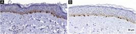
Melan-A images of the TA/KET group. Arrows indicate pendulous melanocytes before (A) and reduced after treatment (B) (Melan-A, ×40).
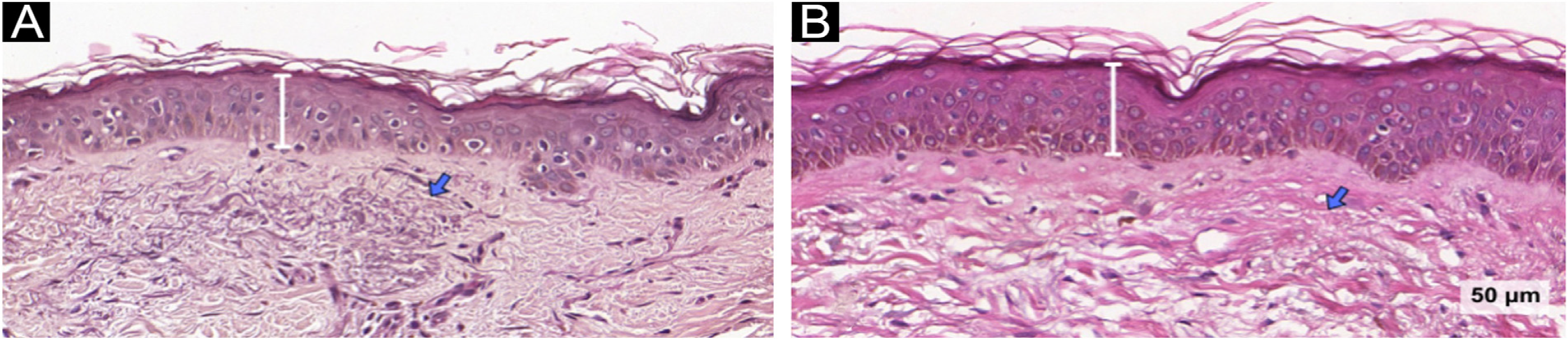
An increase in epidermal thickness was observed exclusively in the TA/KET group at day-90 (14.3; 95% CI: 7.1 to 21.4; p < 0.001). This group also showed a significant reduction in solar elastosis (-0.20; 95% CI: -0.41 to -0.36; p = 0.014), suggesting a favorable remodeling effect in the dermal compartment (Fig. 5). Furthermore, the number of basal membrane disruptions decreased significantly only in the TA/KET group after treatment (-0.73; 95% CI: -1.04 to -0.4; p < 0.001) (Fig. 6).
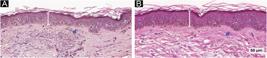
Hematoxylin & eosin images of the TA/KET group: (A) before, (B) after. Bars indicate the increased epidermal thickness; arrows show reduced solar elastosis (Hematoxylin & eosin, ×40).
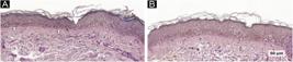
PAS images of the TA/KET group: (A) before, (B) after. Arrows highlight areas of basement membrane discontinuity restored after treatment (40× magnification).
No significant changes were observed between groups or over time in mast cell count (evaluated using toluidine blue staining), stratum corneum thickness and compac-tion (assessed via H&E), or VEGF expression (evaluated by immunostaining).
Discussion
Although no clinical differences were observed between groups after treatment, and a relapse occurred in both groups 30-days after treatment suspension,17 this study revealed distinct histopathological changes following the interventions, suggesting a potential role of mast cell stabi-lizers and antihistamines in the treatment of melasma.
The pathogenesis of melasma involves multiple mech-anisms beyond melanocyte hypertrophy. Alterations in different skin compartments, such as the epidermis, dermis, basement membrane, and cutaneous barrier, are involved in the disease process.7 Histological features such as solar elastosis, increased perivascular mast cells and sebaceous glands, basement membrane disruption, and enhanced vascularization have been consistently demonstrated in melasma-affected skin.25-28 Additionally, a transcriptional analysis of melasma lesions reported the involvement of nearly 300 genes, underscoring the complexity of its patho-physiology, which extends beyond melanocytes to include dermal components as well.29
The reduction in epidermal melanin observed in both the TA and TA/KET groups is consistent with previous evidence supporting the depigmenting effects of TA, even when used without topical lightening agents.10,11 However, the addition of ketotifen did not yield a significantly greater reduction in melanin, possibly due to the dominant effect of TA mask-ing any additional impact. Notably, although between-group differences in melanocyte volume were not statistically significant, the TA/KET group showed a more pronounced reduction after treatment.
While melanophages are commonly increased in hyper-pigmented skin,30,31 their exact role in melasma remains uncertain. Given their absolutely low density compared to epidermal melanin,32 their contribution may be more indicative of chronic photodamage than a primary driver of pigmentation. Nevertheless, melanophages are recognized markers of photoaging and are more prevalent in individuals with darker phototypes.30,31
Although total melanocyte counts remained stable in both groups, a significant reduction in pendulum melanocytes was observed in the TA/KET group. These cells are more prevalent in melasma lesions compared to perilesional skin33 and are supposed to represent inac-tive dendritic melanocytes, as demonstrated by confocal microscopy.26 Lacking epidermal connectivity, they are unlikely to contribute directly to melanogenesis but can be induced by UVA radiation.34 While the precise mechanisms underlying their formation are unclear, evidence suggests that chronic sun exposure may increase MMP-2 expression, promoting basement membrane disruption and pendulum melanocyte formation.35
Mast cell density is typically higher in chronically sun-exposed areas compared to non-exposed regions,36 and they induce melanogenesis via bioactive mediators acting on his-tamine H2 receptors.12 The role of mast cells in photoaging is further supported by experimental studies in UV-irradiated mice, in which ketotifen treatment reduced wrinkle for-mation, epidermal thinning, mast cell accumulation, and matrix degradation.15 Although mast cell counts did not change in the present study, several histological markers of photoaging improved only in the TA/KET group, including reduced solar elastosis, restoration of basement membrane integrity, decreased pendulum melanocyte presence, and increased epidermal thickness.
The maintenance of dermal and epidermal homeosta-sis is critically dependent on the integrity of the basement membrane. In melasma, this structure is often disrupted, facilitating the diffusion of dermal cytokines into the epidermis, which enhances melanogenesis and supports melanocyte protrusion into the dermis. In addition, fibrob-lasts in the melasma dermis exhibit a senescent phenotype with impaired capacity for basement membrane repair.37 In the present study, treatment with TA/KET led to a significant reduction in basement membrane disruptions, an effect not observed in the TA group.
In a cross-sectional study of 50 patients with facial melasma, epidermal atrophy was observed in 90% of lesions versus 28% of perilesional skin samples.32 Similarly, the authors observed increased epidermal thickness after treat-ment in the TA/KET group. A recent study proposed that a thicker epidermis may enable a more uniform distribu-tion of melanin, improving the absorption of solar radiation by photochromophores and reducing direct UV impact on melanocytes, thereby attenuating melanogenesis.6
Solar elastosis, a hallmark of chronic UV exposure, is more prevalent in melasma-affected skin compared to adjacent areas, reinforcing the hypothesis that photoaging contributes to melasma pathogenesis. UVB exposure stimu-lates keratinocytes to promote melanocyte proliferation via secretion of SCF, í3FGF, and a-MSH.5 In this trial, the reduc-tion in solar elastosis observed in the TA/KET group suggests that ketotifen may exert a beneficial remodeling effect on the dermal matrix.
Melasma is associated with increased vascularity, which is not merely a consequence of UV damage but appears to play a central role in disease pathogenesis. VEGF, known to influence melanocyte activity through its receptor, is upreg-ulated in melasma lesions.38 In this study, however, VEGF expression did not decrease in either group, indicating that TA is ineffective to act in this aspect.
The stratum corneum in melasma is typically more compact, and improving its integrity can enhance protection against UV radiation. Treatments such as triple combina-tion creams and procedures like chemical peels or lasers have demonstrated efficacy in reducing stratum corneum compaction.4,34 In the present study, no significant changes in the stratum corneum were observed in either group, potentially due to the absence of adjunctive topical ther-apies or resurfacing interventions.
Finally, as melasma is a multifactorial condition involv-ing multiple skin compartments and pathogenic pathways, the most effective interventions should target multiple fronts to restore skin homeostasis and promote long-lasting results. Mast cells can stimulate melanogenesis through the release of bioactive mediators acting on histamine H2 recep-tors. Ketotifen, a mast cell stabilizer, has demonstrated potential skin-rejuvenating effects, including increased epi-dermal thickness and reductions in solar elastosis, pendulum melanocytes, and basement membrane disruption. These results are consistent with previous studies showing keto-tifen’s ability to prevent UV-induced wrinkle formation in murine models and further support its potential to improve photoaging-related parameters in melasma-affected skin.15 Since the combination of oral TA/KET was not superior to oral TA alone in terms of clinimetric parameters,17 this association should be tested in clinical trials with different regimens before it can be recommended in clinical prac-tice. Notably, the topical application of ketotifen remains largely unexplored, highlighting the need for future studies integrating it into combination treatment strategies.
Limitations
The use of 3 mm biopsies may not represent the whole melasma across the face. The relatively short duration of the trial may also have limited the observation of robust dermal remodulation despite the histopathological changes found. Ketotifen was not assessed without its combination with TA. Toluidine blue, while effective in detecting abun-dant mast cell populations, may compromise the sensitivity for precise quantification of sparse cells. PAS staining of the basement membrane may occasionally produce artifac-tual discontinuities, even when the membrane is structurally intact; however, it did not hinder the detection of changes.
Conclusion
In this trial, both interventions led to a reduction of epi-dermal melanin. The combination of KET with TA produced histopathological changes indicative of dermal and epi-dermal remodeling that surpassed those observed with TA alone, including increased epidermal thickness, reduced solar elastosis, fewer pendulum melanocytes, and improved basement membrane integrity, features associated with photoaging reversal and restoration of skin homeostasis. These findings suggest that targeting mast cell-mediated pathways may offer structural benefits beyond pigment reduction, potentially addressing deeper pathogenic com-ponents of melasma.
-
☆
Study conducted at the Department of Dermatology, Universi-dade Federal de São Paulo, São Paulo, SP, Brazil
-
Financial supportThe trial had major funding from FUNADERSP (Dermatology support fund of the state of São Paulo) under the number 109/22.The donation of sunscreens was made by the company Eucerin and of tranexamic acid by Galena Farmacêutica.
Registration
This trial was registered on Plataforma Brazil (https://plataformabrasil.saude.gov.br) under the number CAAE 57773122.9.0000.5505.
This protocol was registered by the ReBEC platform (Brazilian registry of Clinical Trials) under the number ReBEC: RBR-10jn7f39.
Research data availability
The entire dataset supporting the results of this study was published in this article.
References
- 1 Hexsel D, Lacerda DA, Cavalcante AS, Machado Filho CA, Kalil CL, Ayres EL, et al. Epidemiology of melasma in Brazilian patients: a multicenter study. Int J Dermatol. 2014;53:440-4.
- 2 Dias MO, Minagawa FH, de Abreu AFT, Barbosa MMC, de Amorim RP, Esposito ACC, et al. Prevalence of facial melasma among adult women in a multiracial population. Int J Dermatol. 2024;63:e89-91.
- 3 Handel AC, Miot LDB, Miot HA. Melasma: a clinical and epidemi-ological review. An Bras Dermatol. 2014;89:771-82.
- 4 Espósito AC, Brianezi G, de Souza NP, Miot LDB, Marques MEA, Miot HA. Exploring pathways for sustained melanogenesis in facial melasma: an immunofluorescence study. Int J Cosmet Sci. 2018;40:420-4.
- 5 Cassiano D, Esposito ACC, Hassun K, Lima MMDA, Lima EVA, Miot LDB, et al. Histological changes in facial melasma after treatment with triple combination cream with or without oral tranexamic acid and/or microneedling: a randomised clinical trial. Indian J Dermatol Venereol Leprol. 2022;88:761-70.
- 6 Sheth VM, Pandya AG. Melasma: a comprehensive update: part II. J Am Acad Dermatol. 2011;65:699-714.
- 7 Kwon SH, Hwang YJ, Lee SK, Park KC. Heterogeneous pathol-ogy of melasma and its clinical implications. Int J Mol Sci. 2016;17:824.
- 8 Passeron T, Picardo M. Melasma, a photoaging disorder. Pigment Cell Melanoma Res. 2018;31:461-5.
- 9 Jutley GS, Rajaratnam R, Halpern J, Salim A, Emmett C. System-atic review of randomized controlled trials on interventions for melasma: an abridged Cochrane review. J Am Acad Dermatol. 2014;70:369-73.
- 10 Tan AWM, Sen P, Chua SH, Goh BK. Oral tranexamic acid lightens refractory melasma. Australas J Dermatol. 2017;58:105-8.
- 11 Bala HR, Lee S, Wong C, Pandya AG, Rodrigues M. Oral tranex-amic acid for the treatment of melasma: a review. Dermatol Surg. 2018;44:814-5.
- 12 Yoshida M, Takahashi Y, Inoue S. Histamine induces melanogen-esis and morphologic changes by protein kinase A activation via H2 receptors in human normal melanocytes. J Invest Dermatol. 2000;114:334-42.
- 13 Halova I, Draberova L, Draber P. Mast cell chemotaxis - chemoat-tractants and signaling pathways. Front Immunol. 2012;3:119.
- 14 Palareti G, Legnani C, Cosmi B, Maurer M. Ultraviolet radiation and skin mast cells: effects, mechanisms and relevance for skin diseases. Exp Dermatol. 2018;27:3-8.
- 15 Kim MS, Lee DH, Lee CW, Kim YK, Lee MJ, Shin CY, et al. Mast cell stabilizer, ketotifen, prevents UV-induced wrinkle formation. J Invest Dermatol. 2013;133:1104-7.
- 16 Dias JAF, Lima PB, Cassiano DP, Espósito ACC, Bagatin E, Miot LDB, et al. Oral ketotifen associated with famotidine for the treatment of facial melasma: a randomized, double-blind, placebo-controlled trial. J Eur Acad Dermatol Venereol. 2022;36:e123-5.
- 17 Cruz ACL, Cassiano DP, Bleixuvehl de Brito M, Espósito ACC, Hassun K, Miot HA, et al. Oral ketotifen associated with oral tranexamic acid for the treatment of facial melasma in women:a randomized, double-blind, placebo-controlled trial. J Eur Acad Dermatol Venereol. 2025;39:e39-41.
- 18 Miola AC, Espósito ACC, Miot HA. Techniques for ran-domization and allocation for clinical trials. J Vasc Bras. 2025;23:e20240046.
- 19 Bagatin E, Miot HA. How to design and write a clinical research protocol in cosmetic dermatology. An Bras Dermatol. 2013;88:69-75.
- 20 Schneider CA, Rasband WS, Eliceiri KW. NIH Image to ImageJ: 25 years of image analysis. Nat Methods. 2012;9:671-5.
- 21 Espósito AC, Brianezi G, de Souza NP, Miot LDB, Miot HA. Exploratory study of epidermis, basement membrane zone, upper dermis alterations and Wnt pathway activation in melasma compared to adjacent and retroauricular skin. Ann Dermatol. 2020;32:101-8.
- 22 Miot HA. Assessing normality of data in clinical and experimental trials. J Vasc Bras. 2017;16:88-91.
- 23 Miot HA. Analysis of data with dependent measures in clinical and experimental studies. J Vasc Bras. 2023;22:e20220150.
- 24 Miola AC, Miot HA. P-value and effect-size in clinical and exper-imental studies. J Vasc Bras. 2021;20:e20210038.
- 25 Chung JH, Eun HC. Angiogenesis in skin aging and photoaging. J Dermatol. 2007;34:593-600.
- 26 Kang HY, Bahadoran P, Suzuki I, Zugaj D, Khemis A, Passeron T, et al. In vivo reflectance confocal microscopy detects pig-mentary changes in melasma at a cellular level resolution. Exp Dermatol. 2010;19:228-33.
- 27 Hernandez-Barrera R, Torres-Alvarez B, Castanedo-Cazares JP, Oros-Ovalle C, Moncada B. Solar elastosis and presence of mast cells as key features in the pathogenesis of melasma. Clin Exp Dermatol. 2008;33:305-8.
- 28 Torres-Alvarez B, Mesa-Garza IG, Castanedo-Cazares JP, Fuentes-Ahumada C, Oros-Ovalle C, Navarrete-Solis J, et al. Histochemical and immunohistochemical study in melasma: evi-dence of damage in the basal membrane. Am J Dermatopathol. 2011;33:291-5.
- 29 Kang HY, Suzuki I, Lee DJ, Ha J, Reiniche P, Aubert J, et al. Transcriptional profiling shows altered expression of Wnt pathwayand lipid metabolism-related genes as well as melanogenesis-related genes in melasma. J Invest Dermatol. 2011;131:1692-700.
- 30 Grimes PE, Yamada N, Bhawan J. Light microscopic, immuno-histochemical, and ultrastructural alterations in patients with melasma. Am J Dermatopathol. 2005;27:96-101.
- 31 Kang WH, Yoon KH, Lee ES, Kim J, Lee KB, Yim H, et al. Melasma: histopathological characteristics in 56 korean patients. Br J Der-matol. 2002;146:228-37.
- 32 Kang HY, Ortonne JP. What should be considered in treatment of melasma. Ann Dermatol. 2010;22:373-8.
- 33 Gautam M, Patil S, Nadkarni N, Sandhu M, Godse K, Setia M. Histopathological comparison of lesional and perilesional skin in melasma: a cross-sectional analysis. Indian J Dermatol Venereol Leprol. 2019;85:367-73.
- 34 Bhawan J, Grimes P, Pandya AG, Keady M, Byers HR, Guevara IL, et al. A histological examination for skin atrophy after 6 months of treatment with fluocinolone acetonide 0.01%, hydro-quinone 4%, and tretinoin 0.05% cream. Am J Dermatopathol. 2009;31:794-8.
- 35 Lee DJ, Park KC, Ortonne JP, Kang HY. Pendulous melanocytes: a characteristic feature of melasma and how it may occur. Br J Dermatol. 2012;166:684-6.
- 36 Janssens AS, Heide R, den Hollander JC, Mulder PG, Tank B, Oranje AP. Mast cell distribution in normal adult skin. J Clin Pathol. 2005;58:285-9.
- 37 Espósito AC, Brianezi G, de Souza NP, Santos DC, Miot LDB, Miot HA. Ultrastructural characterization of damage in the basement membrane of facial melasma. Arch Dermatol Res. 2020;312:223-7.
- 38 Kim EH, Kim YC, Lee ES, Kang HY. The vascular characteristics of melasma. J Dermatol Sci. 2007;46:111-6.
Edited by
-
Editor
Luciana P. Fernandes Abbade.