Convolutional Neural Network Applications In Cardiac Imaging
Abstract
Neural networks are computer models that mimic the workings of the human brain, learning from large volumes of data to perform increasingly complex tasks. This is done by means of interconnected artificial neurons that propagate information from raw layers to refined layers, sometimes of a weighted nature, weaving in the classification or identification of non-linear patterns. The Convolutional Neural Network (CNN) is a specialized deep learning architecture designed to extract and analyze spatial patterns within images, enabling applications in diagnostic decision-making and longitudinal clinical monitoring. Its use in Computed Tomography and Magnetic Resonance Imaging through cardiac image segmentation makes it possible to quickly and efficiently define cardiac structures, playing an important role in the reconstruction of three-dimensional images and in supporting preoperative procedures. In addition, compared to other forms of conventional analysis, it guarantees objective interpretations and superior image quality, based on the removal of artifacts from the studied segment. Echocardiograms have shown good results in identifying pathologies such as pulmonary hypertension, amyloidosis, hypertrophic cardiomyopathy and congenital diseases, and studies on the automation of these exams have shown an opening for what could be a dissemination framework in more remote areas where manual operators are not available. Consequently, CNN-based approaches have the potential to streamline cardiovascular imaging workflows, minimize inter-observer variability, and expand diagnostic capabilities to underserved and remote regions with limited access to specialized care.
Keywords
Computer Neural Networks; Artificial Intelligence; Cardiac Imaging Techniques
Resumo
Redes neurais são modelos computacionais que imitam o funcionamento do cérebro humano, aprendendo a partir de grandes volumes de dados para realizar tarefas cada vez mais complexas. Isso é feito por meio de neurônios artificiais interconectados que propagam informações de camadas brutas para camadas refinadas, às vezes com pesos atribuídos, permitindo a classificação ou identificação de padrões não lineares. A Rede Neural Convolucional (CNN, do inglês Convolutional Neural Network) é uma arquitetura especializada de aprendizado profundo (deep learning) projetada para extrair e analisar padrões espaciais em imagens, possibilitando aplicações em decisões diagnósticas e monitoramento clínico longitudinal. Seu uso em tomografia computadorizada e ressonância magnética, por meio da segmentação de imagens cardíacas, permite definir estruturas cardíacas de forma rápida e eficiente, desempenhando um papel importante na reconstrução de imagens tridimensionais e no suporte a procedimentos pré-operatórios. Além disso, em comparação com outras formas de análise convencional, a CNN garante interpretações objetivas e qualidade superior de imagem, com base na remoção de artefatos do segmento estudado. Ecocardiogramas têm mostrado bons resultados na identificação de patologias como hipertensão pulmonar, amiloidose, cardiomiopatia hipertrófica e doenças congênitas, e estudos sobre a automação desses exames apontam para uma possível ampliação do acesso em áreas remotas onde operadores manuais não estão disponíveis. Consequentemente, abordagens baseadas em CNN têm o potencial de otimizar os fluxos de trabalho em imagem cardiovascular, minimizar a variabilidade entre observadores e expandir as capacidades diagnósticas para regiões carentes e remotas com acesso limitado a cuidados especializados.
Palavras-chave
Redes neurais de computação; inteligência artificial; técnicas de imagem cardíaca

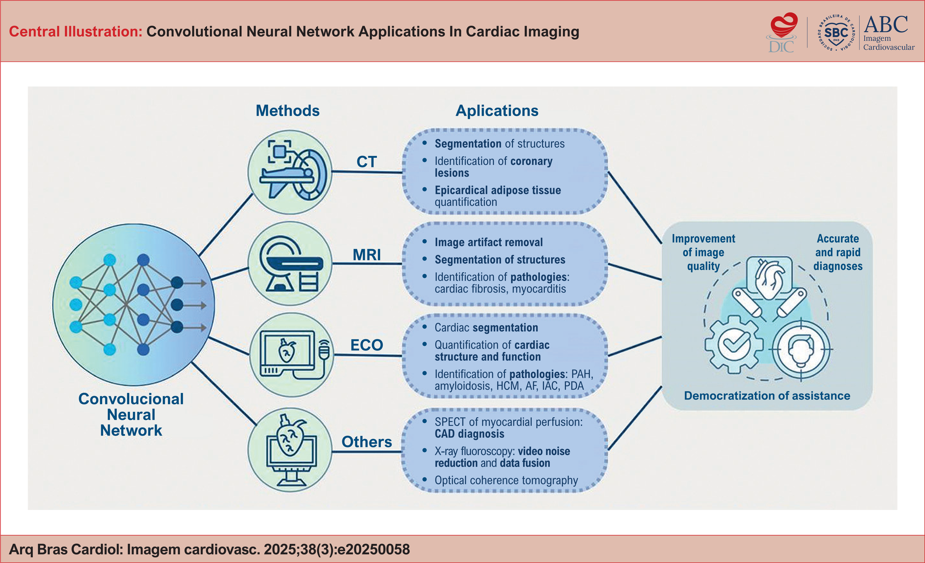
Applications of Convolutional Neural Networks in different types of diagnostic imaging methods in cardiology.
AF: Atrial Fibrilation; CAD: Coronary artery disease; CT: Computed Tomography; ECO: Echocardiography; HCM: Hypertrophic Cardiomyopathy; IAC: Interatrial Communication; MRI: Magnetic Imaging Resonance; OCT: Optical Coherence Tomography; PAH: Pulmonary Arterial Hypertension; PDA: Patent Ductus Arteriosus; SPECT-MPI: Single Photon Emission Computed Tomography – Myocardial Perfusion Imaging.
Introduction
The human nervous system is one of the most efficient ways of capturing and interpreting data, reflecting the stimuli and experiences to which the body is subjected throughout life.1 According to the phenomenon of neuroplasticity, all these interactions dictate brain modifications, being adapted for decision-making and allowing pre-existing knowledge to be learned and act as a basis for the creation of new information circuits.2
Neural networks have emerged as artificial models that try to recover this processing quickly and effectively in order to solve the problems raised, becoming important tools in the dynamic field of artificial intelligence (AI).3 In order to overcome this challenge, networks are trained to identify recognition patterns from thousands or millions of examples, making it possible to build the system's own learning.4 This happens through what is called "input", i.e. any new concept that is inserted into the ordering. The set of inputs generates a response, the "output", which can be influenced in a weighted way, always taking a reference standard as its essence.5
This can be exemplified simply: in the assessment of a patient with suspected atrial fibrillation (AF), when an electrocardiogram is requested for investigation, each voltage in the tracing can take on the role of an input neuron, which can continue to influence the cascade and serve as a new input for the next neuron, solidifying the final response.6 Thus, if the diagnosis is AF when there is no corresponding tracing, the weights that led to that result are reduced. On the other hand, if there is a match, they are strengthened and reused in subsequent analyses.7 In addition, extra notions can be added, such as the patient's age, gender and comorbidities.
The application of AI in the medical field has been widely explored and, in recent years, has become a disruptive technology for interpreting images.8 Within this context, the Convolutional Neural Network (CNN) has emerged as a version of this processing aimed at recognizing and classifying images, reducing the computational effort and improving its robustness.8 The hierarchical arrangement of multiple convolutional layers allows CNNs to progressively transform low-level features – such as pixel intensity gradientes – into increasingly abstract and clinically relevant representations.9 During the training period, the CNN exercises and individualizes its filters according to the task to be performed. In a way, image classification starts by detecting the edges of coarser pixels in the initial layer, using this to detect simple shapes in the second layer and so on, allowing more complex features to be distinguished. Finally, the last layer is responsible for advanced image reading, based on the filters built.10
The advantages of CNN over other neural networks is that it establishes the vital resources for successful prediction, reducing the dimensionality of the resources in the previous layer.11
Accurate segmentation of cardiac images is essential for clinical decision making, and Deep Learning techniques and the use of CNN have shown remarkable results in this area, although their quantification and analysis remains an ongoing challenge due to the complexity of cardiac substructures and existing anatomical variability. The individualization of this analysis is essential to solidify objective evidence of the most varied pathologies investigated, facilitating diagnosis, monitoring and clinical follow-up of the patient.12
In this approach, through the image segmentation method, the convolutional neural network enables fast and efficient responses. This is because in certain situations, the presence of certain features is more important than others. Sometimes recognizing the location of an alteration in the heart chamber is the only essential objective, and it is not necessary to see these structures with high pixel definition.13
The application scenario covers a wide range of imaging exams, from use in echocardiography to calculate end-systolic and end-diastolic volume, through to the ability to assess the ventricular or atrial space in multiple slices, as well as the observation of coronary calcification plaques through CT-guided coronary angiography (CCTA). The range of applications of CNNs in cardiac imaging is highlighted in the Central Illustration. For the most part, these aspects are supported by figures and engravings from atlases or other documents that are included in the model's database. Another interesting point is that there are already studies using the system itself to weave in and ensure the quality of ultrasounds and other tests made available.5
Applications of CNN in CT
The use of convolutional neural networks has excelled in automating the segmentation of cardiac and vascular structures in computed tomography (CT) images, a process by which cardiac structures or pathological lesions are delimited.5,12,13 The segmentation of cardiac images by CT has clinical relevance in the identification of valvular heart disease, evaluation of coronary artery disease, diagnosis of congenital heart disease, preoperative planning and in extracardiac indications.12 The application of CNNs has brought speed and efficiency to the cardiac segmentation process, since it was previously carried out manually and subjectively by radiologists. In a study of the segmentation of cardiac chambers and great vessels, Sharkey et al. presented results indicating that the variability of analysis between observers is similar to the variability between the observer and the CNN.14 These findings support the premise that CNN-based segmentation can deliver reproducible, objective, and diagnostically accurate results, comparable to expert manual assessment.
With regard to the identification of anatomical structures, Chen, et al. showed that the convolutional neural network proved to be an accurate and efficient tool for assessing the volume and three-dimensional reconstruction of the left atrium.15 The calculation of volume by CNN proved to be capable of predicting the risk of recurrence of atrial fibrillation in patients after catheter ablation. Astudillo, et al. used a CNN technique to identify the coronary cusps and the right and left coronary ostia on multi-detector row computed tomography.16 These structures are important reference points for defining the perimeter and area of the aortic annulus and, consequently, the aortic valve prostheses to be implanted by transcatheter means. The method proved to be reproducible and reliable, and could be applied in routine preoperative planning in order to reduce time spent and accuracy in defining reference points.
CNNs have also proved to be excellent tools for identifying pathological images in coronary angiography. Coronary computed tomography angiography is an important method for assessing the severity of arterial stenosis, as well as being highly accurate in quantifying atherosclerosis when compared to intravascular ultrasound. In a multicenter study, a neural network was trained to segment coronary plaques.17 The study showed excellent agreement between the Deep Learning tool and the measurements of experienced readers regarding the volume of the atheroma plaque and the degree of arterial stenosis. There was also agreement with vascular ultrasound measurements, as well as prognostic value in terms of the risk of future acute myocardial infarction.
Quantification of epicardial adipose tissue can also be optimized through the use of CNNs. Epicardial adipose tissue is a metabolically active depot of visceral fat and may be related to visceral obesity and metabolic syndrome.18 Coronary angiography is the gold standard for identifying epicardial adipose tissue, but its manual quantification is laborious and challenging. In this scenario, West et al. developed a CNN to automatically quantify epicardial adipose tissue and identify its clinical association with cardiac and non-cardiac diseases.19 The method proved to be accurate, fast and reproducible for quantifying epicardial adiposity using coronary angiography. The tool was also able to show good prognostic value for mortality in general and for cardiovascular events.
Applications of CNN in MRI
In recent years, magnetic resonance imaging has become an excellent tool for investigating cardiovascular diseases, with a high capacity to provide complex diagnoses.20,21 It is a non-invasive, versatile method free of ionizing radiation, capable of identifying structure, function, flow and characterization of cardiac tissue. Late gadolinium enhancement and T1/T2 mapping have added important information about cardiac viability and pathologies such as tissue infarction and diffuse myocardial fibrosis. The development of flow MRI has also brought major advances by providing blood flow information in several planes, which allows the identification of intracardiac shunts.20 However, the lengthy scanning process and slow image production are disadvantages of MRI. MRI images must take cardiac and respiratory movement into account to avoid artifacts, which must be suppressed by reconstruction algorithms. In this scenario, Deep learning–based reconstruction methods have demonstrated superior performance compared with conventional algorithms, yielding higher-fidelity images, enhanced artifact suppression, and substantially reduced processing times.
Hauptmann, et al. trained a three-dimensional residual CNN in U-Net to remove spatio-temporal artifacts in subsampling real-time MRI data from images of patients with congenital heart disease.22 Artifact removal by the CNN was compared with the conventional compression sensing technique, a tool that requires intense and time-consuming computational reconstruction. The CNN-based method achieved artifact suppression over five times faster than the compressed sensing approach, while delivering superior image quality and more accurate ventricular volumetric measurements. El-Rewaidy et al. also presented important results on the reconstruction of MRI images with delayed gadolinium enhancement using a convolutional neural network.23 Compared to the compression sensing technique, image reconstruction using CNN was 300 times faster.
CNNs have influenced multiple stages of the MRI workflow, from image acquisition planning and real-time reconstruction to post-processing tasks such as quantitative analysis and prognostic modeling. Like CT, MRI also plays an important role in the segmentation and consequent identification of cardiac structures.24 In this process using MRI, the use of CNNs has also been related to greater efficiency.25 Sander et al. used a CNN to perform the segmentation of cardiac structures and another auxiliary CNN to automatically identify areas of segmentation failure.26 The study showed that cardiac image segmentation using CNNs associated with the manual correction of detected segmentation failures resulted in increased performance.
In terms of disease identification, many studies have used conventional image indices as input data for neural networks to diagnose heart disease. For example, cardiac scarring identified on magnetic resonance imaging is associated with unfavorable cardiovascular outcomes. Bekheet, et al., developed a CNN called FibrosisNet to diagnose cardiac fibrosis on MRI, showing high diagnostic accuracy and precision.27 Sharifrazi et al., obtained important results by combining a deep convolutional neural network with the K-means clustering technique, forming CNN-KCL, to diagnose and classify myocarditis based on MRI image data.28 The technique using CNN outperformed other traditional Machine Learning algorithms in classifying myocarditis in terms of accuracy and precision.
Applications of CNN in echocardiography
Echocardiography is the most easily accessible and widely used imaging method for assessing cardiac structure and function. Its advantages include rapid image acquisition and the absence of ionizing radiation, which make it an efficient and democratic method. Echocardiography can be used both to screen asymptomatic patients and to diagnose and follow up cardiac conditions.29,30 As it is an operator-dependent test, it requires a certain degree of experience on the part of the professional, and there can be variations in interpretation.29,31
When it comes to applying automated image interpretation to echocardiography, there are some intrinsic challenges to this method. The echocardiogram consists of generating both static images, as well as videos and Doppler recordings collected from different points of view to obtain the desired parameters. In addition, the values obtained in the measurements can vary greatly due to the intrinsic variability of heartbeats. There is also the variation that already exists when extrapolating measurements from two-dimensional images to measurements of the three-dimensional object.32,33
Despite these impasses and given the complexity of the diagnostic method in question, an automated learning approach would bring many benefits and assist human interpretation, as it would allow automatic identification and classification of views. In fact, the use of Deep Learning (DL), and more specifically Convolutional Neural Networks (CNNs), to train artificial intelligence in the interpretation of echocardiograms would make it possible to reduce costs and, consequently, democratize this test. This technological integration could enable echocardiographic acquisition by operators with limited specialized training, thereby facilitating access to cardiac imaging in geographically remote or resource-limited settings.32
Several studies have shown the effectiveness of using CNNs in echocardiography. Among the applications observed, the majority are for quantifying the structure and function of the heart and for cardiac segmentation.10,29,34–38 It has also been used in the detection of left ventricular hypertrophy,39–41 aortic stenosis,42,43 identification of cardiac phases,44 early detection of acute myocardial infarction45 and classification of cardiac visualizations.46,47 The model used by Ghorbani et al, in addition to accurately identifying elements of cardiac structure and function, trained a CNN to predict systemic phenotypes that modify cardiovascular risk, such as age, gender, height and weight, from echocardiogram images.29
Zhang et al went further and trained a convolutional neural network for fully automated interpretation of echocardiograms, including visualization identification, image segmentation, quantification of heart structure and function, and disease detection. This study demonstrated success in the automated analysis of view type and image segmentation. With regard to the structure and function of the heart, there was some discrepancy between manual and automated measurements, especially of the structure, with a tendency to overestimate the values. Finally, the model was successful in identifying pulmonary arterial hypertension (PAH), amyloidosis and hypertrophic cardiomyopathy, based on analysis of the masses, structure and function of the heart chambers.32
Recent studies have also evaluated the capacity for automatic diagnosis of pulmonary arterial hypertension and,48 according to the results obtained by DuBrock et al,49 the algorithm used is capable of speeding up the diagnosis of PAH by 6 to 18 months, an extremely relevant aspect in a disease where early diagnosis is crucial for effective control of the condition.48,49
Another described pathology that can be identified by echocardiography using CNNs is occult atrial fibrillation (AF). Yuan et al developed a model that was able to distinguish between sinus rhythm and AF, as well as predicting the concomitant presence of paroxysmal AF in those with sinus rhythm. The model performed better than using clinical risk factors, transthoracic echocardiogram measurements, left atrial size or CHA2DS2VASc score.50 Research using DL has also been carried out and obtained favorable results in the area of fetal and neonatal echocardiography, such as for the prenatal diagnosis of atrial septal defect51 and for the identification of patent ductus arteriosus in neonates.52
Application of CNN in other methods
Single Photon Emission Computed Tomography (SPECT) of myocardial perfusion is a test that can identify the presence of myocardial ischemia and therefore coronary artery disease. Betancur et al analyzed the application of deep learning to this diagnostic method, aiming to verify the ability to automatically predict coronary disease through CNNs trained with myocardial perfusion images, compared to the usual method currently used. The result was greater efficacy and better detection sensitivity with the deep learning method.53 Other studies have also evaluated the ability to automatically classify myocardial perfusion images. Papadrianos et al trained CNNs to identify three possible outputs - infarction, ischemia and normal perfusion - and all models evaluated presented positive and reliable results.54 Su et al demonstrated the feasibility and accuracy of CNN-based diagnosis for CAD, with a system that is already used in practice and is capable of considerably reducing the time needed to interpret images.55
X-ray fluoroscopy allows real-time visualization of the cardiac structure, which makes it an indispensable method for performing image-guided procedures, such as cardiac resynchronization therapy, angiography and endovascular aortic repair. Deep learning has also been successfully tested to aid these procedures, either by reducing video noise or by fusing preoperative data with intraoperative x-ray images in order to guide the intervention and reduce contrast exposure.56–59 Studies have also shown better sensitivity and specificity in characterizing coronary lesions using optical coherence tomography when deep learning is applied.60–62
Conclusion
The application of CNN in cardiac image analysis has emerged as a versatile tool, contributing to accurate and rapid diagnoses. Its incorporation into cardiovascular imaging makes it possible to remove the bias of inter-observer variability, reduce image artifacts, improve the automation of measurements and, finally, overcome geographical barriers by allowing the evaluation of images acquired remotely and thus democratizing care. Future perspectives should focus on large-scale validation studies to confirm the generalizability and clinical robustness of CNN-based approaches across diverse patient populations and imaging protocols. Furthermore, integrating these algorithms into routine workflows will require addressing regulatory and interoperability challenges to ensure safe and effective adoption in real-world cardiovascular practice.
-
Sources of funding
There were no external funding sources for this study.
-
Study association
This study is not associated with any thesis or dissertation work.
-
Ethics approval and consent to participate
This article does not contain any studies with human participants or animals performed by any of the authors.
-
Use of Artificial Intelligence
The authors did not use any artificial intelligence tools in the development of this work.
Data Availability
The underlying content of the research text is contained within the manuscript.
References
- 1 Rotta NT, Ohlweiler L, Riesgo RS. Transtornos da Aprendizagem. Porto Alegre: Artmed; 2015.
-
2 Costa RLS. Neuroscience and Learning. Rev Bras Educ. 2023;28:e280010. doi: 10.1590/S1413-24782023280010.
» https://doi.org/10.1590/S1413-24782023280010 -
3 Kriegeskorte N, Golan T. Neural Network Models and Deep Learning. Curr Biol. 2019;29(7):R231-6. doi: 10.1016/j.cub.2019.02.034.
» https://doi.org/10.1016/j.cub.2019.02.034 -
4 Krittanawong C, Zhang H, Wang Z, Aydar M, Kitai T. Artificial Intelligence in Precision Cardiovascular Medicine. J Am Coll Cardiol. 2017;69(21):2657-64. doi: 10.1016/j.jacc.2017.03.571.
» https://doi.org/10.1016/j.jacc.2017.03.571 -
5 Litjens G, Ciompi F, Wolterink JM, de Vos BD, Leiner T, Teuwen J, et al. State-of-the-Art Deep Learning in Cardiovascular Image Analysis. JACC Cardiovasc Imaging. 2019;12(8 Pt 1):1549-65. doi: 10.1016/j.jcmg.2019.06.009.
» https://doi.org/10.1016/j.jcmg.2019.06.009 -
6 Krittanawong C, Johnson KW, Rosenson RS, Wang Z, Aydar M, Baber U, et al. Deep Learning for Cardiovascular Medicine: A Practical Primer. Eur Heart J. 2019;40(25):2058-73. doi: 10.1093/eurheartj/ehz056.
» https://doi.org/10.1093/eurheartj/ehz056 -
7 El-Taraboulsi J, Cabrera CP, Roney C, Aung N. Deep Neural Network Architectures for Cardiac Image Segmentation. Artif Intell Life Sci. 2023;4:100083. doi: 10.1016/j.ailsci.2023.100083.
» https://doi.org/10.1016/j.ailsci.2023.100083 -
8 Johnson KW, Soto JT, Glicksberg BS, Shameer K, Miotto R, Ali M, et al. Artificial Intelligence in Cardiology. J Am Coll Cardiol. 2018;71(23):2668-79. doi: 10.1016/j.jacc.2018.03.521.
» https://doi.org/10.1016/j.jacc.2018.03.521 -
9 Bizopoulos P, Koutsouris D. Deep Learning in Cardiology. IEEE Rev Biomed Eng. 2019;12:168-93. doi: 10.1109/RBME.2018.2885714.
» https://doi.org/10.1109/RBME.2018.2885714 -
10 Lau ES, Di Achille P, Kopparapu K, Andrews CT, Singh P, Reeder C, et al. Deep Learning-Enabled Assessment of Left Heart Structure and Function Predicts Cardiovascular Outcomes. J Am Coll Cardiol. 2023;82(20):1936-48. doi: 10.1016/j.jacc.2023.09.800.
» https://doi.org/10.1016/j.jacc.2023.09.800 -
11 Wang P, Qiao J, Liu N. An Improved Convolutional Neural Network-Based Scene Image Recognition Method. Comput Intell Neurosci. 2022;2022:3464984. doi: 10.1155/2022/3464984.
» https://doi.org/10.1155/2022/3464984 -
12 Alnasser TN, Abdulaal L, Maiter A, Sharkey M, Dwivedi K, Salehi M, et al. Advancements in Cardiac Structures Segmentation: A Comprehensive Systematic Review of Deep Learning in CT Imaging. Front Cardiovasc Med. 2024;11:1323461. doi: 10.3389/fcvm.2024.1323461.
» https://doi.org/10.3389/fcvm.2024.1323461 -
13 Lüscher TF, Wenzl FA, D’Ascenzo F, Friedman PA, Antoniades C. Artificial Intelligence in Cardiovascular Medicine: Clinical Applications. Eur Heart J. 2024;45(40):4291-304. doi: 10.1093/eurheartj/ehae465.
» https://doi.org/10.1093/eurheartj/ehae465 -
14 Sharkey MJ, Taylor JC, Alabed S, Dwivedi K, Karunasaagarar K, Johns CS, et al. Fully Automatic Cardiac Four Chamber and Great Vessel Segmentation on CT Pulmonary Angiography Using Deep Learning. Front Cardiovasc Med. 2022;9:983859. doi: 10.3389/fcvm.2022.983859.
» https://doi.org/10.3389/fcvm.2022.983859 -
15 Chen HH, Liu CM, Chang SL, Chang PY, Chen WS, Pan YM, et al. Automated Extraction of Left Atrial Volumes from Two-Dimensional Computer Tomography Images Using a Deep Learning Technique. Int J Cardiol. 2020;316:272-8. doi: 10.1016/j.ijcard.2020.03.075.
» https://doi.org/10.1016/j.ijcard.2020.03.075 -
16 Astudillo P, Mortier P, Bosmans J, De Backer O, Jaegere P, Iannaccone F, et al. Automatic Detection of the Aortic Annular Plane and Coronary Ostia from Multidetector Computed Tomography. J Interv Cardiol. 2020;2020:9843275. doi: 10.1155/2020/9843275.
» https://doi.org/10.1155/2020/9843275 - 17 Lin A, Park C. HHS Public Access international multicentre study. 2022;4(4):10–1.
-
18 Oikonomou EK, Antoniades C. The Role of Adipose Tissue in Cardiovascular Health and Disease. Nat Rev Cardiol. 2019;16(2):83-99. doi: 10.1038/s41569-018-0097-6.
» https://doi.org/10.1038/s41569-018-0097-6 -
19 West HW, Siddique M, Williams MC, Volpe L, Desai R, Lyasheva M, et al. Deep-Learning for Epicardial Adipose Tissue Assessment with Computed Tomography: Implications for Cardiovascular Risk Prediction. JACC Cardiovasc Imaging. 2023;16(6):800-16. doi: 10.1016/j.jcmg.2022.11.018.
» https://doi.org/10.1016/j.jcmg.2022.11.018 -
20 Oscanoa JA, Middione MJ, Alkan C, Yurt M, Loecher M, Vasanawala SS, et al. Deep Learning-Based Reconstruction for Cardiac MRI: A Review. Bioengineering. 2023;10(3):334. doi: 10.3390/bioengineering10030334.
» https://doi.org/10.3390/bioengineering10030334 -
21 Argentiero A, Muscogiuri G, Rabbat MG, Martini C, Soldato N, Basile P, et al. The Applications of Artificial Intelligence in Cardiovascular Magnetic Resonance-A Comprehensive Review. J Clin Med. 2022;11(10):2866. doi: 10.3390/jcm11102866.
» https://doi.org/10.3390/jcm11102866 -
22 Hauptmann A, Arridge S, Lucka F, Muthurangu V, Steeden JA. Real-Time Cardiovascular MR with Spatio-Temporal Artifact Suppression Using Deep Learning-Proof of Concept in Congenital Heart Disease. Magn Reson Med. 2019;81(2):1143-56. doi: 10.1002/mrm.27480.
» https://doi.org/10.1002/mrm.27480 -
23 El-Rewaidy H, Neisius U, Mancio J, Kucukseymen S, Rodriguez J, Paskavitz A, et al. Deep Complex Convolutional Network for Fast Reconstruction of 3D Late Gadolinium Enhancement Cardiac MRI. NMR Biomed. 2020;33(7):e4312. doi: 10.1002/nbm.4312.
» https://doi.org/10.1002/nbm.4312 -
24 Peng P, Lekadir K, Gooya A, Shao L, Petersen SE, Frangi AF. A Review of Heart Chamber Segmentation for Structural and Functional Analysis Using Cardiac Magnetic Resonance Imaging. MAGMA. 2016;29(2):155-95. doi: 10.1007/s10334-015-0521-4.
» https://doi.org/10.1007/s10334-015-0521-4 -
25 Xiong Z, Fedorov VV, Fu X, Cheng E, Macleod R, Zhao J. Fully Automatic Left Atrium Segmentation from Late Gadolinium Enhanced Magnetic Resonance Imaging Using a Dual Fully Convolutional Neural Network. IEEE Trans Med Imaging. 2019;38(2):515-24. doi: 10.1109/TMI.2018.2866845.
» https://doi.org/10.1109/TMI.2018.2866845 -
26 Sander J, de Vos BD, Išgum I. Automatic Segmentation with Detection of Local Segmentation Failures in Cardiac MRI. Sci Rep. 2020;10(1):21769. doi: 10.1038/s41598-020-77733-4.
» https://doi.org/10.1038/s41598-020-77733-4 -
27 Bekheet M, Sallah M, Alghamdi NS, Rusu-Both R, Elgarayhi A, Elmogy M. Cardiac Fibrosis Automated Diagnosis Based on FibrosisNet Network Using CMR Ischemic Cardiomyopathy. Diagnostics. 2024;14(3):255. doi: 10.3390/diagnostics14030255.
» https://doi.org/10.3390/diagnostics14030255 -
28 Sharifrazi D, Alizadehsani R, Joloudari JH, Band SS, Hussain S, Sani ZA, et al. CNN-KCL: Automatic Myocarditis Diagnosis Using Convolutional Neural Network Combined with K-Means Clustering. Math Biosci Eng. 2022;19(3):2381-402. doi: 10.3934/mbe.2022110.
» https://doi.org/10.3934/mbe.2022110 -
29 Ghorbani A, Ouyang D, Abid A, He B, Chen JH, Harrington RA, et al. Deep Learning Interpretation of Echocardiograms. NPJ Digit Med. 2020;3:10. doi: 10.1038/s41746-019-0216-8.
» https://doi.org/10.1038/s41746-019-0216-8 -
30 American College of Cardiology Foundation Appropriate Use Criteria Task Force; American Society of Echocardiography; American Heart Association; American Society of Nuclear Cardiology; Heart Failure Society of America; Heart Rhythm Society; et al. ACCF/ASE/AHA/ASNC/HFSA/HRS/SCAI/SCCM/SCCT/SCMR 2011 Appropriate Use Criteria for Echocardiography. A Report of the American College of Cardiology Foundation Appropriate Use Criteria Task Force, American Society of Echocardiography, American Heart Association, American Society of Nuclear Cardiology, Heart Failure Society of America, Heart Rhythm Society, Society for Cardiovascular Angiography and Interventions, Society of Critical Care Medicine, Society of Cardiovascular Computed Tomography, Society for Cardiovascular Magnetic Resonance American College of Chest Physicians. J Am Soc Echocardiogr. 2011;24(3):229-67. doi: 10.1016/j.echo.2010.12.008.
» https://doi.org/10.1016/j.echo.2010.12.008 -
31 De Geer L, Oscarsson A, Engvall J. Variability in Echocardiographic Measurements of Left Ventricular Function in Septic Shock Patients. Cardiovasc Ultrasound. 2015;13:19. doi: 10.1186/s12947-015-0015-6.
» https://doi.org/10.1186/s12947-015-0015-6 -
32 Zhang J, Gajjala S, Agrawal P, Tison GH, Hallock LA, Beussink-Nelson L, et al. Fully Automated Echocardiogram Interpretation in Clinical Practice. Circulation. 2018;138(16):1623-35. doi: 10.1161/CIRCULATIONAHA.118.034338.
» https://doi.org/10.1161/CIRCULATIONAHA.118.034338 -
33 Madani A, Arnaout R, Mofrad M, Arnaout R. Fast and Accurate View Classification of Echocardiograms Using Deep Learning. NPJ Digit Med. 2018;1:6. doi: 10.1038/s41746-017-0013-1.
» https://doi.org/10.1038/s41746-017-0013-1 -
34 Shi S, Alimu P, Mahemut P. The Study of Echocardiography of Left Ventricle Segmentation Combining Transformer and Convolutional Neural Networks. Int Heart J. 2024;65(5):889-97. doi: 10.1536/ihj.23-638.
» https://doi.org/10.1536/ihj.23-638 -
35 Jiang J, Deng H, Xue Y, Liao H, Wu S. Detection of Left Atrial Enlargement Using a Convolutional Neural Network-Enabled Electrocardiogram. Front Cardiovasc Med. 2020;7:609976. doi: 10.3389/fcvm.2020.609976.
» https://doi.org/10.3389/fcvm.2020.609976 -
36 Painchaud N, Duchateau N, Bernard O, Jodoin PM. Echocardiography Segmentation with Enforced Temporal Consistency. IEEE Trans Med Imaging. 2022;41(10):2867-78. doi: 10.1109/TMI.2022.3173669.
» https://doi.org/10.1109/TMI.2022.3173669 -
37 Lei Y, Fu Y, Roper J, Higgins K, Bradley JD, Curran WJ, et al. Echocardiographic Image Multi-Structure Segmentation Using Cardiac-SegNet. Med Phys. 2021;48(5):2426-37. doi: 10.1002/mp.14818.
» https://doi.org/10.1002/mp.14818 -
38 Wehbe RM, Katsaggelos AK, Hammond KJ, Hong H, Ahmad FS, Ouyang D, et al. Deep Learning for Cardiovascular Imaging: A Review. JAMA Cardiol. 2023;8(11):1089-98. doi: 10.1001/jamacardio.2023.3142.
» https://doi.org/10.1001/jamacardio.2023.3142 -
39 Jian Z, Wang X, Zhang J, Wang X, Deng Y. Diagnosis of Left Ventricular Hypertrophy Using Convolutional Neural Network. BMC Med Inform Decis Mak. 2020;20(1):243. doi: 10.1186/s12911-020-01255-2.
» https://doi.org/10.1186/s12911-020-01255-2 -
40 Nazar W, Nazar K, Daniłowicz-Szymanowicz L. Machine Learning and Deep Learning Methods for Fast and Accurate Assessment of Transthoracic Echocardiogram Image Quality. Life. 2024;14(6):761. doi: 10.3390/life14060761.
» https://doi.org/10.3390/life14060761 -
41 Duffy G, Cheng PP, Yuan N, He B, Kwan AC, Shun-Shin MJ, et al. High-Throughput Precision Phenotyping of Left Ventricular Hypertrophy with Cardiovascular Deep Learning. JAMA Cardiol. 2022;7(4):386-95. doi: 10.1001/jamacardio.2021.6059.
» https://doi.org/10.1001/jamacardio.2021.6059 -
42 Holste G, Oikonomou EK, Mortazavi BJ, Coppi A, Faridi KF, Miller EJ, et al. Severe Aortic Stenosis Detection by Deep Learning Applied to Echocardiography. Eur Heart J. 2023;44(43):4592-604. doi: 10.1093/eurheartj/ehad456.
» https://doi.org/10.1093/eurheartj/ehad456 -
43 Wessler BS, Huang Z, Long GM Jr, Pacifici S, Prashar N, Karmiy S, et al. Automated Detection of Aortic Stenosis Using Machine Learning. J Am Soc Echocardiogr. 2023;36(4):411-20. doi: 10.1016/j.echo.2023.01.006.
» https://doi.org/10.1016/j.echo.2023.01.006 -
44 Farhad M, Masud MM, Beg A, Ahmad A, Ahmed LA, Memon S. Cardiac Phase Detection in Echocardiography Using Convolutional Neural Networks. Sci Rep. 2023;13(1):8908. doi: 10.1038/s41598-023-36047-x.
» https://doi.org/10.1038/s41598-023-36047-x -
45 Muraki R, Teramoto A, Sugimoto K, Sugimoto K, Yamada A, Watanabe E. Automated Detection Scheme for Acute Myocardial Infarction Using Convolutional Neural Network and Long Short-Term Memory. PLoS One. 2022;17(2):e0264002. doi: 10.1371/journal.pone.0264002.
» https://doi.org/10.1371/journal.pone.0264002 -
46 Naser JA, Lee E, Pislaru SV, Tsaban G, Malins JG, Jackson JI, et al. Artificial Intelligence-Based Classification of Echocardiographic Views. Eur Heart J Digit Health. 2024;5(3):260-9. doi: 10.1093/ehjdh/ztae015.
» https://doi.org/10.1093/ehjdh/ztae015 -
47 Østvik A, Smistad E, Aase SA, Haugen BO, Lovstakken L. Real-Time Standard View Classification in Transthoracic Echocardiography Using Convolutional Neural Networks. Ultrasound Med Biol. 2019;45(2):374-84. doi: 10.1016/j.ultrasmedbio.2018.07.024.
» https://doi.org/10.1016/j.ultrasmedbio.2018.07.024 -
48 Sun D, Hu Y, Li Y, Yu X, Chen X, Shen P, et al. Chamber Attention Network (CAN): Towards Interpretable Diagnosis of Pulmonary Artery Hypertension Using Echocardiography. J Adv Res. 2024;63:103-15. doi: 10.1016/j.jare.2023.10.013.
» https://doi.org/10.1016/j.jare.2023.10.013 -
49 DuBrock HM, Wagner TE, Carlson K, Carpenter CL, Awasthi S, Attia ZI, et al. An Electrocardiogram-Based AI Algorithm for Early Detection of Pulmonary Hypertension. Eur Respir J. 2024;64(1):2400192. doi: 10.1183/13993003.00192-2024.
» https://doi.org/10.1183/13993003.00192-2024 -
50 Yuan N, Stein NR, Duffy G, Sandhu RK, Chugh SS, Chen PS, et al. Deep Learning Evaluation of Echocardiograms to Identify Occult Atrial Fibrillation. NPJ Digit Med. 2024;7(1):96. doi: 10.1038/s41746-024-01090-z.
» https://doi.org/10.1038/s41746-024-01090-z -
51 Zhou X, Yang T, Ruan Y, Zhang Y, Liu X, Zhao Y, et al. Application of Neural Networks in Prenatal Diagnosis of Atrioventricular Septal Defect. Transl Pediatr. 2024;13(1):26-37. doi: 10.21037/tp-23-394.
» https://doi.org/10.21037/tp-23-394 -
52 Erno J, Gomes T, Baltimore C, Lineberger JP, Smith DH, Baker GH. Automated Identification of Patent Ductus Arteriosus Using a Computer Vision Model. J Ultrasound Med. 2023;42(12):2707-13. doi: 10.1002/jum.16305.
» https://doi.org/10.1002/jum.16305 -
53 Betancur J, Commandeur F, Motlagh M, Sharir T, Einstein AJ, Bokhari S, et al. Deep Learning for Prediction of Obstructive Disease from Fast Myocardial Perfusion SPECT: A Multicenter Study. JACC Cardiovasc Imaging. 2018;11(11):1654-63. doi: 10.1016/j.jcmg.2018.01.020.
» https://doi.org/10.1016/j.jcmg.2018.01.020 -
54 Papandrianos NI, Feleki A, Papageorgiou EI, Martini C. Deep Learning-Based Automated Diagnosis for Coronary Artery Disease Using SPECT-MPI Images. J Clin Med. 2022;11(13):3918. doi: 10.3390/jcm11133918.
» https://doi.org/10.3390/jcm11133918 -
55 Su TY, Chen JJ, Chen WS, Chang YH, Lu HH. Deep Learning for Myocardial Ischemia Auxiliary Diagnosis Using CZT SPECT Myocardial Perfusion Imaging. J Chin Med Assoc. 2023;86(1):122-30. doi: 10.1097/JCMA.0000000000000833.
» https://doi.org/10.1097/JCMA.0000000000000833 -
56 Toth D, Miao S, Kurzendorfer T, Rinaldi CA, Liao R, Mansi T, et al. 3D/2D Model-to-Image Registration by Imitation Learning for Cardiac Procedures. Int J Comput Assist Radiol Surg. 2018;13(8):1141-9. doi: 10.1007/s11548-018-1774-y.
» https://doi.org/10.1007/s11548-018-1774-y -
57 Breininger K, Albarqouni S, Kurzendorfer T, Pfister M, Kowarschik M, Maier A. Intraoperative Stent Segmentation in X-Ray Fluoroscopy for Endovascular Aortic Repair. Int J Comput Assist Radiol Surg. 2018;13(8):1221-31. doi: 10.1007/s11548-018-1779-6.
» https://doi.org/10.1007/s11548-018-1779-6 -
58 Sadda P, Qarni T. Real-Time Medical Video Denoising with Deep Learning: Application to Angiography. Int J Appl Inf Syst. 2018;12(13):22-8. doi: 10.5120/ijais2018451755.
» https://doi.org/10.5120/ijais2018451755 -
59 Luo Y, Ma Y, O’ Brien H, Jiang K, Kohli V, Maidelin S, et al. Edge-Enhancement Densenet for X-Ray Fluoroscopy Image Denoising in Cardiac Electrophysiology Procedures. Med Phys. 2022;49(2):1262-75. doi: 10.1002/mp.15426.
» https://doi.org/10.1002/mp.15426 -
60 Abdolmanafi A, Duong L, Dahdah N, Adib IR, Cheriet F. Characterization of Coronary Artery Pathological Formations from OCT Imaging Using Deep Learning. Biomed Opt Express. 2018;9(10):4936-60. doi: 10.1364/BOE.9.004936.
» https://doi.org/10.1364/BOE.9.004936 -
61 Abdolmanafi A, Cheriet F, Duong L, Ibrahim R, Dahdah N. An Automatic Diagnostic System of Coronary Artery Lesions in Kawasaki Disease Using Intravascular Optical Coherence Tomography Imaging. J Biophotonics. 2020;13(1):e201900112. doi: 10.1002/jbio.201900112.
» https://doi.org/10.1002/jbio.201900112 -
62 Gessert N, Lutz M, Heyder M, Latus S, Leistner DM, Abdelwahed YS, et al. Automatic Plaque Detection in IVOCT Pullbacks Using Convolutional Neural Networks. IEEE Trans Med Imaging. 2019;38(2):426-34. doi: 10.1109/TMI.2018.2865659.
» https://doi.org/10.1109/TMI.2018.2865659
Edited by
-
Editor responsible for the review:
Marcelo Tavares