Important Mitral Regurgitation and Ventricular Dysfunction in Hypereosinophilic Syndrome: A Case Report
Keywords
Mitral Valve Insufficiency; Heart Failure; Hypereosinophilic Syndrome
Palavras-chave
Insuficiência da Valva Mitral; Insuficiência Cardíaca; Síndrome Hipereosinofílica
Keywords
Mitral Valve Insufficiency; Heart Failure; Hypereosinophilic Syndrome
Palavras-chave
Insuficiência da Valva Mitral; Insuficiência Cardíaca; Síndrome Hipereosinofílica
Introduction
Hypereosinophilic syndrome (HES) represents a group of rare hematologic diseases characterized by an increased number of eosinophils in the peripheral blood. Once activated, these eosinophils can cause damage to host tissues and organs. Patients with HES generally present with dermatological, respiratory, and gastrointestinal signs and symptoms. Involvement of the cardiovascular system is uncommon and can lead to serious consequences such as heart failure, arrhythmias, and thromboembolic phenomena.
This article describes a case of eosinophilic endomyocardial disease, also known as Loeffler syndrome, a manifestation of HES characterized by eosinophil-mediated cardiac damage. Despite adequate treatment and control of the hematologic disease, the patient developed biventricular systolic dysfunction and important mitral regurgitation (MR).
Case report
A 29-year-old male patient, with no comorbidities, history of fever, night sweats, and 17-kg weight loss in the last 4 months, sought medical care with fatigue upon mild exertion, pain in the left hypochondrium, and increased abdominal volume. Physical examination revealed pale skin, tachycardia, hepatosplenomegaly, and edema of the lower limbs.
Laboratory tests showed hemoglobin 7.0 g/dL, hematocrit 21.2%, leukocytes 161,000/mm3, neutrophils 120,000/mm3, eosinophils 35,000/mm3, and platelets 87,000/mm3. Given the hypothesis of HES, a bone marrow biopsy was performed, which showed absolute granulocytic predominance and eosinophilia in all maturation stages, suggesting chronic myeloproliferative disease.
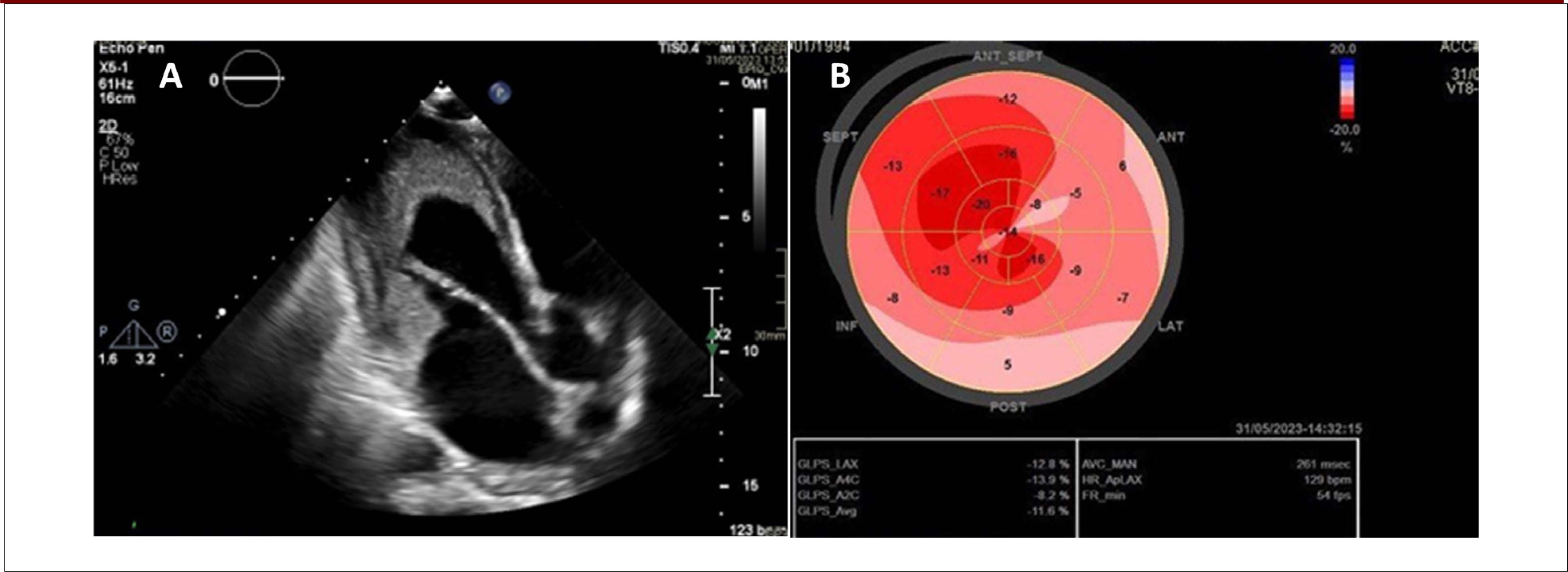
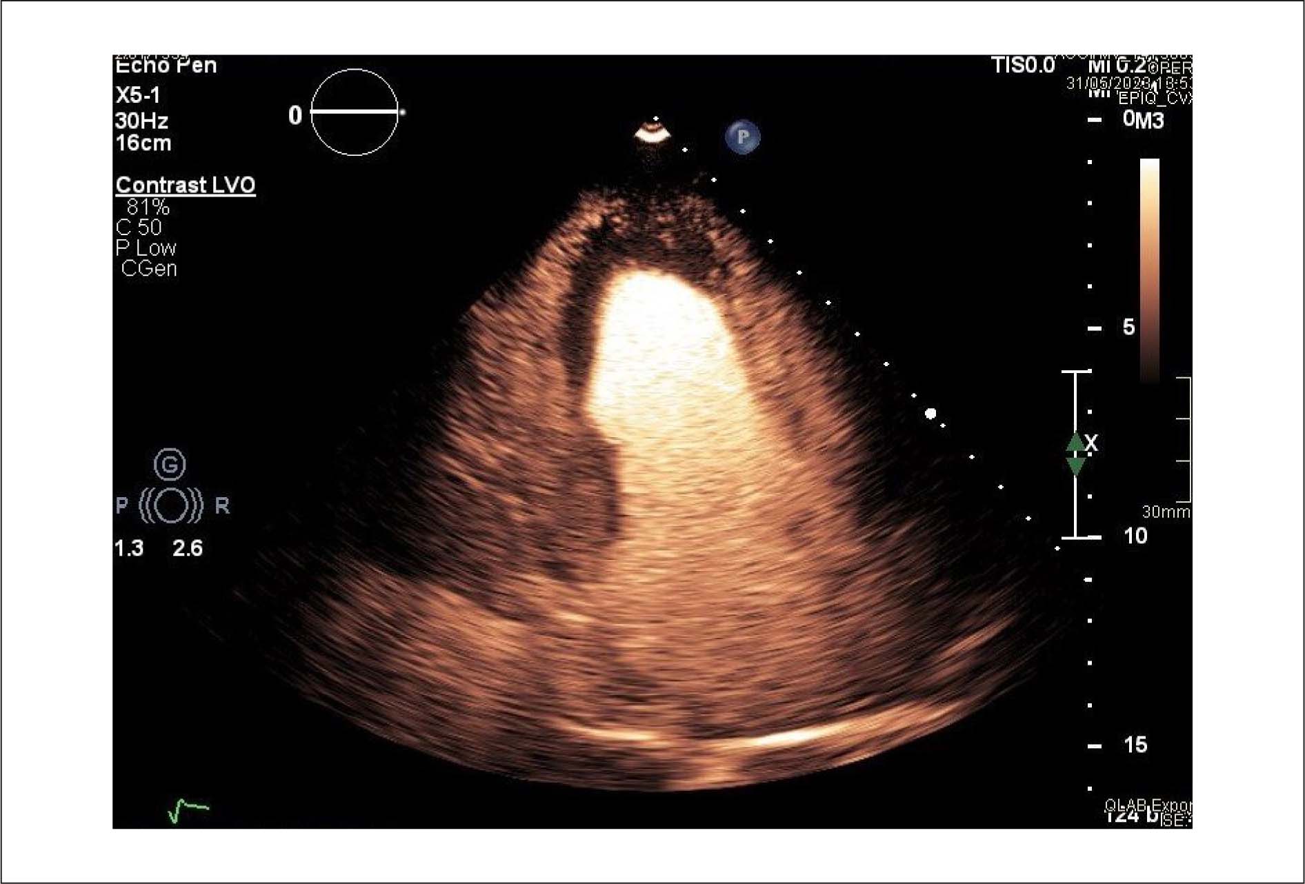
An echocardiogram was performed, showing diffuse endocardial thickening in the left ventricle (LV), which was more pronounced in the apical region, extending to the base of the posterior leaflet of the mitral valve, preserved systolic function, ejection fraction of 68%, and reduced global longitudinal strain (GLS) of −11.6 ( Figure 1 ). There was an echogenic intracavitary image covering the apex and the anterolateral wall, and an ultrasound contrast agent (Sonovue) was infused, suggesting an intracavitary thrombus ( Figure 2 ). The right ventricle did not show any abnormalities, and analysis of diastolic function was impaired by tachycardia. The mitral valve had restricted movement of the posterior leaflet and moderate eccentric MR.

(A) Diffuse endocardial thickening, more pronounced in the apical region and the papillary muscle.(B) Bull's-eye plot showing reduced GLS.
Ultrasound contrast agent showing an echogenic image compatible with a thrombus in the apical region of the LV.
Intravenous furosemide and full anticoagulation with enoxaparin were initiated, due to the clinical condition and the intracardiac thrombus. The patient showed improved congestion symptoms, followed treatment with hematology, and was discharged from hospital 30 days later.
The patient was asymptomatic from a cardiovascular perspective when he returned for follow-up echocardiography 1 year after the onset of the disease. The examination showed mild systolic dysfunction of the LV, ejection fraction of 50%, inferior hypokinesia, and rectified septum. The right ventricle showed mild to moderate systolic dysfunction (tricuspid annular plane systolic excursion 13 mm and fractional area change 30%). Assessment of diastolic function was impaired by significant mitral reflux, and the mitral valve showed reduced mobility and important MR filling the entire left atrium ( Figure 3 ).
(A) Moderate MR jet (yellow arrow) before antineoplastic treatment. (B) Important MR jet, even after treatment. LV: left ventricle.
Discussion
HES represents a group of rare diseases defined by peripheral blood eosinophil counts greater than 1.5 × 10⁹/L. It can be classified as primary (myeloid or lymphocytic neoplasm), secondary (infectious, autoimmune, drug-related, allergic, and metabolic causes), and idiopathic.
HES generally affects individuals between 20 and 50 years of age, and it is more common in men, especially HES of myeloproliferative origin. It can affect different tissues and organs, causing signs and symptoms such as skin lesions, diarrhea, abdominal pain, fever, and weight loss. When it affects the cardiovascular system, it represents a severe form of the disease with high morbidity and mortality.2
Loeffler syndrome is the manifestation of HES characterized by eosinophil-mediated cardiac damage. In its early stages, it is asymptomatic, but it causes inflammation and necrosis of the endocardium with formation of intracardiac thrombi and fibrosis. It can cause heart failure (systolic and diastolic dysfunction), thromboembolic phenomena, arrhythmias, and valvular diseases, mainly regurgitation of the atrioventricular valves, resulting from valvular involvement due to fibrosis.
It is worth highlighting that MR can decrease after treatment of the underlying disease with corticosteroids., Nonetheless, in this case, MR worsened, even after treatment. Furthermore, the patient developed biventricular systolic dysfunction, and he had already presented reduced GLS since diagnosis of the disease, reinforcing the ability of GLS to detect ventricular systolic dysfunction early.
Echocardiography is essential in the evaluation of patients with Loeffler syndrome. In the initial (inflammatory) phase, increased echogenicity of the subendocardium is observed. Subsequently, intracardiac thrombi may appear, which are typically located in the apex of the LV and the subvalvular regions of atrioventricular valves. The use of an ultrasound contrast agents increases the sensitivity of thrombus detection, in addition to allowing differential diagnosis of other conditions, such as apical hypertrophic cardiomyopathy and noncompaction of the myocardium.
Following identification of ventricular dysfunction on echocardiography, cardiac magnetic resonance imaging was performed for further evaluation, as this is the most accurate exam for tissue characterization, helping to define inflammation, edema, and fibrosis in all disease stages ( Figure 4 ).

(A) Cine magnetic resonance imaging showing attachment of the papillary muscles to the lateral wall of the LV with loss of trabeculation and mitral (orange arrow) and tricuspid (yellow arrow) regurgitation jets. (B) Delayed enhancement showing diffuse circumferential subendocardial fibrosis, without corresponding to a coronary territory (white arrows) and associated with thrombus covering the left ventricular subendocardium (asterisks). (C) Native T1 mapping showing diffusely increased T1, mainly in the septal wall, indicating edema and fibrosis, associated with the thrombus (asterisk).
Endomyocardial biopsy is the gold standard for diagnosis, providing evidence of eosinophilic infiltrate in the cardiac tissue.4 However, due to the potential for complications, it is only performed in select cases.
Treatment depends on the stage of the disease and the degree of fibrosis. The main goals are to reduce hypereosinophilia and to prevent the progression of tissue damage and thrombus formation. Corticosteroids are the most commonly used medication, but many patients do not respond well. Other options include interferon-alpha, hydroxyurea, imatinib, mepolizumab, and immunomodulatory drugs.1 – 3
The prognosis of HES has improved in recent years due to a better understanding of the disease and the emergence of new therapeutic modalities. However, cardiac dysfunction remains the leading cause of mortality.8
Conclusion
We have reported a case of Loeffler syndrome secondary to chronic myeloproliferative disease. Despite adequate diagnosis and treatment, the patient developed biventricular systolic dysfunction and important MR. We emphasize the importance of early diagnosis, treatment, and long-term follow-up as means of reducing the morbidity and mortality of this disease.
-
Sources of Funding
There were no external funding sources for this study.
-
Study Association
This study is not associated with any thesis or dissertation work.
-
Ethics Approval and Consent to Participate
This study was approved by the Ethics Committee of the Hospital das Clínicas da Faculdade de Medina da USP under the protocol number 7.469.009. All the procedures in this study were in accordance with the 1975 Helsinki Declaration, updated in 2013. Informed consent was obtained from all participants included in the study.
-
Use of Artificial Intelligence
The authors did not use any artificial intelligence tools in the development of this work.
-
Availability of Research Data
The underlying content of the research text is contained within the manuscript.
References
-
1 Shomali W, Gotlib J. World Health Organization and International Consensus Classification of Eosinophilic Disorders: 2024 Update on Diagnosis, Risk Stratification, and Management. Am J Hematol. 2024;99(5):946-68. doi: 10.1002/ajh.27287.
» https://doi.org/10.1002/ajh.27287 -
2 Requena G, van den Bosch J, Akuthota P, Kovalszki A, Steinfeld J, Kwon N, et al. Clinical Profile and Treatment in Hypereosinophilic Syndrome Variants: A Pragmatic Review. J Allergy Clin Immunol Pract. 2022;10(8):2125-34. doi: 10.1016/j.jaip.2022.03.034.
» https://doi.org/10.1016/j.jaip.2022.03.034 -
3 Takla A, Shah P, Sbenghe M, Baibhav B, Feitell S. Incessant Ventricular Tachycardia: An Atypical Presentation of Chronic Eosinophilic Leukemia. JACC Case Rep. 2024;29(16):102461. doi: 10.1016/j.jaccas.2024.102461.
» https://doi.org/10.1016/j.jaccas.2024.102461 -
4 Bondue A, Carpentier C, Roufosse F. Hypereosinophilic Syndrome: Considerations for the Cardiologist. Heart. 2022;108(3):164-71. doi: 10.1136/heartjnl-2020-317202.
» https://doi.org/10.1136/heartjnl-2020-317202 -
5 Inoue T, Watanabe C, Ayukawa H, Nadahama T, Hosokawa A, Beppu K, et al. Biopsy-Proven Loeffler Endocarditis Successfully Treated with Steroids. Circulation. 2015;131(8):e353-4. doi: 10.1161/CIRCULATIONAHA.114.012976.
» https://doi.org/10.1161/CIRCULATIONAHA.114.012976 -
6 van Dongen IM, van Kraaij DJW, Schalla S, La Rocca HPB, Driessen RGH. Severe Mitral Regurgitation Caused by Eosinophilic Endocarditis. J Cardiol Cases. 2014;10(3):108-10. doi: 10.1016/j.jccase.2014.05.012.
» https://doi.org/10.1016/j.jccase.2014.05.012 -
7 Jin X, Ma C, Wang Y, Yang J. A Case of Loeffler Endocarditis That Showed Endomyocardial Systolic Dysfunction Detected by Layer Specific Strain Analysis. Int Heart J. 2017;58(6):1001-3. doi: 10.1536/ihj.16-422.
» https://doi.org/10.1536/ihj.16-422 -
8 Polito MV, Hagendorff A, Citro R, Prota C, Silverio A, De Angelis E, et al. Loeffler's Endocarditis: An Integrated Multimodality Approach. J Am Soc Echocardiogr. 2020;33(12):1427-41. doi: 10.1016/j.echo.2020.09.002.
» https://doi.org/10.1016/j.echo.2020.09.002 -
9 Bonanad C, Monmeneu JV, López-Lereu MP. Loeffler Endocarditis of the Left Ventricle: Cardiac Magnetic Resonance Findings. Heart Lung Circ. 2013;22(12):1056-7. doi: 10.1016/j.hlc.2013.03.087.
» https://doi.org/10.1016/j.hlc.2013.03.087
Edited by
-
Editor responsible for the review:
Marcelo Tavares
The underlying content of the research text is contained within the manuscript.