Assessment of Acute Chest Pain in the Emergency Department: How Point-of-Care Ultrasound Can Assist the Cardiologist
Abstract
The use of point-of-care ultrasound (POCUS) in emergency settings is now well established as part of the initial assessment of critically ill patients. In cases of acute chest pain, POCUS plays a significant role in differential diagnosis, helping to reduce diagnostic errors and, most importantly, preventing the discharge of patients with potentially life-threatening conditions. In this context, the main diagnostic considerations include acute coronary syndrome (ACS), pericarditis, pulmonary thromboembolism (PTE), and acute aortic syndromes (AAS). POCUS has proven effective in all of these scenarios and can be widely applied with proper training, both by novice physicians and experienced echocardiographers. This article aims to discuss the key ways in which POCUS can specifically aid in the differential diagnosis of chest pain.
Keywords:
Emergencies; Acute Coronary Syndrome; Pulmonary Embolism; Aortic Dissection

Assessment of Acute Chest Pain in the Emergency Department: How Point-of-Care Ultrasound Can Assist the Cardiologist
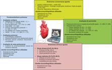
Use of POCUS in the differential diagnosis of acute chest pain. IVC: inferior vena cava; LVEF: left ventricular ejection fraction; LV: left ventricle; RA: right atrium; RV: right ventricle; RWMA: regional wall motion abnormality; Se: sensitivity; Sp: specificity; TAPSE: tricuspid annular plane systolic excursion.
Resumo
O uso da ultrassonografia point-of-care (POCUS) nas unidades de emergência já está consolidado como parte da avaliação inicial de pacientes críticos. Em casos de dor torácica aguda, o POCUS contribui significativamente para o diagnóstico diferencial, reduzindo a taxa de erros diagnósticos e, sobretudo, evitando a alta hospitalar de pacientes com doenças potencialmente fatais. Nessa situação, os principais diagnósticos são a síndrome coronariana aguda (SCA), a pericardite, a tromboembolia pulmonar (TEP) e as síndromes aórticas agudas (SAA). Em todos esses cenários, o POCUS já demonstrou sua eficácia e pode ser amplamente aplicado com o treinamento adequado, tanto por médicos iniciantes quanto por ecocardiografistas experientes. Este artigo tem como objetivo discutir os principais aspectos em que o POCUS pode auxiliar, de forma direcionada, no diagnóstico diferencial da dor torácica.
Palavras-chave:
Emergência; Síndrome Coronariana Aguda; Embolia Pulmonar; Dissecção Aórtica
Avaliação da Dor Torácica Aguda no Pronto-Socorro: Como o Ultrassom Point-of-Care Pode Auxiliar o Cardiologista
Utilização do POCUS no diagnóstico diferencial da dor torácica aguda. AD: átrio direito; ACS: alteração da contratilidade segmentar; E: especificidade; FEVE: fração de ejeção do ventrículo esquerdo; S: sensibilidade; TAPSE: tricuspid annular plane systolic excursion; VCI: veia cava inferior; VD: ventrículo direito; VE: ventrículo esquerdo.
Introduction
The use of point-of-care ultrasound (POCUS) in emergency settings is now well established as an integral part of the initial assessment of critically ill patients. In cases of acute chest pain, POCUS has proven to be a valuable tool in differential diagnosis, helping reduce diagnostic errors and, most importantly, preventing the discharge of patients with potentially life-threatening conditions.1,2 Among the main differential diagnoses in this scenario are acute coronary syndrome (ACS), pericarditis, pulmonary thromboembolism (PTE), and acute aortic syndromes (AAS).
a) ACS
The diagnosis of ACS is primarily based on clinical history, electrocardiography (ECG), and troponin levels. Although these tools are simple and widely used, the increasing adoption of POCUS raises questions about its potential to improve diagnostic accuracy, shorten time to diagnosis, assist in risk stratification, and detect complications associated with ACS.1,2
Following clinical assessment, patients with suspected ACS in the emergency department (ED) can be pragmatically classified into three groups:1,2
-
Definitive or highly probable diagnosis of ACS (e.g., typical clinical presentation with ST-segment elevation and/or troponin elevation);
-
Ruled-out diagnosis (e.g., low-suspicion presentation with normal ECG and troponin levels);
-
Indeterminate diagnosis.
In confirmed cases, POCUS can be helpful in identifying complications in unstable patients, such as acute pulmonary edema or cardiogenic shock. When the diagnosis of ACS is ruled out, POCUS may assist in the differential diagnosis, such as AAS, pericarditis, or PTE.1,2
The indeterminate diagnosis group includes, for instance, patients with initially negative troponins but at intermediate risk for adverse events (e.g., a History, ECG, Age, Risk factors, Troponin [HEART] score between 4 and 6, or a 0/1-hour algorithm from the European Society of Cardiology [ESC] falling into the observation zone). In such cases, POCUS may reveal signs of poor prognosis and support the decision to conduct an earlier inpatient investigation. This group also includes patients presenting to the ED with a clinical or electrocardiographic presentation suggestive of acute coronary occlusion (ACO). If uncertainty remains, POCUS may help to increase or decrease the pretest probability of ACO.1-3
In recent years, there has been a growing recognition of the limitations of using ST-segment elevation as a dichotomous criterion to define ACO. Consequently, greater importance is being placed on clinical presentation and other ECG findings. In this context, POCUS emerges as a valuable complementary tool for detecting regional wall motion abnormalities (RWMA). As with ECG findings, echocardiographic findings should not be interpreted in a binary manner but rather as part of a broader process of clinical reasoning.3
A recent study evaluated whether the presence of RWMA on POCUS could predict obstructive coronary artery disease (CAD) in patients presenting to the ED with chest pain. Among the 657 participants with suspected ACS, approximately 11% showed RWMA. These findings were significantly more frequent in those who required revascularization (26.2% vs. 7.6%), with odds ratios ranging from 3.16 to 3.68 depending on the statistical adjustment. The analysis included all three risk groups of the ESC 0/1-hour algorithm (rule-in, observe, and rule-out), demonstrating that the presence or absence of RWMA influenced CAD risk across all categories. These findings reinforce that POCUS can help identify higher-risk patients, even without offering a definitive diagnosis. However, the accuracy of the method in detecting acute myocardial infarction (AMI) varies depending on the population studied.4
Another retrospective study examined whether the detection of RWMA on POCUS could accelerate the indication for revascularization in patients with possible ACO. The study included 234 patients presenting to the ED with chest pain and negative troponins, excluding those with ST-segment elevation. Among the 23 patients who underwent revascularization, 14 had RWMA on POCUS, which was performed on average 36 minutes before the first troponin result. The mean time to revascularization was 432 minutes in patients with RWMA, compared to 2,158 minutes in those without. Despite methodological limitations, the study suggests that POCUS may provide relevant information in less time than troponin testing.5
RWMA may represent either an acute coronary event or sequelae of prior events. Some studies on the topic choose to exclude patients with a history of AMI from their analyses. Although this distinction is not always possible based solely on clinical history, the presence of RWMA supports clinical reasoning and risk stratification by identifying patients with a higher likelihood of adverse outcomes. These patients should therefore be considered for hospital admission and further investigation, taking into account other factors that contribute to pretest probability.4
Several studies have investigated the value of POCUS performed by emergency physicians. A study published in 2020 compared trained emergency physicians with experienced echocardiographers, evaluating the accuracy of the former in detecting RWMA in patients with chest pain in the ED. A total of 89 patients were assessed, and the emergency physicians achieved a sensitivity of 76.9% and a specificity of 92.1%. The professionals evaluated were three in-hospital physicians with approximately 5 years of POCUS experience who underwent a specific 3-hour training on RWMA detection conducted by a cardiologist.6
Another similar study compared RWMA assessments performed by emergency medicine residents with formally conducted echocardiograms in the context of ST-elevation AMI. The study included five first-year residents, three second-year residents, and one third-year resident, all of whom underwent video-based training focused on RWMA identification. These residents had prior experience with at least 25 cardiac POCUS exams and 150 general POCUS exams, although they had no specific training in RWMA detection before the study. A total of 75 patients were included, 62% of whom showed RWMA on formal echocardiography. POCUS performed by the residents achieved a sensitivity of 88% and a specificity of 92% compared to the gold standard used in the study (formal echocardiography or, in some cases, ventriculography).7
These studies highlight several important points. First, it appears feasible to train physicians who are not echocardiography specialists to assess RWMA using POCUS. However, the accuracy achieved by these professionals tends to be lower than that of experienced echocardiographers. Moreover, findings observed in studies using echocardiograms performed by specialists are likely not fully reproducible when POCUS is used by less experienced operators. The level of agreement, as well as the sensitivity and specificity of the exam, can vary significantly depending on the clinical context and the provider’s level of training. Prospective studies incorporating POCUS into clinical decision-making may help clarify its true impact on the assessment of patients with chest pain.6,7
Finally, in unstable patients — such as those with acute pulmonary edema or cardiogenic shock — POCUS can aid in the differential diagnosis by enabling the assessment of biventricular function, volume status, and the identification of mechanical complications such as mitral regurgitation and/or pericardial effusion.2
b) Pericardial assessment
In the systematic evaluation of the heart using POCUS, visualization of the pericardium plays a key role in hemodynamic characterization and the differential diagnosis of chest pain.8,9
In particular, when managing pericardial effusions, POCUS offers healthcare professionals a fast, accurate, and bedside-available diagnostic tool. Its use enables not only precise estimation of effusion volume but also early detection of cardiac tamponade and appropriate guidance for interventions such as pericardiocentesis, significantly contributing to improved clinical outcomes.8,9
Under physiological conditions, the pericardial space contains approximately 15 to 50 mL of fluid, a volume that is generally undetectable by simpler ultrasound machines. A pericardial effusion is defined as the abnormal accumulation of fluid in this space, becoming more easily visualized on ultrasound examination.8,9
Among the differential diagnoses of chest pain, pericarditis accounts for approximately 5% of ED visits. Its clinical criteria include chest pain with features typical of pericardial involvement, suggestive ECG findings, presence of a pericardial friction rub, and evidence of pericardial effusion or an increase in the volume of a previously known effusion.9
Performing POCUS at the bedside facilitates the diagnosis of both pericarditis and pericardial effusion, as even small abnormal fluid collections in the pericardial space can be detected and considered diagnostic. The method has high accuracy, with sensitivity reaching up to 96% and specificity up to 98% when performed by adequately trained emergency physicians — not necessarily cardiologists.10
Pericardial effusion can be measured using several basic echocardiographic windows, such as parasternal long and short axes, apical four-chamber, and subcostal views. Based on the observed thickness of the fluid layer, the effusion can be classified as minimal, small, moderate, or large (Table 1).10
The presence of abnormal fluid in the pericardial space — its volume and rate of accumulation — may exert extrinsic compression on the cardiac chambers, directly affecting atrial and ventricular relaxation. This restriction depends not only on the absolute fluid volume but also on the pericardium’s elasticity to accommodate it. In cases of rapid accumulation, even small volumes may significantly increase intrapericardial pressure. On the other hand, in cases of slow and progressive accumulation, the pericardium can adapt due to its distensibility, accommodating volumes exceeding 500 mL.10
The echocardiographic findings most commonly identified on POCUS in patients with cardiac tamponade include:8-10
b.1) Late diastolic collapse of the right atrium (RA)
Diastolic collapse of the RA is one of the most important and earliest echocardiographic signs of cardiac tamponade.9 This phenomenon occurs during atrial relaxation, when RA volume is reduced and intrapericardial pressure peaks, causing the atrial wall to invaginate into the chamber. It is a highly sensitive finding, particularly when the collapse lasts for more than one-third of the cardiac cycle. However, its specificity is moderate (approximately 82%), as brief RA collapses can also occur in other conditions, such as elevated intrathoracic pressure. Nevertheless, sustained RA collapse is a strong indicator of cardiac tamponade.9,10
b.2) Diastolic collapse of the right ventricle (RV)
Diastolic collapse of the RV is typically observed during early diastole, when the chamber volume is still reduced. Although its sensitivity is slightly lower than that of RA collapse (approximately 93%), this finding is highly specific for tamponade. It is important to note that in certain clinical scenarios—such as RV hypertrophy or elevated diastolic pressure in the RV (e.g., acute or chronic cor pulmonale)—this collapse may be absent even in the presence of tamponade.9,10
b.3) Dilated inferior vena cava (IVC) with minimal or absent respiratory variation
In cases of cardiac tamponade, the RA becomes unable to adequately accommodate venous return due to the compression caused by increased intrapericardial pressure. As a result, the IVC remains dilated, with minimal or absent respiratory variation. This finding has high sensitivity for cardiac tamponade (95–97%) and a high negative predictive value (NPV). However, its specificity is low (around 40%), as it can also be seen in other conditions such as chronic lung disease, heart failure, tricuspid regurgitation, and other cardiopathies.9,10
The IVC is typically visualized in a sagittal subcostal view, just below the xiphoid process. The diameter should be measured approximately 2 to 3 cm before its junction with the RA, near the confluence with the hepatic vein. In some cases, M-mode can be used to improve measurement accuracy. The IVC is considered abnormal when its diameter exceeds 2.1 cm and inspiratory variation is less than 50%.9,10
b.4) Swinging heart
This finding is characteristic of large pericardial effusions and refers to the pendular movement of the heart within the pericardial sac.
The integration of POCUS into routine clinical practice has been shown to improve diagnostic accuracy and enable timely interventions, optimizing care for patients with pericardial involvement. However, it is essential to recognize the limitations of this modality. The effectiveness of POCUS is highly dependent on the operator’s experience and training, which can lead to variability in results. In addition, limited access to ultrasound equipment and trained personnel remains a significant barrier, particularly in resource-constrained settings.9,10
c) PTE
PTE is one of the leading causes of chest pain in ED s, accounting for 5% to 10% of cases. Without treatment, its mortality rate is approximately 30%, but this can be reduced to 3%–8% with early therapy.1,11 Early diagnosis is essential but challenging. POCUS has proven to be a valuable bedside tool, allowing for rapid and integrated evaluation of cardiac, pulmonary, and vascular findings, with reported specificity as high as 96.7%.12
The ultrasound assessment of patients with suspected PTE should include three main regions: the heart, lungs, and deep venous system of the lower limbs. The combination of findings across these areas increases diagnostic accuracy and can support early therapeutic decisions, including the initiation of reperfusion therapy even in the absence of a pulmonary CT angiogram.
c.1) Cardiac assessment
In the context of PTE, cardiac POCUS aims to identify signs of acute RV strain and dysfunction. In the absence of such findings, the exam can help rule out PTE in hemodynamically unstable patients and assist in identifying alternative causes of instability, such as left ventricle (LV) dysfunction or valvular disease. The main echocardiographic findings associated with PTE include:
-
RV dilation: RV/LV ratio > 1 in apical or subxiphoid views and/or end-diastolic diameter > 30 mm in the parasternal long-axis view. Sensitivity: 31%–72%; specificity: 87%–96%.1,12
-
McConnell’s sign: hypokinesia of the RV free wall with preserved apical motion. Sensitivity: 77%; specificity: 94%.12
-
Interventricular septal flattening (“D-sign”): characterized by flattening of the septum toward the LV, predominantly during diastole, observed in the parasternal short-axis view.1
-
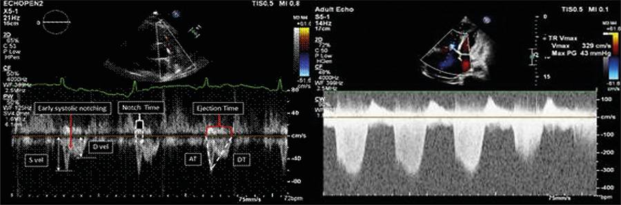
60/60 sign: pulmonary artery acceleration time (PAT) < 60 ms combined with tricuspid regurgitation gradient (TRG) < 60 mmHg. This pattern is suggestive of acute PTE, with sensitivity up to 98% when associated with RV overload (Figure 1).12,13
-
Pulmonary midsystolic notch: a Doppler finding in the RV outflow tract, characterized by a pronounced notch after a sharp initial peak, followed by a dome-shaped waveform. Alerhand et al. reported 92% sensitivity and 99% specificity for massive or submassive PTE; for PTE in general, sensitivity was 34% and specificity 97%.13
-
Dilated IVC with inspiratory collapse < 50%.1
-
Tricuspid annular plane systolic excursion (TAPSE) < 17 mm, indicating RV systolic dysfunction. Sensitivity: 88%; specificity: 77%.1,13
c.2) Pulmonary assessment
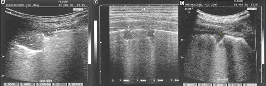
Pulmonary POCUS can be performed using either a convex or linear transducer, scanning the intercostal spaces in the anterior, lateral, and posterior regions of the chest. The main findings suggestive of pulmonary infarction secondary to PTE include (Figure 2):
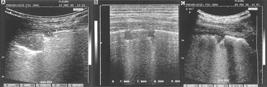
– Pulmonary consolidations: A – triangular; B – rounded; C – polygonal with adjacent pleural effusion.
-
Triangular subpleural consolidations (the most common, seen in approximately 85% of cases) or, less frequently, rounded or polygonal consolidations (3%).1
-
Presence of A-lines associated with pleural sliding.
-
Pleural effusion, usually unilateral and associated with the area of consolidation.1
When combined with cardiac findings and evidence of deep vein thrombosis (DVT), these ultrasound patterns significantly increase clinical suspicion for PTE, with specificity reaching up to 99%.13
c.3) Deep vein assessment
Ultrasound evaluation of the deep venous system of the lower limbs is a key component of the POCUS protocol in patients with suspected PTE. The absence of venous compressibility is highly suggestive of DVT. When identified in patients with compatible clinical signs or in those with suggestive cardiac and pulmonary POCUS findings, it may justify the immediate initiation of anticoagulant therapy.1
The multi-organ POCUS approach allows for the integration of both direct and indirect signs of PTE, providing a rapid assessment with high sensitivity and specificity.8 The simultaneous presence of RV dysfunction, pulmonary consolidations, and DVT carries high predictive value. Recent studies highlight the growing role of POCUS, especially in emergency settings and resource-limited environments. In addition, POCUS enables the exclusion of other critical differential diagnoses, such as pneumothorax, cardiac tamponade, and aortic dissection. Thus, POCUS is increasingly recognized as an essential tool in the evaluation of patients with chest pain and suspected PTE — particularly when pulmonary CT angiography is not available.1
d) Aortic assessment
AAS should always be considered key differential diagnoses in the evaluation of chest pain in emergency settings. As with other conditions discussed, visualization of the ascending aorta should be part of the initial POCUS assessment, forming an integral part of the diagnostic algorithm for patients presenting with chest pain.14,15
The ADvISED study (Diagnostic Accuracy of the Aortic Dissection Detection Risk Score Plus D-Dimer for Acute Aortic Syndromes), which evaluated patients with chest pain in the ED, applied a protocol combining the Aortic Dissection Detection Risk Score (ADD-RS) with D-dimer testing. The results demonstrated a negative predictive value (NPV) of 99.7% for ruling out AAS, although the specificity of the model alone was limited (57.3%) for positive diagnosis.16
A subanalysis of that study incorporated POCUS into the algorithm and showed that the presence of direct signs of AAS raised the diagnostic specificity to 97.4%. Even indirect signs performed well, with specificity of 74.5%. The inclusion of POCUS increased the NPV to 100% and improved overall diagnostic accuracy, with the area under the curve (AUC) rising from 0.77 to 0.88.17
The PROFUNDUS study, conducted prospectively with over 1,900 patients, also evaluated the use of POCUS combined with D-dimer for AAS investigation in emergency settings. In that study, the presence of direct signs on POCUS showed 98.4% specificity for diagnosing AAS, while indirect signs reached specificity between 88% and 96.5%. As with ADvISED, the combined use of POCUS and D-dimer achieved an NPV of 100%.18
In clinical practice, the key question becomes: “Which echocardiographic views should be used, and what findings should be sought?” The preferred views include:
-
Parasternal long-axis view: allows assessment of the LV outflow tract and ascending aorta;
-
Suprasternal view (at the suprasternal notch): provides visualization of the aortic arch and descending thoracic aorta.14,15
During the evaluation, physicians should actively search for direct and indirect signs of AAS, as outlined below:
Direct signs:
-
Presence of an intimal flap (dissection of the tunica intima);
-
Aortic wall hematoma;
-
Penetrating aortic ulcer.
Indirect signs:
-
Aortic dilation;
-
Pericardial effusion;
-
Aortic valve insufficiency.
References
-
1 Ha YR, Toh HC. Clinically Integrated Multi-Organ Point-Of-Care Ultrasound for Undifferentiated Respiratory Difficulty, Chest Pain, or Shock: A Critical Analytic Review. J Intensive Care. 2016;4:54. doi: 10.1186/s40560-016-0172-1.
» https://doi.org/10.1186/s40560-016-0172-1 -
2 Byrne RA, Rossello X, Coughlan JJ, Barbato E, Berry C, Chieffo A, et al. 2023 ESC Guidelines for the Management of Acute Coronary Syndromes. Eur Heart J. 2023;44(38):3720-826. doi: 10.1093/eurheartj/ehad191.
» https://doi.org/10.1093/eurheartj/ehad191 -
3 McLaren J, Alencar JN, Aslanger EK, Meyers HP, Smith SW. From ST-Segment Elevation MI to Occlusion MI: The New Paradigm Shift in Acute Myocardial Infarction. JACC Adv. 2024;3(11):101314. doi: 10.1016/j.jacadv.2024.101314.
» https://doi.org/10.1016/j.jacadv.2024.101314 -
4 Xu C, Melendez A, Nguyen T, Ellenberg J, Anand A, Delgado J, et al. Point-of-Care Ultrasound May Expedite Diagnosis and Revascularization of Occult Occlusive Myocardial Infarction. Am J Emerg Med. 2022;58:186-91. doi: 10.1016/j.ajem.2022.06.010.
» https://doi.org/10.1016/j.ajem.2022.06.010 -
5 Roggel A, Jehn S, Dykun I, Balcer B, Al-Rashid F, Totzeck M, et al. Regional Wall Motion Abnormalities on Focused Transthoracic Echocardiography in Patients Presenting with Acute Chest Pain: A Predefined Post Hoc Analysis of the Prospective Single-Centre Observational EPIC-ACS study. BMJ Open. 2024;14(9):e085677. doi: 10.1136/bmjopen-2024-085677.
» https://doi.org/10.1136/bmjopen-2024-085677 -
6 Croft PE, Strout TD, Kring RM, Director L, Vasaiwala SC, Mackenzie DC. WAMAMI: Emergency Physicians Can Accurately Identify Wall Motion Abnormalities in Acute Myocardial Infarction. Am J Emerg Med. 2019;37(12):2224-8. doi: 10.1016/j.ajem.2019.03.037.
» https://doi.org/10.1016/j.ajem.2019.03.037 -
7 Saglam C, Ünlüer EE, Yamanoglu NGÇ, Kara PH, Ediboglu E, Bektasli R, et al. Accuracy of Emergency Physicians for Detection of Regional Wall Motion Abnormalities in Patients with Chest Pain Without ST-Elevation Myocardial Infarction. J Ultrasound Med. 2021;40(7):1335-42. doi: 10.1002/jum.15513.
» https://doi.org/10.1002/jum.15513 -
8 Mandavia DP, Hoffner RJ, Mahaney K, Henderson SO. Bedside Echocardiography by Emergency Physicians. Ann Emerg Med. 2001;38(4):377-82. doi: 10.1067/mem.2001.118224.
» https://doi.org/10.1067/mem.2001.118224 -
9 Imazio M, De Ferrari GM. Cardiac Tamponade: An Educational Review. Eur Heart J Acute Cardiovasc Care. 2021;10(1):102-9. doi: 10.1177/2048872620939341.
» https://doi.org/10.1177/2048872620939341 -
10 Chiabrando JG, Bonaventura A, Vecchié A, Wohlford GF, Mauro AG, Jordan JH, et al. Management of Acute and Recurrent Pericarditis: JACC State-of-the-Art Review. J Am Coll Cardiol. 2020;75(1):76-92. doi: 10.1016/j.jacc.2019.11.021.
» https://doi.org/10.1016/j.jacc.2019.11.021 -
11 Konstantinides SV, Meyer G, Becattini C, Bueno H, Geersing GJ, Harjola VP, et al. 2019 ESC Guidelines for the Diagnosis and Management of Acute Pulmonary Embolism Developed in Collaboration with the European Respiratory Society (ERS). Eur Heart J. 2020;41(4):543-603. doi: 10.1093/eurheartj/ehz405.
» https://doi.org/10.1093/eurheartj/ehz405 -
12 Daley JI, Dwyer KH, Grunwald Z, Shaw DL, Stone MB, Schick A, et al. Increased Sensitivity of Focused Cardiac Ultrasound for Pulmonary Embolism in Emergency Department Patients with Abnormal Vital Signs. Acad Emerg Med. 2019;26(11):1211-20. doi: 10.1111/acem.13774.
» https://doi.org/10.1111/acem.13774 -
13 Albricker ACL, Freire CMV, Santos SND, Alcantara ML, Saleh MH, Cantisano AL, et al. Joint Guideline on Venous Thromboembolism - 2022. Arq Bras Cardiol. 2022;118(4):797-857. doi: 10.36660/abc.20220213.
» https://doi.org/10.36660/abc.20220213 -
14 Bernett J, Strony R. Diagnosing Acute Aortic Dissection with Aneurysmal Degeneration with Point of Care Ultrasound. Am J Emerg Med. 2017;35(9):1384.e3-1384.e4. doi: 10.1016/j.ajem.2017.05.052.
» https://doi.org/10.1016/j.ajem.2017.05.052 -
15 Lee PY, Saad K, Hossain A, Lieu I, Allencherril J. Initial Evaluation and Management of Patients Presenting with Acute Chest Pain in the Emergency Department. Curr Cardiol Rep. 2023;25(12):1677-86. doi: 10.1007/s11886-023-01984-6.
» https://doi.org/10.1007/s11886-023-01984-6 -
16 Nazerian P, Mueller C, Soeiro AM, Leidel BA, Salvadeo SAT, Giachino F, et al. Diagnostic Accuracy of the Aortic Dissection Detection Risk Score Plus D-Dimer for Acute Aortic Syndromes: The ADvISED Prospective Multicenter Study. Circulation. 2018;137(3):250-8. doi: 10.1161/CIRCULATIONAHA.117.029457.
» https://doi.org/10.1161/CIRCULATIONAHA.117.029457 -
17 Nazerian P, Mueller C, Vanni S, Soeiro AM, Leidel BA, Cerini G, et al. Integration of Transthoracic Focused Cardiac Ultrasound in the Diagnostic Algorithm for Suspected Acute Aortic Syndromes. Eur Heart J. 2019;40(24):1952-60. doi: 10.1093/eurheartj/ehz207.
» https://doi.org/10.1093/eurheartj/ehz207 -
18 Morello F, Bima P, Castelli M, Capretti E, Soeiro AM, Cipriano A, et al. Diagnosis of Acute Aortic Syndromes with Ultrasound and D-Dimer: The PROFUNDUS Study. Eur J Intern Med. 2024;128:94-103. doi: 10.1016/j.ejim.2024.05.029.
» https://doi.org/10.1016/j.ejim.2024.05.029
-
Study Association:
This study is not associated with any thesis or dissertation work.
-
Ethics Approval and Consent to Participate:
This article does not contain any studies with human participants or animals performed by any of the authors.
-
Use of Artificial Intelligence:
The authors did not use any artificial intelligence tools in the development of this work.
-
Availability of Research Data:
The underlying content of the research text is contained within the manuscript.
-
Sources of Funding:
There were no external funding sources for this study.
Edited by
-
Editor responsible for the review:
Marcelo Tavares
The underlying content of the research text is contained within the manuscript.