
 My Approach to Diagnose Fabry Disease
Abstract
Fabry disease (FD) is an inherited lysosomal storage disorder caused by a deficiency of the enzyme alpha-galactosidase A (α-Gal A). This enzymatic defect leads to the cytoplasmic lysosomal accumulation of globotriaosylceramides (GB3 and LysoGB3), resulting in multisystemic clinical manifestations. Cardiovascular involvement, often mimicking hypertrophic cardiomyopathy, is the main determinant of morbidity and mortality, due to the development of arrhythmia, myocardial ischemia, and heart failure. Although FD is a rare condition in the general population, the availability of specific enzyme replacement therapy, which can alter the natural course of the disease, underscores the importance of including FD as a key differential diagnosis among storage cardiomyopathies. Diagnostic evaluation should encompass a thorough clinical assessment, with particular attention to patient history and physical examination, complemented by laboratory testing and imaging studies, such as electrocardiography and echocardiography. Cardiac magnetic resonance imaging, including late gadolinium enhancement and T1 and T2 parametric mapping, provides additional diagnostic and prognostic information and should ideally be performed at the time of initial diagnosis. Definitive diagnosis is established by genetic sequencing of the GLA gene, located on the long arm of the X chromosome, enabling the selection of the most appropriate therapeutic strategy for each patient.
Keywords
Fabry Disease; Glycosphingolipids; Hypertrophic Cardiomyopathy
Resumo
A doença de Fabry (DAF) é um erro inato do metabolismo dos glicoesfingolipídeos, causado pela deficiência da enzima alfa-galactosidase A (alfa-Gal A). Como consequência, ocorre acúmulo lisossomal citoplasmático de globotriaosilceramidas (GB3 e LysoGB3), resultando em manifestações clínicas multissistêmicas. O envolvimento cardiovascular, que pode mimetizar uma cardiomiopatia hipertrófica, representa o principal determinante de morbimortalidade, devido ao desenvolvimento de arritmias, isquemia miocárdica e insuficiência cardíaca. Embora a prevalência da doença seja baixa na população geral, a disponibilidade de terapia de reposição enzimática específica, capaz de modificar sua evolução, destaca a DAF como diagnóstico diferencial relevante entre as cardiomiopatias por depósito. A investigação diagnóstica deve incluir uma avaliação clínica minuciosa, com ênfase na anamnese e no exame físico, além da realização de exames laboratoriais e de imagem, como eletrocardiograma e ecocardiograma. A ressonância magnética cardíaca, utilizando realce tardio com gadolínio e mapas paramétricos T1 e T2, oferece informações adicionais diagnósticas e prognósticas, devendo ser preferencialmente realizada no momento do diagnóstico inicial. A confirmação definitiva é obtida por meio do sequenciamento genético do gene GLA, localizado no braço longo do cromossomo X, permitindo, assim, a definição da abordagem terapêutica mais adequada paracada paciente.
Palavras-chave
Doença de Fabry; Glicoesfingolipídeos; Cardiomiopatia Hipertrófica
Introduction
Fabry disease (FD; OMIM 301500) is a rare, X-linked lysosomal storage disorder. Pathogenic variants in the GLA gene, located on the long arm of the X chromosome, lead to a deficiency of the enzyme alpha-galactosidase A (α-Gal A), which is responsible for the catabolism of glycosphingolipids, such as globotriaosylceramide (GB3) and its deacetylated form, Lyso-GB3, within lysosomes. The accumulation of these substrates begins in the intrauterine period and triggers an intense tissue inflammatory response, coupled with local oxidative stress. This cascade promotes cellular injury, apoptosis, and, progressively, organ dysfunction and failure.1
Cardiovascular involvement, which can mimic hypertrophic cardiomyopathy (phenocopy), is the leading cause of morbidity and mortality among Brazilian patients, primarily due to the development of arrhythmias, myocardial ischemia, and heart failure.2 Despite its low prevalence in the general population, the availability of disease-specific enzyme replacement therapy, capable of modifying the disease course, highlights FD as a key differential diagnosis among storage cardiomyopathies.
The diagnostic approach to FD-associated cardiomyopathy should include a comprehensive clinical evaluation, encompassing a detailed medical history and physical examination, as well as accessible laboratory and imaging studies, such as electrocardiography and echocardiography. Cardiac magnetic resonance imaging provides additional diagnostic and prognostic insights and should preferably be performed during the initial diagnostic workup. Definitive diagnosis is established through genetic sequencing of the GLA gene, which also guides the selection of the most appropriate therapeutic strategy for each patient.3
My approach to
Diagnosing a rare cardiomyopathy poses a significant challenge for cardiologists, particularly when dealing with phenocopies. The estimated prevalence of FD in the general population is approximately 1 in 117,000 cases.4 However, this figure may reach as high as 1 in 3,100 when considering neonatal screening, a strategy that has been progressively incorporated into the Brazilian public health care system, as already implemented in certain regions, such as the Distrito Federal.5
The low prevalence of FD in the general population highlights two key considerations. First, it emphasizes the importance of including FD in the differential diagnosis of cardiomyopathies, given the availability of specific treatment through enzyme replacement therapy, which has been recently incorporated into Brazil's Unified Health System ( Sistema Único de Saúde , SUS). This therapy has the potential to positively alter the natural history of the disease and improve patients’ symptom-free survival. Second, it reinforces the need to thoroughly identify and manage more prevalent conditions — such as systemic hypertension, valvular heart disease, and, in the Brazilian context, rheumatic heart disease — before considering FD as the underlying cause of the presenting symptoms.
The diagnostic workup for FD should begin with a thorough clinical history. Figure 1 outlines the key elements that may raise clinical suspicion of FD. Patient age and sex should also be considered, as the clinical expression of the disease varies depending on the type of pathogenic variant identified in the GLA gene.
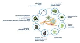
In the classic form of FD, cardiovascular manifestations — such as palpitations and exercise intolerance — may appear as early as childhood. These symptoms are often accompanied by other early manifestations, including characteristic neuropathic pain (Fabry crises) triggered by exposure to extreme temperatures, hypohidrosis or anhidrosis with impaired thermoregulation, gastrointestinal disturbances with alternating constipation and diarrhea, tinnitus or hearing loss, as well as cornea verticillata and angiokeratomas typically located in the pelvic region and/or mucosal surfaces.6
In contrast, in the nonclassic form of FD, also referred to as late-onset FD, these early systemic manifestations are often absent, with clinical presentation predominantly characterized by cardiac or renal involvement. These may occasionally be associated with central nervous system ischemic events, such as strokes occurring at relatively young ages.7
A careful assessment of the patient's personal history of symptoms and prior disorders is also essential, as is gathering information on first-degree relatives. Commonly reported symptoms include exertional chest pain, palpitations, exercise intolerance, orthostatic hypotension, and/or syncope, which may be associated with renal dysfunction, ischemic neurological events, or polyneuropathy. Family history should include targeted questions regarding known cases of FD, cardiomyopathies, strokes, sudden cardiac death, and/or renal failure requiring dialysis.8 Based on these data, it is advisable to construct a family pedigree, ideally encompassing at least three generations, in order to identify the familial inheritance pattern, which in the case of FD is X-linked.9
Once the clinical data have been collected, electrocardiography is the first ancillary tool available in the evaluation of FD-associated cardiomyopathy. The most frequent electrocardiographic findings are summarized in Table 1 . Although PR interval shortening combined with signs of left ventricular overload is not specific to FD, its presence may suggest glycosphingolipid deposition in cardiac tissues. These findings can aid in differential diagnosis with other cardiomyopathies presenting with a hypertrophic phenotype, such as sarcomeric cardiomyopathy or cardiac amyloidosis.
Early conduction abnormalities may be detected as early as childhood; however, they typically become clinically apparent during adolescence, manifesting as palpitations or exercise intolerance. Additional findings include corrected QT interval abnormalities, electrocardiographic signs suggestive of myocardial ischemia secondary to microvascular dysfunction (MINOCA), as well as ventricular or supraventricular arrhythmias, the latter carrying a potential risk of adverse outcomes, such as embolic events or sudden cardiac death at a young age.10 , 11
More advanced diagnostic tools are required for phenotypic characterization of the disease, contributing to the definition of the therapeutic strategy and the planning of long-term follow-up. In this context, echocardiography stands out as a widely accessible, low-cost, easy-to-perform imaging modality. The most common echocardiographic findings are presented in Table 1 .
The earliest sign of cardiac involvement is diastolic dysfunction, which cannot be attributed to pressure or volume overload, nor to conditions such as systemic hypertension, valvular heart disease, or diabetes mellitus. Another characteristic finding is increased left ventricular wall thickness, with lower cutoff values than those established for sarcomeric hypertrophic cardiomyopathy: ≥11 mm in women and ≥12 mm in men. This distinction is justified by the fact that, in storage diseases, wall thickness equal to or greater than 15 mm is often associated with myocardial fibrosis, which serves as an arrhythmogenic substrate and a marker of poor prognosis.
Concentric hypertrophy is the most common presentation; however, eccentric or isolated apical patterns have also been described. Glycosphingolipid deposits also affect the chordae tendineae and papillary muscles, leading to significant thickening, as well as the endocardium, resulting in the so-called "binary sign." Biventricular ejection fraction tends to remain preserved throughout the natural course of the disease, except in the presence of myocardial fibrosis, which increases the risk of progressive systolic dysfunction.
Emerging techniques, such as global longitudinal strain (GLS) analysis, provide additional valuable information for the differential diagnosis with other causes of hypertrophy and with the athlete's heart. GLS reduction, particularly due to impaired strain in the basal segment of the inferolateral wall, is strongly correlated with the presence of myocardial fibrosis detected by cardiac magnetic resonance imaging. Moreover, mild valvular regurgitation — especially of the mitral and aortic valves — as well as proximal thoracic aortic ectasia, are additional echocardiographic findings commonly observed in FD with cardiac involvement.12 , 13
Cardiac magnetic resonance imaging is considered the gold standard for the diagnosis of FD-associated cardiomyopathy, given the comprehensive diagnostic and prognostic information it provides. Late gadolinium enhancement, typically located in the mid-myocardial layer of the basal inferolateral wall, has significant prognostic value due to its association with an increased risk of heart failure and arrhythmia, as well as serving as a marker of chronic cardiac involvement.14
Native T1 parametric mapping typically shows characteristically reduced values in FD, which may be altered even before the development of ventricular hypertrophy. The only other condition that shares this pattern of reduced T1 values is cardiac-involved hemochromatosis, which can be differentiated through serum iron metabolism markers and liver ultrasound. Conversely, T2 mapping in FD reveals characteristically elevated values, contributing — together with T1 mapping — to the early diagnosis of cardiomyopathy as well as aiding in the differential diagnosis with other etiologies of cardiomyopathy.15 , 16
Cardiac biomarkers, such as high-sensitivity troponin and NT-proBNP, are elevated in the presence of FD-associated cardiomyopathy. Lyso-GB3 is considered the most specific biomarker for the disease, with markedly elevated plasma levels, particularly in the classic form. Following the initiation of enzyme replacement therapy, a reduction in Lyso-GB3 levels is observed, making it a useful marker for monitoring therapeutic effectiveness. In contrast, GB3 levels in plasma and urine are not routinely assessed in clinical practice, especially in women, as they may present altered levels even in individuals without the disease, thus limiting their diagnostic utility.17 , 18
Definitive diagnosis of FD is established by identifying a pathogenic variant in the GLA gene through genetic testing. As illustrated in Central Illustration, in male individuals, genetic testing may be preceded by measurement of enzymatic activity, which typically shows markedly low levels in this population. In females, however, due to the presence of two X chromosomes, enzymatic activity may be normal or only slightly reduced, limiting its diagnostic utility. Moreover, to confirm the diagnosis, at least one of the following criteria must be present: signs or symptoms suggestive of FD (e.g., neuropathic pain, cornea verticillata, or angiokeratomas); elevated plasma Lyso-GB3 levels; or a family history of FD confirmed by the same pathogenic variant.19 , 20
This diagnostic pathway is crucial for the appropriate therapeutic management of patients with FD-associated cardiomyopathy, especially considering the recent approval by the Brazilian National Committee for the Incorporation of Technologies into the Unified Health System of specific therapies capable of modifying the prognosis and natural course of the disease. Enzyme replacement therapies are currently available through the SUS high-cost pharmacy network nationwide. Therefore, it is the cardiologist's responsibility to actively ensure early access to the most appropriate and available treatment for these patients.
-
Sources of Funding
There were no external funding sources for this study.
-
Study Association
This study is not associated with any thesis or dissertation work.
-
Ethics Approval and Consent to Participate
This article does not contain any studies with human participants or animals performed by any of the authors.
-
Use of Artificial Intelligence
The authors did not use any artificial intelligence tools in the development of this work.
-
Availability of Research Data
The underlying content of the research text is contained within the manuscript.
References
- 1 Pastores GM, Lien YH. Biochemical and Molecular Genetic Basis of Fabry Disease. J Am Soc Nephrol. 2002;13(Suppl 2):S130-3.
-
2 Martins AM, Kyosen SO, Garrote J, Marques FM, Guilhem JG, Macedo E, et al. Demographic Characterization of Brazilian Patients Enrolled in the Fabry Registry. Genet Mol Res. 2013;12(1):136-42. doi: 10.4238/2013.January.24.5.
» https://doi.org/10.4238/2013.January.24.5 -
3 Ortiz A, Germain DP, Desnick RJ, Politei J, Mauer M, Burlina A, et al. Fabry Disease Revisited: Management and Treatment Recommendations for Adult Patients. Mol Genet Metab. 2018;123(4):416-27. doi: 10.1016/j.ymgme.2018.02.014.
» https://doi.org/10.1016/j.ymgme.2018.02.014 -
4 Desnick RJ. Fabry Disease: α-Galactosidase A Deficiency. Academic Press. 2025;1:695-708. doi: 10.1016/B978-0-443-19041-4.00079-0.
» https://doi.org/10.1016/B978-0-443-19041-4.00079-0 -
5 Spada M, Pagliardini S, Yasuda M, Tukel T, Thiagarajan G, Sakuraba H, et al. High Incidence of Later-Onset Fabry Disease Revealed by Newborn Screening. Am J Hum Genet. 2006;79(1):31-40. doi: 10.1086/504601.
» https://doi.org/10.1086/504601 -
6 Hopkin RJ, Jefferies JL, Laney DA, Lawson VH, Mauer M, Taylor MR, et al. The Management and Treatment of Children with Fabry Disease: A United States-Based Perspective. Mol Genet Metab. 2016;117(2):104-13. doi: 10.1016/j.ymgme.2015.10.007.
» https://doi.org/10.1016/j.ymgme.2015.10.007 -
7 Desnick RJ, Brady R, Barranger J, Collins AJ, Germain DP, Goldman M, et al. Fabry Disease, an Under-Recognized Multisystemic Disorder: Expert Recommendations for Diagnosis, Management, and Enzyme Replacement Therapy. Ann Intern Med. 2003;138(4):338-46. doi: 10.7326/0003-4819-138-4-200302180-00014.
» https://doi.org/10.7326/0003-4819-138-4-200302180-00014 -
8 Yogasundaram H, Kim D, Oudit O, Thompson RB, Weidemann F, Oudit GY. Clinical Features, Diagnosis, and Management of Patients with Anderson-Fabry Cardiomyopathy. Can J Cardiol. 2017;33(7):883-97. doi: 10.1016/j.cjca.2017.04.015.
» https://doi.org/10.1016/j.cjca.2017.04.015 -
9 Bennett RL, Hart KA, O’Rourke E, Barranger JA, Johnson J, MacDermot KD, et al. Fabry Disease in Genetic Counseling Practice: Recommendations of the National Society of Genetic Counselors. J Genet Couns. 2002;11(2):121-46. doi: 10.1023/a:1014545521753.
» https://doi.org/10.1023/a:1014545521753 -
10 Zada M, Lo Q, Trivedi SJ, Harapoz M, Boyd AC, Devine K, et al. Electrocardiographic Characteristics and Their Correlation with Echocardiographic Alterations in Fabry Disease. J Cardiovasc Dev Dis. 2022;9(1):11. doi: 10.3390/jcdd9010011.
» https://doi.org/10.3390/jcdd9010011 -
11 Namdar M, Steffel J, Vidovic M, Brunckhorst CB, Holzmeister J, Lüscher TF, et al. Electrocardiographic Changes in Early Recognition of Fabry Disease. Heart. 2011;97(6):485-90. doi: 10.1136/hrt.2010.211789.
» https://doi.org/10.1136/hrt.2010.211789 -
12 Yeung DF, Sirrs S, Tsang MYC, Gin K, Luong C, Jue J, et al. Echocardiographic Assessment of Patients with Fabry Disease. J Am Soc Echocardiogr. 2018;31(6):639-49.e2. doi: 10.1016/j.echo.2018.01.016.
» https://doi.org/10.1016/j.echo.2018.01.016 -
13 Pieroni M, Moon JC, Arbustini E, Barriales-Villa R, Camporeale A, Vujkovac AC, et al. Cardiac Involvement in Fabry Disease: JACC Review Topic of the Week. J Am Coll Cardiol. 2021;77(7):922-36. doi: 10.1016/j.jacc.2020.12.024.
» https://doi.org/10.1016/j.jacc.2020.12.024 -
14 Nordin S, Kozor R, Bulluck H, Castelletti S, Rosmini S, Abdel-Gadir A, et al. Cardiac Fabry Disease with Late Gadolinium Enhancement is a Chronic Inflammatory Cardiomyopathy. J Am Coll Cardiol. 2016;68(15):1707-8. doi: 10.1016/j.jacc.2016.07.741.
» https://doi.org/10.1016/j.jacc.2016.07.741 -
15 Augusto JB, Johner N, Shah D, Nordin S, Knott KD, Rosmini S, et al. The Myocardial Phenotype of Fabry Disease Pre-Hypertrophy and Pre-Detectable Storage. Eur Heart J Cardiovasc Imaging. 2021;22(7):790-9. doi: 10.1093/ehjci/jeaa101.
» https://doi.org/10.1093/ehjci/jeaa101 -
16 Perry R, Shah R, Saiedi M, Patil S, Ganesan A, Linhart A, et al. The Role of Cardiac Imaging in the Diagnosis and Management of Anderson-Fabry Disease. JACC Cardiovasc Imaging. 2019;12(7 Pt 1):1230-42. doi: 10.1016/j.jcmg.2018.11.039.
» https://doi.org/10.1016/j.jcmg.2018.11.039 -
17 Yogasundaram H, Nikhanj A, Putko BN, Boutin M, Jain-Ghai S, Khan A, et al. Elevated Inflammatory Plasma Biomarkers in Patients with Fabry Disease: A Critical Link to Heart Failure with Preserved Ejection Fraction. J Am Heart Assoc. 2018;7(21):e009098. doi: 10.1161/JAHA.118.009098.
» https://doi.org/10.1161/JAHA.118.009098 -
18 Carnicer-Cáceres C, Arranz-Amo JA, Cea-Arestin C, Camprodon-Gomez M, Moreno-Martinez D, Lucas-Del-Pozo S, et al. Biomarkers in Fabry Disease. Implications for Clinical Diagnosis and Follow-up. J Clin Med. 2021;10(8):1664. doi: 10.3390/jcm10081664.
» https://doi.org/10.3390/jcm10081664 -
19 Sirrs S, Bichet DG, Iwanochko RM, Khan A, Moore D, Oudit G, et al. 2017 Canadian Fabry Disease Guidelines Canadian Fabry Disease Treatment Guidelines 2017 [Internet]. Toronto: The Canadian Fabry Association; 2017 [cited 2025 Mar 3]. Available from: https://www.fabrycanada.com/content/uploads/Final-Can-FD-Treatment-Guidelines-2017Oct18.pdf
» https://www.fabrycanada.com/content/uploads/Final-Can-FD-Treatment-Guidelines-2017Oct18.pdf -
20 Arbelo E, Protonotarios A, Gimeno JR, Arbustini E, Barriales-Villa R, Basso C, et al. 2023 ESC Guidelines for the Management of Cardiomyopathies. Eur Heart J. 2023;44(37):3503-626. doi: 10.1093/eurheartj/ehad194.
» https://doi.org/10.1093/eurheartj/ehad194
Edited by
-
Editor responsible for the review:
Marcelo Tavares
The underlying content of the research text is contained within the manuscript.

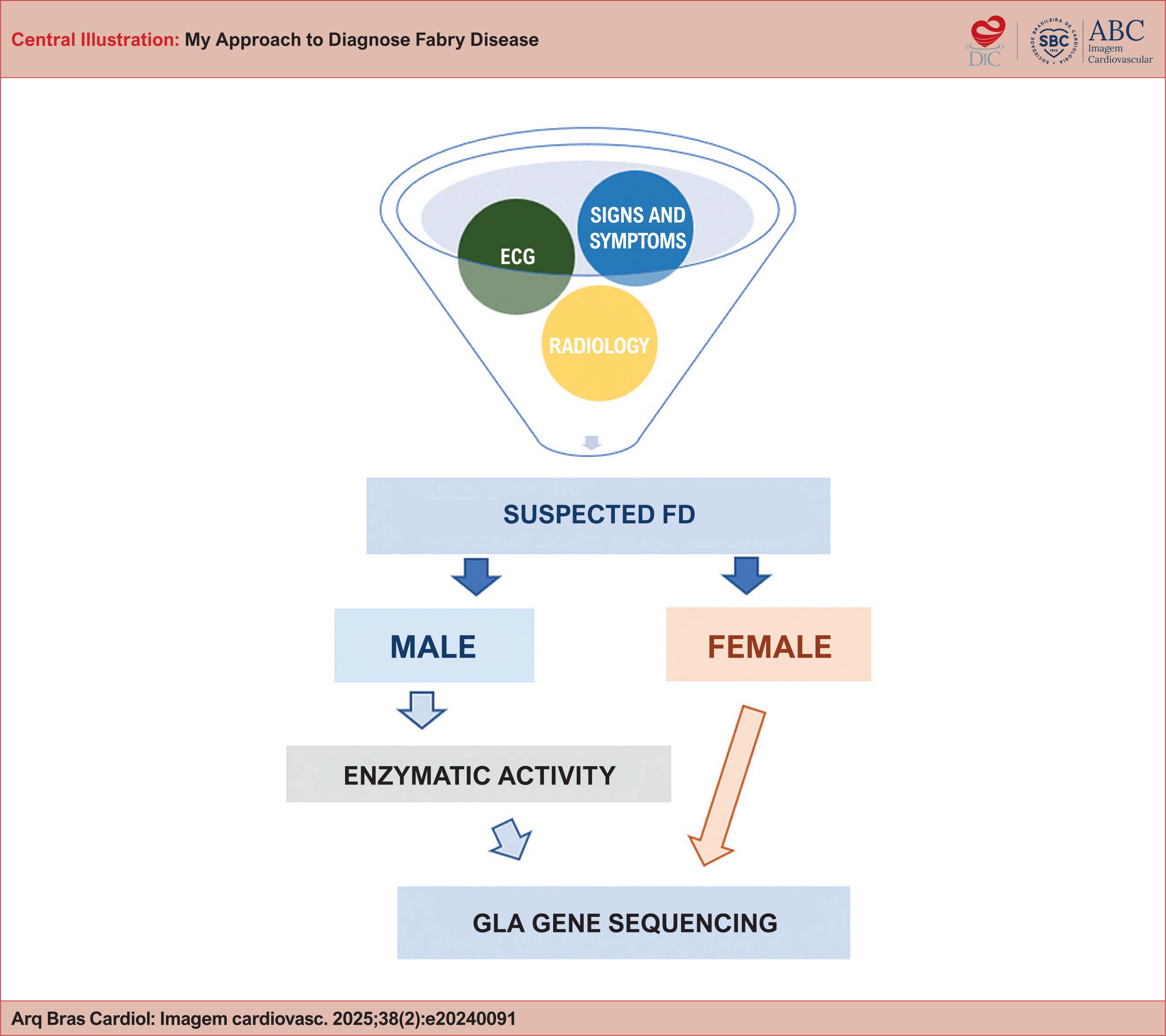 Diagnostic sequence of FD: ancillary tests. ECG: electrocardiogram; GLA: gene associated with FD (X chromosome); FD: Fabry
