Assessment of Coronary Flow Reserve by Myocardial Perfusion Scintigraphy using CZT Equipment
Abstract
This case report presents a myocardial perfusion analysis in a 67-year-old asymptomatic male patient with known coronary artery disease (CAD) and multiple risk factors. While myocardial perfusion Imaging (MPI) with single-photon emission computed tomography (SPECT) revealed a 5% area of ischemia, the myocardial blood flow quantification assessment in a camera equipped with cadmium-zinc-telluride (CZT) detectors demonstrated a reduced global myocardial flow reserve (MFR) of 1.57. Coronary angiography confirmed extensive multivessel disease, including an 80% stenosis in the left anterior descending artery (LAD) and an occlusion of the circumflex artery (LCx), findings not fully identified by the semi-quantitative SPECT MPI. This case underscores the importance of MFR assessment, particularly using CZT technology, in detecting significant CAD that may be underestimated by traditional SPECT methods. It highlights the potential of dynamic CZT SPECT to provide more comprehensive functional information, enhancing risk stratification and potentially influencing patient management in complex cases of suspected or known CAD.
Keywords
Coronary Artery Disease; Single-Photon Emission-Computed Tomography; Myocardial Ischemia
Resumo
O presente relato de caso apresenta análise da perfusão do miocárdio em um paciente masculino assintomático de 67 anos com doença arterial coronariana (DAC) conhecida e múltiplos fatores de risco. Enquanto a perfusão do miocárdio por tomografia computadorizada por emissão de fóton único (SPECT) revelou área de isquemia do miocárdio de 5%, a gama câmara utilizando detectores de telureto de cádmio e zinco (CZT) revelou uma reserva de fluxo miocárdico (RFM) global reduzida (1,57). A cinecoronariografia coronária confirmou doença multiarterial extensa, incluindo uma lesão de 80% na artéria descendente anterior (ADA) e uma oclusão da artéria circunflexa (ACX), achados não totalmente reconhecidos pela perfusão do miocárdio por SPECT. O presente caso demonstra a importância da avaliação da RFM, particularmente usando a tecnologia CZT, na identificação de DAC significativa que pode ser subestimada pelos métodos tradicionais de SPECT. Isso destaca o potencial da CZT para fornecer informações funcionais mais abrangentes, melhorando a estratificação de risco e potencialmente influenciando o manejo do paciente em casos complexos de DAC suspeita ou conhecida.
Palavras-chave
Doença da Artéria Coronariana; Tomografia Computadorizada de Emissão de Fóton Único; Isquemia Miocárdica
Introduction
Myocardial perfusion imaging (MPI) using single-photon emission computed tomography (SPECT) plays a crucial role in the diagnosis, risk stratification, and prognostic evaluation of coronary artery disease (CAD), with its clinical value being well established.1 , 2 Although SPECT provides valuable information, it also has certain limitations.2 , 3 Recent advancements in novel cameras using cadmium-zinc-telluride (CZT) detectors offer improved spatial and temporal resolution, enabling dynamic imaging and assessment of myocardial flow reserve (MFR).2 – 4 MFR serves as an indicator of microvascular function and has been shown to provide incremental information beyond perfusion analysis alone.2 Studies have demonstrated good correlation between CZT SPECT and positron emission tomography (PET) for myocardial blood flow (MBF) quantification.3 , 5
This case report highlights the limitations of SPECT without myocardial blood flow quantification in a patient with suspected CAD and underscores the importance of MFR assessment using CZT SPECT. It demonstrates how quantitative flow data can reveal critical information that is not readily apparent through traditional qualitative or semi-quantitative SPECT analysis, with potential implications for clinical decision-making and patient management.
Case report
A 67-year-old asymptomatic male patient with a medical history of systemic arterial hypertension, dyslipidemia, type 2 diabetes mellitus, sedentary lifestyle, and former smoking. He reported undergoing coronary angioplasty 10 years ago in the right coronary artery (RCA). The patient was on beta-blockers, statins, calcium channel blockers, spironolactone, antiplatelet agents, and proton pump inhibitors. He underwent myocardial perfusion scintigraphy at rest and after pharmacologic stress with dipyridamole, using 12 mCi and 36 mCi, respectively, of the radiopharmaceutical 99mTc-sestamibi on a D-SPECT CARDIO system (Spectrum Dynamics Medical) for the evaluation of myocardial ischemia and coronary flow reserve (CFR). Pharmacologic stress was performed using dipyridamole (0.568 mg/kg) over 4 minutes, with 99mTc-sestamibi administered at the 7th minute for acquisition of both dynamic and perfusion images.
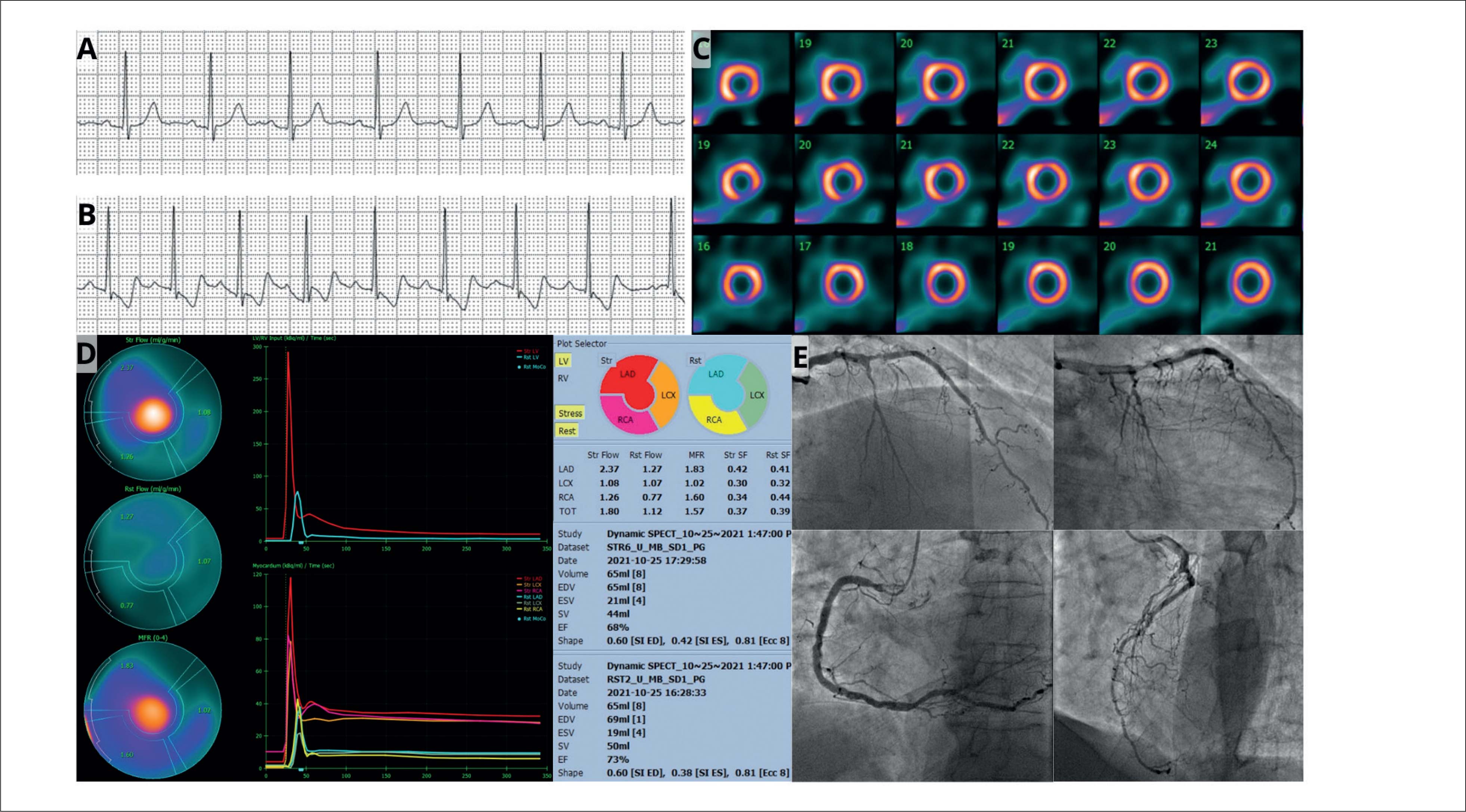
Perfusion analysis revealed an estimated ischemic defect involving 5% of the left ventricle, affecting the mid anterior, mid anterolateral, and mid inferior segments. The global MFR was reduced, with an estimated value of 1.57 (normal > 2.0). Segmental MFR values (stress/rest) were estimated at 1.60 in the RCA, 1.02 in the circumflex artery (LCx), and 1.83 in the left anterior descending artery (LAD) ( Figure 1 ). The patient subsequently underwent coronary angiography, which showed a patent previous stent, a calcified 80% stenosis in the LAD, 50% in the RCA, 90% in the posterior descending artery (PDA), and total occlusion of the LCx ( Figure 1 ).

A) Electrocardiogram (resting): sinus rhythm, nonspecific ventricular repolarization abnormalities. B) Electrocardiogram: Peak pharmacological stress showing ST-segment depression. C) Perfusion analysis relative to stress-induced myocardial ischemia in the anterior (middle), anterolateral (middle), and inferior (middle) segments. Estimated ischemic defect area in 5% of the left ventricle. Fixed defect in the inferior segment (apical). D) Coronary flow reserve: There was no adequate increase in blood flow in the myocardium under stress. Global MBF reserve = 1.57. Reserve (stress/rest) in the LAD = 1.83; LCX = 1.02; RCA = 1.60. Post-stress ejection fraction was estimated at 68%. E) Coronary angiography: Obstructive three-vessel atherosclerotic CAD. Left coronary artery: left coronary artery trunk with diffuse non-obstructive irregularities and calcifications. LAD with 70% lesions in the middle third and 80% in the distal third. LCX occluded in the proximal third. RCA with 50% lesion in the proximal third, 30% in the distal third. Posterior descending artery with 90% lesion in the middle third. Posterior ventricular branch with 40% lesion in the proximal third intrastent. LAD: left anterior descending artery; LCX: left circumflex artery; RCA: right coronary artery.
Discussion
This case report highlights how myocardial perfusion scintigraphy can underestimate the extent of myocardial ischemia. It underscores the importance of evaluating coronary flow reserve (CFR). CFR provides a more comprehensive assessment of myocardial perfusion through the absolute quantification of blood flow.6 Although the patient was asymptomatic, with known coronary artery disease (CAD) and a low ischemic burden, the reduced CFR indicates a high risk of extensive CAD and future cardiac events.1 The coronary microvasculature plays a crucial role in myocardial perfusion, and its assessment improves risk stratification.7 , 8
CZT cameras offer higher resolution, 3D dynamic acquisition capability, and accurate myocardial flow reserve (MFR) assessments, which enhance the detection of ischemic regions—particularly in patients with obstructive coronary artery disease (CAD).9 , 10 In this case, a D-SPECT system (CZT camera) was used, revealing a discrepancy between the findings of semi-quantitative SPECT and the blood flow quantification data obtained through dynamic CZT SPECT imaging. The detection of a globally impaired flow reserve is associated with more extensive anatomic coronary disease, and the territory of the circumflex artery—where a coronary occlusion was identified—showed the most pronounced reduction in regional MFR, demonstrating the superior diagnostic accuracy of CZT technology.
Conclusion
The reported case highlights the importance of considering myocardial flow reserve (MFR) and the potential use of CZT gamma cameras in the evaluation of patients with suspected or known coronary artery disease (CAD), especially when myocardial perfusion findings are inconclusive or inconsistent with the clinical presentation.
-
Sources of Funding
There were no external funding sources for this study.
-
Study Association
This study is not associated with any thesis or dissertation work.
-
Ethics Approval and Consent to Participate
This study was approved by the Ethics Committee of the Hospital Pró-Cardíaco under the protocol number 80960324.0.0000.5533. All the procedures in this study were in accordance with the 1975 Helsinki Declaration, updated in 2013. Informed consent was obtained from all participants included in the study.
-
Use of Artificial Intelligence
The authors did not use any artificial intelligence tools in the development of this work.
-
Availability of Research Data and Other Materials
The underlying content of the research text is contained within the manuscript.
References
-
1 Zavadovsky KV, Mochula AV, Maltseva AN, Boshchenko AA, Baev AE, Andreev SL, et al. The Diagnostic Value of SPECT CZT Quantitative Myocardial Blood Flow in High-Risk Patients. J Nucl Cardiol. 2022;29(3):1051-63. doi: 10.1007/s12350-020-02395-8.
» https://doi.org/10.1007/s12350-020-02395-8 -
2 Wang J, Li S, Chen W, Chen Y, Pang Z, Li J. Diagnostic Efficiency of Quantification of Myocardial Blood Flow and Coronary Flow Reserve with CZT Dynamic SPECT Imaging for Patients with Suspected Coronary Artery Disease: A Comparative Study with Traditional Semi-Quantitative Evaluation. Cardiovasc Diagn Ther. 2021;11(1):56-67. doi: 10.21037/cdt-20-728.
» https://doi.org/10.21037/cdt-20-728 -
3 Bailly M, Thibault F, Courtehoux M, Metrard G, Angoulvant D, Ribeiro MJ. Myocardial Flow Reserve Measurement During CZT-SPECT Perfusion Imaging for Coronary Artery Disease Screening: Correlation with Clinical Findings and Invasive Coronary Angiography-The CFR-OR Study. Front Med. 2021;8:691893. doi: 10.3389/fmed.2021.691893.
» https://doi.org/10.3389/fmed.2021.691893 -
4 Souza ACDAH, Gonçalves BKD, Tedeschi A, Lima RSL. Quantification of Coronary Flow Reserve with CZT Gamma Camera in the Evaluation of Multivessel Coronary Disease. Arq Bras Cardiol. 2018;111(4):635-37. doi: 10.5935/abc.20180196.
» https://doi.org/10.5935/abc.20180196 -
5 Mesquita C, Palazzo I, Pacheco J, Ker W, Sabra MMM, Chambi A, et al. Assessing Myocardial Flow Reserve Using CZT SPECT Perfusion Imaging: A Step Towards Clinical Application. Eur Heart J Cardiovasc Imaging. 2024;25(Suppl_1):jeae142.073. doi: 10.1093/ehjci/jeae142.073.
» https://doi.org/10.1093/ehjci/jeae142.073 -
6 Acampa W, Zampella E, Assante R, Genova A, Simini G, Mannarino T, et al. Quantification of Myocardial Perfusion Reserve by CZT-SPECT: A Head to Head Comparison with 82Rubidium PET Imaging. J Nucl Cardiol. 2021;28(6):2827-39. doi: 10.1007/s12350-020-02129-w.
» https://doi.org/10.1007/s12350-020-02129-w -
7 Sakuma H, Ishida M. Advances in Myocardial Perfusion MR Imaging: Physiological Implications, the Importance of Quantitative Analysis, and Impact on Patient Care in Coronary Artery Disease. Magn Reson Med Sci. 2022;21(1):195-211. doi: 10.2463/mrms.rev.2021-0033.
» https://doi.org/10.2463/mrms.rev.2021-0033 -
8 Assante R, Zampella E, Cantoni V, Green R, D’Antonio A, Mannarino T, et al. Prognostic Value of Myocardial Perfusion Imaging by Cadmium Zinc Telluride Single-Photon Emission Computed Tomography in Patients with Suspected or Known Coronary Artery Disease: A Systematic Review and Meta-Analysis. Eur J Nucl Med Mol Imaging. 2023;50(12):3647-58. doi: 10.1007/s00259-023-06344-8.
» https://doi.org/10.1007/s00259-023-06344-8 -
9 Kelshiker MA, Seligman H, Howard JP, Rahman H, Foley M, Nowbar AN, et al. Coronary Flow Reserve and Cardiovascular Outcomes: A Systematic Review and Meta-Analysis. Eur Heart J. 2022;43(16):1582-93. doi: 10.1093/eurheartj/ehab775.
» https://doi.org/10.1093/eurheartj/ehab775 -
10 Lima R, Bezerra ALF, Daibes M, Domenico C, Lorenzo A. Clinical Impact of Assessment of Myocardial Flow Reserve in Identifying the Cause of Chest Discomfort. Arq Bras Cardiol. 2024;121(6):e20230700. doi: 10.36660/abc.20230700.
» https://doi.org/10.36660/abc.20230700
Edited by
-
Editor responsible for the review:
Marcelo Tavares
The underlying content of the research text is contained within the manuscript.