 Prevention, surveillance, and early detection of hepatocellular carcinoma – a Brazilian multidisciplinary consensus
ABSTRACT
Background: Hepatocellular carcinoma is a major cause of cancer-related mortality and represents a growing public health challenge.
Aims: To develop multidisciplinary evidence-based recommendations for the prevention and early detection of hepatocellular carcinoma.
Methods: This consensus was coordinated by the Brazilian Society of Surgical Oncology, with the participation of 13 additional national societies. A steering committee defined the key clinical questions addressing prevention, surveillance, and early detection strategies. Recommendation statements were informed by a comprehensive, non-systematic review of the literature conducted using PubMed, Scopus, and the Cochrane Library.
Results: Eighteen recommendations reached consensus, covering three main domains: 1. Primary prevention, including HBV vaccination, timely antiviral therapy for viral hepatitis, and lifestyle interventions for metabolic dysfunction. 2. Secondary prevention, outlining risk-based surveillance strategies for patients with advanced fibrosis or cirrhosis due to hepatitis B virus, hepatitis C virus, or metabolic dysfunction–associated steatotic liver disease. 3. Operational standards for surveillance, emphasizing ultrasound as the cornerstone of monitoring, the importance of structured reporting, management of small nodules, and the selective use of cross-sectional imaging.
Conclusions: These multidisciplinary recommendations provide practical, adaptable guidance for the prevention, surveillance, and early detection of hepatocellular carcinoma. This guidance document aims to reduce the burden of hepatocellular carcinoma and support better clinical outcomes across diverse healthcare settings.
Keywords:
Liver; Carcinoma, Hepatocellular; Consensus; Guidelines as Topic; Early Detection of Cancer; Epidemiology
ARTICLE HIGHLIGHTS
This is the first article in a four-part series of the Brazilian multidisciplinary consensus on hepatocellular carcinoma.
This consensus provides evidence-based recommendations on prevention, surveillance, and early detection.
It integrates the available scientific literature with national clinical expertise to support and guide clinical decision-making.

CENTRAL MESSAGE Prevention and early detection save lives in liver cancer. This Brazilian multidisciplinary consensus integrates the best scientific evidence and the expertise of specialists from across the country to more effectively guide the prevention, identification, and surveillance of hepatocellular carcinoma. By answering key clinical questions and offering practical, easy-to-use recommendations, this document helps physicians and healthcare teams provide better care to people at risk.
PERSPECTIVES This consensus provides practical guidance to improve the prevention and early detection of liver cancer in daily clinical care. The recommendations emphasize raising awareness of hepatocellular carcinoma, stimulating the development of broader surveillance programs, promoting earlier diagnosis, and improving outcomes for patients at risk for developing primary liver cancer.
RESUMO
Racional: O carcinoma hepatocelular (CHC) é uma das principais causas de mortalidade relacionada ao câncer e representa um crescente desafio para a saúde pública.
Objetivos: Desenvolver recomendações multidisciplinares baseadas em evidências para a prevenção e detecção precoce do CHC.
Métodos: Este consenso foi coordenado pela Sociedade Brasileira de Oncologia Cirúrgica, com a participação de outras 13 sociedades nacionais. Um comitê diretivo definiu as principais questões clínicas abordando estratégias de prevenção, vigilância e detecção precoce. As recomendações foram elaboradas com base em uma revisão abrangente e não sistemática da literatura, realizada utilizando PubMed, Scopus e a Biblioteca Cochrane.
Resultados: Dezoito recomendações foram obtidas por consenso, abrangendo três domínios principais: (1) Prevenção primária, incluindo vacinação contra o VHB, terapia antiviral oportuna para hepatite viral e intervenções no estilo de vida para disfunção metabólica. (2) Prevenção secundária, delineando estratégias de vigilância baseadas em risco para pacientes com fibrose avançada ou cirrose devido a VHB, VHC ou MASLD. (3) Padrões operacionais para vigilância, enfatizando a ultrassonografia como a base do monitoramento, a importância da elaboração de relatórios estruturados, o manejo de pequenos nódulos e o uso seletivo de exames de imagem seccionais.
Conclusões: Estas recomendações multidisciplinares fornecem orientações práticas e adaptáveis para a prevenção, vigilância e detecção precoce do CHC. Este documento de orientação visa reduzir a carga do CHC e apoiar melhores resultados clínicos em diversos contextos de saúde.
Palavras-chave:
Fígado; Carcinoma Hepatocelular; Consenso; Guias como Assunto; Detecção Precoce de Câncer; Epidemiologia
INTRODUCTION
Liver cancer represents a major global health challenge. According to GLOBOCAN 2020, it is the seventh most common malignancy and the second leading cause of cancerrelated death worldwide124. Hepatocellular carcinoma (HCC) constitutes 75–85% of primary liver tumors87.
In Brazil, although epidemiological information remains limited, data from the National Cancer Institute of Brazil (INCA) indicate that liver cancer ranks 15th in incidence, with the highest rates observed in the South region for both sexes95. Between 2001 and 2015, more than 120,000 deaths were recorded due to malignant liver neoplasms, predominantly in men (56.9%)29. Mortality varies regionally, with the North presenting the highest rates overall, while female mortality peaks in the Northeast and male mortality in the South29.
In Brazil, viral hepatitis remains the leading cause of HCC. Nearly 50% of cases are linked to the hepatitis B virus (HBV) and around 25% to the hepatitis C virus (HCV)41,44. HCV-infected individuals have a 17-fold higher risk of developing HCC48, and coinfection with HBV accelerates progression to cirrhosis and hepatic decompensation19. Infection by the hepatitis D virus, which relies on HBV for replication, further increases the risk of HCC when compared with HBV alone57,101.
Metabolic dysfunction–associated risk factors are becoming increasingly relevant45. Diabetes, overweight, and obesity have an association with the development of metabolic dysfunction-associated steatotic liver disease (MASLD) and a two-fold increase in HCC risk, even in the absence of cirrhosis33,92. With the rapid rise of obesity and diabetes, MASLD is projected to become a leading cause of HCC in the future7. Excessive alcohol consumption (>40–60 g/day) is also a major contributor28, displaying a linear dose-response relationship and doubling HCC risk in individuals with HCV coinfection42. Environmental exposures, especially aflatoxin contamination of poorly stored grains and nuts, remain a dominant etiologic factor in parts of Asia and Africa, due to carcinogenic mutations such as those affecting TP5311,28. Additionally, rare hereditary metabolic and cholestatic disorders are associated with an increased risk58.
Given the diversity of etiologic profiles, regional disparities in incidence and mortality, and varying levels of access to surveillance and treatment, context-specific guidance is crucial. In this setting, a Brazilian multidisciplinary consensus is essential to translate high-quality scientific evidence into standardized recommendations. The objective of this consensus is to synthesize the most current national and international literature on the prevention, surveillance, and early detection of HCC, to provide structured, multidisciplinary guidance, and to serve as the first article in a four-part series developed by this Brazilian multidisciplinary consensus addressing the full continuum of HCC care.
METHODS
Study design
This consensus was developed under the leadership of the Brazilian Society of Surgical Oncology, in collaboration with 13 additional Brazilian medical societies, thereby ensuring comprehensive, multidisciplinary representation (Table 1). The initiative focused on developing evidence-based recommendations to address the prevention, surveillance, and early detection of HCC.
Expert panel
The expert panel consisted of 43 specialists appointed by the participating societies (Figure 1). Panel members were selected based on their expertise in hepatology, oncology, hepatopancreatobiliary surgery, liver transplantation, radiology, gastroenterology, and related fields. The panel was responsible for evaluating scientific evidence, supporting the development of rationales, and issuing recommendations based on the strength of the evidence.

Geographic distribution of Brazilian cities participating in the Hepatocellular carcinoma consensus. Circle size is proportional to the frequency of participating experts in each city.
Formulation of clinical questions
Initially, a scientific steering committee identified key topics and designed clinically relevant questions about HCC surveillance strategies, surveillance criteria, and preventive interventions. These questions were organized to reflect clinical decisionmaking needs across different settings and were refined through preliminary meetings with representatives of all participating societies to guarantee a broad, cross-disciplinary perspective.
Literature review
A targeted narrative review of the literature was conducted to inform the development of the clinical questions and recommendations. Searches were performed in PubMed, Scopus, and the Cochrane Library, focusing on studies published over the past 30 years. The search aimed to identify clinically relevant evidence.
Eligible sources included clinical studies in humans, such as observational studies, randomized and non-randomized trials, systematic reviews, practice guidelines, and prior consensus statements relevant to HCC prevention, surveillance, and early detection. Preclinical, experimental, and animal studies were not prioritized unless considered essential for contextual understanding.
Publications were selected based on their relevance to the predefined topics and clinical applicability. The steering committee reviewed and synthesized the evidence qualitatively, ensuring consistency of interpretation and alignment between the supporting data and the final recommendations.
Consensus process and voting rounds
Following expert deliberations, the synthesized clinical questions and draft recommendations were circulated to representatives of the 14 participating societies through an electronic voting platform as part of an adapted Delphi process94. Participants independently rated each item and provided written feedback. After the first round, aggregated results and anonymized comments were discussed in structured online meetings, and statements were revised to enhance clarity, scientific accuracy, and clinical applicability.
A second round of electronic voting was then conducted using the revised items. Consensus was defined a priori as an approval rate of ≥80%, based on the response options “completely accept” or “accept with reservations.” Statements reaching this threshold in the second round were considered to have achieved consensus and were incorporated into the final document, whereas those that did not meet the predefined criterion were excluded from the final recommendations..
Evidence level and recommendation grading
The quality of evidence and the strength of recommendations were assessed using the UpToDate® grading system136. Evidence quality reflects confidence in effect estimates and is categorized as high, moderate, or low. Evidence quality reflects confidence in estimates and is categorized as high, moderate, or low, ranging from consistent results derived from well-conducted randomized controlled trials, systematic reviews, evidence based primarily on observational studies or trials with significant methodological limitations. Recommendations are classified as strong or weak according to the balance between benefits and risks, the quality of supporting evidence, and the degree of certainty surrounding outcomes. Strong recommendations indicate that benefits clearly outweigh risks and apply to most patients in most clinical scenarios. In contrast, weak recommendations reflect uncertainty, suggesting that alternative approaches may be equally reasonable (Table 2)
RESULTS
Expert characteristics
A total of 43 Brazilian experts participated in this national multidisciplinary consensus on HCC. The expert panel developed 18 evidence-based recommendations addressing key aspects of prevention, surveillance, and early diagnosis of HCC. The final recommendations are presented in Table 3.
What is the importance of universal hepatitis B vaccination in preventing hepatocellular carcinoma?
Chronic HBV infection is a well-established risk factor for HCC, accounting for a substantial proportion of global cases, particularly in endemic regions16. Although antiviral therapy can reduce viral replication and lower HCC risk, HBV eradication remains difficult, with functional cure achieved in a minority of patients76,77. Most individuals require long-term or lifelong treatment.
In contrast, universal HBV vaccination, particularly when administered at birth, prevents chronic infection and is a highly effective primary prevention measure. Longitudinal data from Taiwan demonstrate a marked decline in childhood HCC incidence within a decade of implementing a national vaccination program16,64, findings that have been consistently replicated in other populations20,52,60,88,102,140. These results highlight the long-term population-level impact of vaccination on reducing HCC burden.
Recommendation 1
Universal HBV vaccination — preferably initiated at birth — should be implemented as a primary prevention strategy to reduce the incidence of HBV-related HCC. Evidence level: High. Recommendation grade: Strong.
What is the importance and impact of early treatment of hepatitis B virus infection on preventing Hepatocellular carcinoma?
In patients with chronic HBV, several factors are consistently associated with an increased risk of HCC and disease progression, including high serum HBV DNA levels (>104 copies/mL or >2,000 IU/mL), elevated alanine aminotransferase (ALT), hepatitis B e antigen (HBeAg) positivity, and the presence of cirrhosis53. In this context, long-term suppression of viral replication with nucleotide analogs has been shown to significantly reduce the incidence of HCC and overall mortality in patients with immune-active disease or cirrhosis21,79.
The role of antiviral therapy in early disease stages, particularly in immune-tolerant patients with normal ALT levels and no significant fibrosis, remains controversial. Retrospective cohort studies from Asia have suggested higher long-term risks of HCC and liver-related mortality among untreated immune-tolerant individuals69,118. In contrast, prospective data from the Hepatitis B Research Network cohort in North America demonstrated a very low incidence of adverse clinical outcomes among carefully selected immune-tolerant patients under close surveillance80. These divergent findings highlight ongoing uncertainty regarding the benefit of antiviral treatment in this population for HCC prevention.
Recommendation 2
Antiviral treatment with nucleotide analogs should be initiated in patients with chronic HBV who have high serum HBV DNA levels (>104 copies/mL or >2,000 IU/mL), elevated alanine aminotransferase, HBeAg positivity, and/or cirrhosis, as long-term viral suppression reduces the risk of HCC and disease progression. There is no recommendation for routine antiviral treatment in patients in the immune-tolerant phase solely to prevent HCC.Evidence level: Moderate. Recommendation grade: Weak.
What is the importance and impact of early treatment of hepatitis C virus infection on preventing hepatocellular carcinoma?
Chronic HCV infection is a well-established cause of HCC, and the risk increases with the progression of liver fibrosis132. The advent of direct-acting antivirals (DAAs) has significantly transformed the management of HCV, offering sustained virologic response (SVR) rates of 95–100% with favorable safety profiles.
Achieving SVR with DAA therapy is associated with a substantial reduction in the incidence of HCC, particularly when treatment occurs before the development of advanced fibrosis or cirrhosis55,62.
While early retrospective studies raised concerns about a possible association between DAAs and increased HCC risk or recurrence, subsequent large-scale analyses and prospective cohorts refuted this hypothesis and demonstrated the protective role of antiviral therapy63,134. A pivotal French cohort study14 confirmed that patients treated with DAAs had reduced risks of all-cause mortality and HCC, including those with cirrhosis. Notably, this benefit was seen only among patients who achieved SVR, while those without virological cure maintained a high risk of liver related outcomes.
Recommendation 3
Early treatment of chronic HCV infection with DAA is recommended to prevent the progression to advanced fibrosis or cirrhosis and reduce the risk of HCC. The benefit is most evident in patients who achieve sustained virologic response. Evidence level: High. Recommendation grade: Strong.
What is the impact of hepatitis C virus infection treatment on preventing recurrence of hepatocellular carcinoma treated with curative intent (tertiary prevention related to hepatitis C virus)?
A prospective cohort study by Cabibbo et al. showed that DAAs significantly improved overall survival and reduced the risk of hepatic decompensation in 163 patients with compensated HCV-related cirrhosis and successfully treated HCC with curative resection or ablation. However, the risk of HCC recurrence was not significantly reduced (HR 0.7; 95%CI 0.44–1.13; p=0.15)13,57.
Recommendation 4
DAA therapy improves the quality of life and helps prevent hepatic decompensation in patients with cirrhosis. However, the impact of DAAs on reducing the risk of HCC recurrence remains incompletely elucidated, as does the appropriate timing for initiating antiviral treatment. Antiviral therapy may be offered to patients with HCC who have previously been treated with curative intent, given the uncertain impact on recurrence risk, while recognizing the clear benefits of viral eradication in maintaining liver function. While current evidence does not conclusively demonstrate a reduction in recurrence risk, delaying antiviral therapy solely due to concerns about recurrence is not supported. Evidence level: Moderate. Recommendation grade: Weak.
What is the recommended posttreatment surveillance strategy for patients with hepatitis C virus related hepatocellular carcinoma treated with curative intent?
Even after achieving an SVR through DAA therapy, patients with HCV-related cirrhosis remain at risk for HCC recurrence due to the persistent effects of chronic liver injury and the concept of a “field cancerization” effect in cirrhotic livers55,132. Studies have shown that the risk of recurrence after curative-intent treatment, such as resection or ablation, remains high, particularly in the first two years post-treatment13,103.
The persistence of recurrence risk underscores the need for long-term surveillance to enable early detection of potentially treatable lesions. However, the optimal surveillance intervals remain a subject of ongoing debate.
Recommendation 5
Patients with HCV-related HCC treated with curative intent should undergo indefinite surveillance with dynamic contrast-enhanced imaging (either multiphasic abdominal MRI or triphasic CT) every 3 to 6 months, given the persistent risk of recurrence related to underlying liver disease and the field effect of hepatocarcinogenesis.Evidence level: Moderate. Recommendation grade: Weak.
What is the impact of hepatitis B virus infection treatment on preventing hepatocellular carcinoma recurrence (tertiary prevention related to hepatitis B virus)?
In patients with HBV-related HCC, high HBV DNA levels and greater hepatic inflammatory activity are among the strongest predictors of postoperative recurrence18,119. Several retrospective and prospective studies support the role of antiviral therapy with nucleos(t)ide analogs in reducing the risk of recurrence and improving survival following curative-intent treatments.
Early retrospective studies demonstrated that patients with high HBV DNA levels benefited from antiviral therapy after curative liver resection22,138. Subsequent randomized clinical trials confirmed these findings. In one trial, Yin et al.141 showed that postoperative antiviral therapy improved recurrence-free and overall survival. Similarly, Huang et al.50 found that adefovir reduced late recurrence in patients with HBV DNA >2,000 IU/mL after resection. More recently, telbivudine-based therapy was shown to reduce recurrence and improve 5-year overall survival (64 vs. 44%)51.
In the liver transplant setting, HBV recurrence is more common among recipients with HCC than those without (2–35 vs. 1–9.7%) and has been associated with increased risk of tumor recurrence. Therefore, antiviral prophylaxis is critical to minimize both viral and oncologic relapse30,130.
Recommendation 6
Postoperative antiviral therapy is recommended for all patients with HBV-related HCC undergoing curative-intent treatment, including liver resection or transplantation, regardless of baseline viral load.Evidence level: High. Recommendation grade: Strong.
What is the importance of diabetes control in patients with fatty liver disease for the prevention of hepatocellular carcinoma?
The association between diabetes mellitus and HCC has been consistently demonstrated across multiple epidemiological studies5,17,32,46,54,58,71,74,110,1. Yang et al.139, analyzing 14 prospective cohort studies, found that diabetes increased the risk of HCC by 90% (RR 1.9; 95%CI 1.2–2.3). Wang et al.133 reported a pooled relative risk of 2.2 (95%CI 1.7–3.0) for HCC among diabetic patients in a systematic review.
Despite this strong epidemiological association, no randomized controlled trials have evaluated whether intensive glycemic control directly reduces the risk of HCC. Nonetheless, given the significant overlap between diabetes, metabolic syndrome, fatty liver disease, and progression to cirrhosis, glycemic control remains a fundamental component of comprehensive risk reduction strategies in patients with fatty liver disease.
Recommendation 7
Although no randomized controlled trials have confirmed a direct benefit of glycemic control in preventing HCC, diabetes management is strongly recommended in patients with fatty liver disease, especially when additional risk factors for cirrhosis are present. Evidence level: Low. Recommendation grade: Strong.
What is the role of lifestyle interventions and weight loss in reducing hepatocellular carcinoma risk in patients with nonalcoholic fatty liver disease?
MASLD is increasingly recognized as a relevant risk factor for HCC, including in non-cirrhotic individuals7,91. The mechanisms of hepatocarcinogenesis in MASLD are multifactorial and not yet fully understood, but metabolic dysfunction — particularly in the presence of obesity, type 2 diabetes, dyslipidemia, and hypertension — plays a central role72,100,122.
Although the direct impact of weight loss on HCC incidence remains to be definitively established, multiple studies have demonstrated that lifestyle interventions, especially those leading to meaningful weight reduction, improve hepatic steatosis and can promote histological resolution of MASLD and regression of fibrosis73,100,131. In the Cuban cohort by Vilar-Gomez et al.131, ≥10% weight loss led to MASLD resolution in 90% of patients and fibrosis regression in 45%. Additionally, the Singapore Chinese Health Study associated healthy lifestyle habits — such as physical activity, Mediterranean diet, nonsmoking, low alcohol intake, and adequate sleep — with a reduced risk of HCC82. These findings support a preventive role for structured lifestyle modifications, particularly in patients with MASLD and metabolic risk factors.
Recommendation 8
For overweight or obese patients with nonalcoholic fatty liver disease, lifestyle interventions, such as dietary changes and regular physical activity, are recommended to promote weight loss, improve metabolic parameters, and potentially reduce the risk of HCC. Evidence level: High. Recommendation grade: Strong.
How important is reducing alcohol consumption in preventing hepatocellular carcinoma?
Excessive alcohol intake is a well-established risk factor for liver disease progression, particularly in individuals with underlying metabolic dysfunction or viral hepatitis. In patients with MASLD, excessive alcohol consumption has been independently associated with increased hepatic steatosis, liver inflammation, and accelerated fibrosis progression31,72,97. In a Swedish cohort, excessive alcohol use (defined as >60 g/day for men and >48 g/day for women) was linked to significantly higher rates of fibrosis progression in MASLD patients (47 vs. 11%)31.
Evidence also suggests that even light to moderate alcohol consumption may negatively impact liver health in at-risk populations. Ajmera et al.2 and VanWagner et al.129 showed that modest alcohol intake worsened steatosis, increased liver enzyme levels, and reduced the likelihood of MASLD resolution. Additionally, synergistic effects have been documented between alcohol and other HCC risk factors, such as viral hepatitis, diabetes, and obesity28,47,81.
Although prospective studies evaluating HCC incidence in light/moderate drinkers are lacking, the available evidence supports alcohol reduction — especially in individuals with underlying liver disease — as a key preventive strategy.
Recommendation 9
To reduce the risk of HCC, it is recommended to avoid excessive alcohol consumption. Excessive alcohol use is associated with hepatic steatosis, fibrosis progression, and HCC. Evidence level: High. Recommendation grade: Strong.
What is the role of statins, metformin, and coffee consumption in hepatocellular carcinoma prevention?
Observational studies have suggested a protective association between the use of statins and the risk of HCC in patients with chronic liver disease, particularly those with viral hepatitis. Five recent meta-analyses37,56,59,75,143 consistently reported a reduction in HCC incidence among statin users. In the largest analysis, Facciorusso et al.37 evaluated more than 1.9 million individuals and found that lipophilic statins were associated with a 51% reduction in HCC risk (HR 0.49; 95%CI 0.39–0.62). Subgroup analyses confirmed this benefit across patients with or without HBV/HCV infection, diabetes, cirrhosis, and regardless of age or sex. However, all supporting studies are observational, and no randomized controlled trials have yet confirmed a causal relationship. Therefore, the literature still lacks high-quality studies that adequately weigh the benefits against potential adverse effects and does not yet provide strong evidence to support the use of statins solely for chemoprevention.
Metformin has also been associated with a reduced risk of HCC in diabetic patients with chronic liver disease. Several meta-analyses, including Zhou et al.144, have shown that metformin use significantly reduces HCC risk compared with insulin or sulfonylureas (RR 0.49; 95%CI 0.25–0.97). A meta-analysis by Ma et al.83, involving over 550,000 individuals with diabetes, also showed improved survival among HCC patients treated with metformin (OR 0.52; 95%CI 0.40–0.68; p<0.001). Given these findings and the overall benefit in glycemic control, metformin should be prioritized in antidiabetic regimens for these patients.
Coffee consumption has been consistently associated with a dose-dependent reduction in HCC risk, regardless of underlying liver disease etiology. Kennedy et al.67 analyzed 18 cohort studies (over 2 million participants and 2,905 cases) and found that each additional two cups of coffee per day reduced HCC risk by 35% (RR 0.65; 95%CI 0.59–0.72). Similar findings were observed across subgroups with chronic liver disease, alcohol use, obesity, and diabetes. While data on decaffeinated coffee are inconclusive8,67, evidence supports recommending regular coffee consumption as a low-cost, accessible, and potentially protective measure in individuals with chronic liver disease15,35.
Recommendation 10
Coffee consumption should be encouraged in patients with chronic liver disease as an accessible and potentially protective strategy against HCC. Metformin use is recommended in diabetic patients with chronic liver disease for its glycemic benefits and potential reduction in HCC risk. Despite promising observational data, statins should not be routinely used solely for HCC prevention until higher-quality evidence confirms their benefit in this setting. Evidence level: Moderate. Recommendation grade: Weak.
Which patients with viral hepatitis should undergo hepatocellular carcinoma surveillance?
Chronic viral hepatitis infection carries a high likelihood of progression to chronic hepatitis, cirrhosis, and ultimately HCC in a substantial proportion of cases. Longitudinal cohort data suggest that 20–25% of chronic hepatitis patients develop cirrhosis over 20 to 30 years, with an annual risk of HCC ranging from 1 to 2% in cirrhotic patients99. In chronic HBV infection acquired in adulthood, approximately 5–10% of patients progress to cirrhosis, with a subsequent risk of developing HCC84.
In theory, counseling for surveillance and monitoring of patients with chronic viral hepatitis and potential risk for HCC should be prioritized for those with more advanced stages of liver fibrosis (F3–F4) and/or cirrhosis. However, a minority of patients with chronic viral hepatitis — particularly those with HBV infection and/or additional risk factors — can develop HCC even in the presence of only mild or moderate fibrosis. These risk factors include diabetes, metabolic syndrome, HIV or other viral coinfections, family history of HCC, iron overload, and older age. In such cases, surveillance strategies may be individualized, especially when multiple risk factors overlap. In HBV specifically, certain populations — including males over 40, individuals of Asian or African descent, patients with high HBV DNA levels, and those with a family history of HCC — are considered at elevated risk and may benefit from HCC surveillance regardless of fibrosis stage39.
Recommendation 11
HCC surveillance is recommended for all patients with advanced fibrosis (F3) or cirrhosis (F4), regardless of etiology. In chronic HBV, surveillance is also indicated in F0–F2 patients with high-risk features (e.g., older age, male sex, high HBV DNA, family history, or Asian/African ancestry). In selected F0–F2 cases with overlapping risks, individualized surveillance may be considered. Evidence level: High. Recommendation grade: Strong.
Should patients with metabolic dysfunction-associated steatotic liver disease and significant fibrosis undergo hepatocellular carcinoma surveillance?
Brazilian data support the close relationship between metabolic dysfunction and HCC. In a cohort of 110 patients with HCC attributed to MASLD, obesity was present in 52.7%, diabetes in 73.6%, dyslipidemia in 41%, and metabolic syndrome in 57.2%25. Among those with histological confirmation, 61.5% had MASLD-related cirrhosis and 27% showed fibrosis stage 1–3, while a minority exhibited HCC in the absence of fibrosis. In a large U.S. cohort of patients with MASLD, the incidence of HCC reached 1.06 per 100 person-years114. Importantly, hepatic fibrosis is the key prognostic determinant, with advanced fibrosis independently associated with liver-related events and overall mortality. In an Asian cohort of more than 6,500 patients with ultrasound-defined MASLD, an APRI >1.5 significantly predicted HCC risk over a median follow-up of 5.6 years66.
Although only 10–30% of MASLD cases progress to cirrhosis, HCC can occasionally develop in the absence of advanced fibrosis49. Nonetheless, the incidence of HCC in non-cirrhotic MASLD remains extremely low — approximately 0.008 per 100 person-years3,63 — which does not justify routine surveillance in this subgroup. In contrast, individuals with advanced fibrosis (F3) or cirrhosis (F4), whether diagnosed through biopsy or non-invasive tools, carry a substantially higher risk of HCC and are appropriate candidates for surveillance.
Various alternative methods have been proposed to stage MASLD, including biomarkers (e.g., CK-18 fragments), genetic polymorphisms (e.g., PNPLA3 I148M), and metabolomic/lipidomic-based models, alongside advanced imaging techniques34. To reduce diagnostic uncertainty, current recommendations support combining two complementary fibrosis assessments — a serologic and an imaging-based. When both suggest advanced fibrosis or cirrhosis, HCC surveillance is considered justified. Conversely, in the absence of advanced fibrosis, surveillance is not routinely indicated due to the low incidence of HCC63.
Recommendation 12
HCC surveillance is recommended for patients with MASLD who have biopsy-proven or non-invasively diagnosed advanced fibrosis (F3) or cirrhosis (F4). Evidence level: High. Recommendation grade: Strong.
What is the most appropriate imaging modality for hepatocellular carcinoma surveillance?
Over the years, several cohort studies10,23,24,111,137 and costeffectiveness analyses6,78,113 have assessed the benefit of ultrasound-based HCC surveillance and helped define the target populations. A meta-analysis published in 2009 reported that abdominal ultrasound had a sensitivity ranging from 60–80% and a specificity of over 90% for early-stage HCC detection116. More recently, a 2018 meta-analysis including 32 studies from 1990 to 2016 estimated a pooled sensitivity of 84% (95%CI 76–92%) for HCC, but only 47% (95%CI 33–61%) for earlystage tumors. Among the nine studies that directly evaluated early-stage detection, sensitivity was 53% (95%CI 35–70%) and specificity was 91% (95%CI 86–94%)128. The reduced sensitivity of ultrasound surveillance for HCC detection is closely associated with underlying liver conditions that impair acoustic penetration and lesion conspicuity, particularly hepatic steatosis related to MASLD and alcohol-related liver disease. In these settings, increased parenchymal heterogeneity substantially limits image quality and diagnostic performance, contributing to early-stage tumors being missed40.
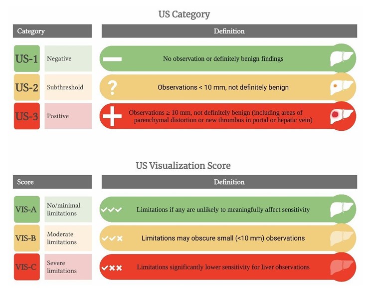
One of the main disadvantages of ultrasound is its marked operator dependence, which can lead to substantial variability in examination quality and diagnostic performance. To improve reporting standardization and communication between imaging specialists and referring clinicians, the ultrasound Liver Imaging Reporting and Data System (US LI-RADS) was introduced. In addition to categorizing focal liver observations, this framework has a visualization score that reflects the overall adequacy of the examination for surveillance purposes. Patients are classified as having no or minimal limitations (score A), moderate limitations (score B), or severe limitations (score C), the latter indicating that ultrasound surveillance may be unreliable. A large meta-analysis including more than 25,000 examinations reported that moderate or severe visualization limitations occur in a substantial proportion of studies, particularly among patients with cirrhosis related to metabolic liver disease and obesity61. Importantly, these limitations are largely patient-related rather than operator-related alone, underscoring that even technically well-performed examinations may be inadequate in certain populations. These findings highlight that, in a meaningful subset of at-risk individuals, ultrasound may be intrinsically suboptimal for surveillance and that such limitations should be explicitly documented in the imaging report to guide subsequent clinical decision-making.
The only large randomized controlled trial on HCC surveillance was conducted in China in 2004 and enrolled nearly 20,000 patients with chronic HBV infection. Participants were randomized to semiannual surveillance (ultrasound plus alpha-fetoprotein (AFP) test) or no surveillance. Despite suboptimal adherence (<60%) in the surveillance arm, significant survival benefits were observed: 66% at 1 year, 53% at 3 years, and 46% at 5 years, compared to 31, 7, and 0%, respectively, in the control group142. Given these striking results, similar studies in Western populations are now considered ethically infeasible. Ultrasound remains the most accessible, cost-effective, and widely used tool for routine monitoring in chronic liver disease98. Despite its advantages, the real-world effectiveness of ultrasound is often compromised by inconsistent surveillance intervals, poor-quality equipment, and limited operator expertise. These limitations contribute to the underdiagnosis of early-stage HCC in at-risk patients27,107,117,128.
Alternative imaging modalities, such as computed tomography (CT) scan or magnetic resonance imaging (MRI), have been proposed for HCC surveillance70. A Korean study demonstrated that MRI achieved substantially higher sensitivity than ultrasound (84.8 vs. 27.3%) and a significantly greater positive predictive value for detecting lesions smaller than 2 cm, while also yielding a markedly lower false-positive rate70. Similar findings were reported in a prospective French study, which likewise showed superior performance of MRI compared with ultrasound for early HCC detection93. However, cost, radiation exposure (in CT), and logistical barriers limit their widespread application.
Abbreviated MRI protocols have gained attention as a potential surveillance option in higher-risk populations — particularly patients with parenchymal heterogeneity due to obesity or advanced fibrosis, which can impair ultrasound accuracy106,123. However, robust evidence supporting their routine use is still lacking, and it is prudent to await the results of ongoing prospective clinical trials (NCT05828446; NCT05716620; NCT05657249; NCT05486572; NCT05095714; NCT04455932; NCT04288323), which are expected to provide more definitive data regarding diagnostic accuracy, clinical effectiveness, and cost-effectiveness before broad implementation in clinical practice can be recommended.
Recommendation 13
Abdominal ultrasound is the first-line imaging modality for HCC surveillance. Evidence level: Moderate. Recommendation grade: Strong.
Should alpha-fetoprotein be used for hepatocellular carcinoma surveillance?
AFP was historically the only available serological biomarker for HCC surveillance. However, when used alone, AFP has suboptimal sensitivity for early-stage tumors, as levels may remain within normal range in a significant proportion of patients38. Although its impact on overall survival remains uncertain, major guidelines currently endorse semiannual surveillance using abdominal ultrasound, with or without AFP, in high-risk populations40,85,96,104.
Recent evidence has explored the accuracy of this combined approach. A 2018 meta-analysis128 including 32 cohort studies (1990–2016) reported that while ultrasound alone had a sensitivity of 84% (95%CI 76–92%) for detecting HCC at any stage, it dropped to 47% (95%CI 3361%) for early-stage tumors. In comparative analyses, ultrasound combined with AFP showed higher sensitivity for any-stage HCC than ultrasound alone (RR 0.88; 95%CI 0.83–0.93). The advantage was even more pronounced for early-stage tumors, where sensitivity improved from 45% (95%CI 30–62%) with ultrasound alone to 63% (95%CI 48–75%) with AFP (p=0.002). However, this increase in sensitivity came at the cost of slightly lower specificity (RR 1.08; 95%CI 1.05–1.09), indicating a potential increase in false positives.
A second meta-analysis, published in 2021 by Colli et al.24, used a bivariate model to evaluate six studies that assessed ultrasound in combination with AFP using a 20 ng/mL cut point. The pooled sensitivity was 96% (95%CI 88–98%), and specificity was 85% (95%CI 73–93%). However, the authors highlighted that all the studies included were at high risk of bias, resulting in low-certainty evidence despite promising diagnostic performance.
Taken together, these findings suggest that while AFP should not be used as a standalone surveillance test, its addition to ultrasound modestly improves early-stage HCC detection and may be particularly helpful in high-risk patients.
Recommendation 14
Serum alpha-fetoprotein should not be used as a standalone method for HCC surveillance. However, its combination with abdominal ultrasound may increase sensitivity for early tumor detection and can be considered, particularly in high-risk patients. Evidence level: Moderate. Recommendation grade: Weak.
What is the ideal interval for hepatocellular carcinoma surveillance?
Two key studies help define the optimal interval. The first, conducted by an Italian group, analyzed 649 cirrhotic patients (Child-Pugh A and B) diagnosed with HCC between 1987 and 2006 during surveillance at 6- or 12-month intervals. Patients who underwent semiannual surveillance (78.6% of the cohort) were diagnosed with earlier-stage HCC, with smaller tumors (≤2 cm), and had significantly better outcomes, including a higher rate of curative treatment and improved overall survival (45 months vs. 30 months; p=0.001) compared to those screened annually112. The second study, a French multicenter trial, included 1,200 patients with cirrhosis who were randomized to 3- or 6-month ultrasound surveillance. The study found no survival benefit with the shorter interval126. Additionally, cost-effectiveness analyses have consistently supported the 6-month interval over both shorter and longer surveillance schedules4.
Recommendation 15
HCC surveillance should be performed every 6 months. Evidence level: Moderate. Recommendation grade: Strong.
How should ultrasound reports be standardized for hepatocellular carcinoma surveillance?
Standardization of ultrasound reporting is essential to ensure consistent quality in HCC surveillance. The American College of Radiology formalized a dedicated ultrasound surveillance system for populations at risk of HCC in 2017 with the introduction of LI-RADS US LI-RADS®. A randomized prospective study conducted in Brazil26 evaluated the performance of conventional ultrasound versus a structured liver protocol based on US LI-RADS®. Among the patients assessed by trained physicians, the structured approach significantly improved detection: 10% (23 nodules/230 patients) vs. 1.3% (3 nodules/235 patients) with standard ultrasound (p<0.001). More recently, the American College of Radiology released the 2024 update of the LI-RADS ultrasound surveillance system60, summarized in Figure 2.

A systematic search for focal liver lesions is mandatory, with detailed characterization of each finding, including the size and morphology35,38,85,108. In addition, reports should explicitly document any acoustic limitations that may impair adequate visualization of the liver and compromise lesion detection26,90,120,125.
In addition to lesion detection, the report should include a Doppler assessment of hepatic vascular structures. Evaluation of the hepatic veins and portosplenic-mesenteric venous system is recommended to detect thrombosis, signs of portal hypertension (such as venous dilation or altered flow direction), splenic size and echotexture changes, and collateral circulation in the left gastric and perisplenic regions. The presence or absence of paraumbilical vein recanalization should also be documented1,86.
Recommendation 16
Ultrasound reports used for HCC surveillance in highrisk patients should be structured and standardized as LIRADS US®, explicitly documenting any acoustic limitations that may impair liver visualization and lesion detection. The report must clearly state the presence or absence of focal hepatic lesions and describe nodules with indeterminate morphology, specifying whether they are smaller or larger than 1 cm. In high-risk patients, ultrasound alone should not be used to establish a definitive diagnosis of hepatic hemangioma, and additional imaging evaluation is required. Color and spectral Doppler assessment of hepatic and portal vascular structures should be routinely included to evaluate for portal hypertension and to identify possible tumor-related or hematologic thrombosis.Evidence level: Low. Recommendation grade: Strong.
What is the recommended approach for nodules smaller than 1 cm detected during hepatocellular carcinoma surveillance?
The detection of subcentimeter nodules on ultrasound in cirrhotic patients remains a significant diagnostic challenge. While liver biopsy might seem a logical step, it has significant limitations in this context. In addition to cost and invasiveness, there is a low risk of complications, including bleeding and tumor seeding along the needle tract9,89,115,135. Moreover, due to the small size of these lesions, sampling errors are common, and histological interpretation may be inconclusive. Even for nodules under 2 cm, false-negative biopsy results may reach 30%38.
The 1 cm threshold was established because such lesions have a low probability of HCC68. These lesions tend to grow slowly, and available evidence does not indicate worse clinical outcomes for an 8 mm nodule compared with a 12 mm nodule12. Although nodules <1 cm may demonstrate arterial phase hyperenhancement on dynamic imaging, the positive predictive value and specificity for HCC at this size are relatively low. For this reason, LI-RADS is applied to lesions <1 cm, and these nodules may be categorized up to LR-4, but not LR-5, reflecting the limited diagnostic certainty in this size range. Dynamic imaging for nodules <1 cm may increase sensitivity but reduces specificity, potentially leading to false-positive diagnoses. Attempts to lower diagnostic thresholds in oncology to enable earlier treatment often lead to overdiagnosis without proven clinical benefit105.
Given the low probability of malignancy and the limitations of biopsy and imaging, short-interval ultrasound follow up is recommended.
Recommendation 17
In patients with nodules smaller than 1 cm detected on ultrasound, follow-up should be performed at 3-4-month intervals for the first 2 years. If the nodule size remains stable after this period, follow-up can be resumed every 6 months. Evidence level: Low. Recommendation grade: Strong.
In which situations should abdominal magnetic resonance imaging or computed tomography be used for hepatocellular carcinoma surveillance?
Although MRI and CT offer superior sensitivity and specificity compared to ultrasound, they are not routinely recommended for HCC surveillance in patients with cirrhosis. These tests have limited availability, higher cost, and potential adverse effects109. Additionally, although the risk of false positives is lower than with ultrasound, it remains a concern in population-based surveillance, and the overall cost-effectiveness is unfavorable for routine use69.
Nonetheless, MRI or CT may be considered in selected cases where ultrasound has significant technical limitations — such as in patients with obesity, nodular liver surface, excessive bowel gas, thoracic wall deformities, or in those awaiting liver transplantation, where accurate lesion detection is critical36.
Recommendation 18
In patients with cirrhosis, MRI or CT may be considered as a surveillance modality for HCC when ultrasound has technical limitations that compromise its diagnostic performance. Evidence level: Low. Recommendation grade: Strong.
DISCUSSION
This consensus represents a coordinated effort by multiple Brazilian medical societies to translate the best available scientific evidence into practical recommendations for the prevention and early detection of HCC.
Rather than serving as a prescriptive rulebook, a consensus document should be interpreted as a clinical framework designed to support informed decision-making127. Preventive and surveillance strategies must be individualized, considering each patient’s clinical profile, social circumstances, and values, as well as the realities of local healthcare delivery. Variations in resource availability, access to imaging modalities, regional expertise, and patient preferences may require adaptation of these recommendations to ensure that care remains safe, equitable, and centered on individual needs.
Although viral hepatitis still accounts for a significant portion of HCC cases, the widespread adoption of universal HBV vaccination and the increasing availability of highly effective DAA for HCV have started to change the epidemiologic landscape. Simultaneously, the rising prevalence of obesity, diabetes, and metabolic syndrome has made MASLD an increasingly important cause of HCC, especially in the Western world, including Brazil43. This epidemiologic shift suggests that much of the historical evidence — mostly based on cohorts dominated by viral hepatitis — may not fully represent the characteristics of future HCC populations. Therefore, prevention strategies are likely to evolve beyond solely controlling viral infections, with a greater focus on modifying metabolic risk factors. As the MASLD burden continues to grow, weightloss strategies, bariatric surgery, and modern pharmacologic treatments for obesity, including agents targeting the GLP-1 and GIP pathways, are expected to become key components of comprehensive HCC prevention65,121.
Despite important advances, substantial evidence gaps persist worldwide and are particularly pronounced in Brazil. Randomized clinical trials addressing HCC prevention remain scarce, and many recommendations necessarily rely on observational data that may be subject to residual confounding. Several clinically relevant questions remain unresolved, including the timing of antiviral therapy and the most appropriate risk-based surveillance strategies for patients with noncirrhotic MASLD. In addition, cost-effectiveness analyses of surveillance modalities tailored to the low- and middle-income countries (LMICs) are scarce, despite marked regional differences in infrastructure and resource availability.
Implementation challenges in low- and middle-income countries
The translation of evidence-based recommendations into routine clinical practice is particularly challenging in LMICs, where healthcare systems often face structural constraints, uneven resource distribution, and limited access to specialized care. In such settings, disparities in diagnostic capacity, availability of trained personnel, and referral networks may hinder timely surveillance and early detection of HCC. Even when national guidelines exist, implementation may be inconsistent across regions due to differences in infrastructure, funding mechanisms, and healthcare organizations.
Equity of access to healthcare is a critical prerequisite for the meaningful implementation of any evidence-based recommendation. Well-formulated guidelines have limited practical value if clinicians lack access to the diagnostic tools, treatments, or referral pathways required to apply them. Technical limitations of imaging-based surveillance further compound this challenge. Ultrasound performance is influenced by operator expertise, liver morphology, obesity, and equipment quality, all of which may compromise early tumor detection in routine practice. These limitations underscore the need for standardized reporting and targeted training initiatives.
Finally, it is essential to acknowledge that the evidence underpinning most current HCC prevention and surveillance strategies is derived mainly from studies conducted in high-income countries. The relative paucity of data generated in low- and middle-income settings limits the ability to fully account for regional differences in demographics, genetics, environmental exposures, socioeconomic conditions, and healthcare system organization, which may not be adequately represented in existing cohorts. Expanding research capacity and generating high-quality data from diverse global settings are therefore essential to improve the external validity and global applicability of preventive and surveillance strategies, and to ensure that future consensus recommendations are responsive to the heterogeneity of HCC care across different health systems and populations worldwide.
CONCLUSIONS
This multidisciplinary Brazilian consensus provides recommendations for the prevention and surveillance of HCC, combining current evidence and clinical expertise. By reinforcing prevention and early detection strategies, this guidance aims to reduce the burden of HCC and support improved clinical outcomes across diverse healthcare settings.
DATA AVAILABILITY
The datasets generated and/or analyzed during the current study are available from the corresponding author upon reasonable request.
HOW TO CITE THIS ARTICLE
Coimbra FJF, Godoy AL, Fernandes PHS, Castro Neto HF, Pinheiro RN, Boin IFSF, et al. Prevention, surveillance, and early detection of hepatocellular carcinoma – a Brazilian multidisciplinary consensus ABCD Arq Bras Cir Dig. 2026;39:e1947. https://doi.org/10.1590/0102-672020260000018e1947
REFERENCES
-
1 Ahmad AK, Atzori S, Maurice J, Taylor-Robinson SD, Lim AK. Non-invasive splenic parameters of portal hypertension: assessment and utility. World J Hepatol. 2020;12(11):1055-66. https://doi.org/10.4254/wjh.v12.i11.1055
» https://doi.org/10.4254/wjh.v12.i11.1055 -
2 Ajmera V, Belt P, Wilson LA, Gill RM, Loomba R, Kleiner DE, et al. Nonalcoholic steatohepatitis clinical research network. among patients with nonalcoholic fatty liver disease, modest alcohol use is associated with less improvement in histologic steatosis and steatohepatitis. Clin Gastroenterol Hepatol. 2018;16(9):1511-20.e5. https://doi.org/10.1016/j.cgh.2018.01.026
» https://doi.org/10.1016/j.cgh.2018.01.026 -
3 Alexander M, Loomis AK, van der Lei J, Duarte-Salles T, Prieto-Alhambra D, Ansell D, et al. Risks and clinical predictors of cirrhosis and hepatocellular carcinoma diagnoses in adults with diagnosed NAFLD: real-world study of 18 million patients in four European cohorts. BMC Med. 2019;17(1):95. https://doi.org/10.1186/s12916-019-1321-x
» https://doi.org/10.1186/s12916-019-1321-x -
4 Andersson KL, Salomon JA, Goldie SJ, Chung RT. Cost effectiveness of alternative surveillance strategies for hepatocellular carcinoma in patients with cirrhosis. Clin Gastroenterol Hepatol. 2008;6(12):1418-24. https://doi.org/10.1016/j.cgh.2008.08.005
» https://doi.org/10.1016/j.cgh.2008.08.005 -
5 Arase Y, Kobayashi M, Suzuki F, Suzuki Y, Kawamura Y, Akuta N, et al. Effect of type 2 diabetes on risk for malignancies includes hepatocellular carcinoma in chronic hepatitis C. Hepatology. 2013;57(3):964-73. https://doi.org/10.1002/hep.26087
» https://doi.org/10.1002/hep.26087 -
6 Arguedas MR, Chen VK, Eloubeidi MA, Fallon MB. Screening for hepatocellular carcinoma in patients with hepatitis C cirrhosis: a cost-utility analysis. Am J Gastroenterol. 2003;98(3):679-90. https://doi.org/10.1111/j.1572-0241.2003.07327.x
» https://doi.org/10.1111/j.1572-0241.2003.07327.x -
7 Balakrishnan M, El-Serag HB. Editorial: NAFLD-related hepatocellular carcinoma - increasing or not? With or without cirrhosis? Aliment Pharmacol Ther. 2018;47(3):437-38. https://doi.org/10.1111/apt.14464
» https://doi.org/10.1111/apt.14464 -
8 Bamia C, Lagiou P, Jenab M, Trichopoulou A, Fedirko V, Aleksandrova K, et al. Coffee, tea and decaffeinated coffee in relation to hepatocellular carcinoma in a European population: multicentre, prospective cohort study. Int J Cancer. 2015;136(8):1899-908. https://doi.org/10.1002/ijc.29214
» https://doi.org/10.1002/ijc.29214 -
9 Blechacz B, Mishra L. Biopsy for liver cancer: how to balance research needs with evidence-based clinical practice. Hepatology. 2015;62(5):1645. https://doi.org/10.1002/hep.27746
» https://doi.org/10.1002/hep.27746 -
10 Bolondi L, Sofia S, Siringo S, Gaiani S, Casali A, Zironi G, et al. Surveillance programme of cirrhotic patients for early diagnosis and treatment of hepatocellular carcinoma: a cost effectiveness analysis. Gut. 2001;48(2):251-9. https://doi.org/10.1136/gut.48.2.251
» https://doi.org/10.1136/gut.48.2.251 -
11 Bressac B, Kew M, Wands J, Ozturk M. Selective G to T mutations of p53 gene in hepatocellular carcinoma from southern Africa. Nature. 1991;350(6317):429-31. https://doi.org/10.1038/350429a0
» https://doi.org/10.1038/350429a0 -
12 Bruix J, Ayuso C. Diagnosis of hepatic nodules in patients at risk for hepatocellular carcinoma: LI-RADS probability versus certainty. Gastroenterology. 2019;156(4):860-2. https://doi.org/10.1053/j.gastro.2019.02.008
» https://doi.org/10.1053/j.gastro.2019.02.008 -
13 Cabibbo G, Celsa C, Calvaruso V, Petta S, Cacciola I, Cannavò MR, et al. Direct-acting antivirals after successful treatment of early hepatocellular carcinoma improve survival in HCV-cirrhotic patients. J Hepatol. 2019;71(2):265-73. https://doi.org/10.1016/j.jhep.2019.03.027
» https://doi.org/10.1016/j.jhep.2019.03.027 -
14 Carrat F, Fontaine H, Dorival C, Simony M, Diallo A, Hezode C, et al. Clinical outcomes in patients with chronic hepatitis C after direct-acting antiviral treatment: a prospective cohort study. Lancet. 2019;393(10179):1453-64. https://doi.org/10.1016/S0140-6736(18)32111-1
» https://doi.org/10.1016/S0140-6736(18)32111-1 -
15 Chagas AL, Mattos AA, Carrilho FJ, Bittencourt PL, Chagas AL, Mattos AA, et al. Brazilian Society of Hepatology updated recommendations for diagnosis and treatment of hepatocellular carcinoma. Arq Gastroenterol. 2020;57(suppl 1):1-20. https://doi.org/10.1590/S0004-2803.202000000-20
» https://doi.org/10.1590/S0004-2803.202000000-20 -
16 Chang MH, Chen CJ, Lai MS, Hsu HM, Wu TC, Kong MS, et al. Universal hepatitis B vaccination in Taiwan and the incidence of hepatocellular carcinoma in children. Taiwan Childhood Hepatoma Study Group. N Engl J Med. 1997;336(26):1855-9. https://doi.org/10.1056/NEJM199706263362602
» https://doi.org/10.1056/NEJM199706263362602 -
17 Chen HP, Shieh JJ, Chang CC, Chen TT, Lin JT, Wu MS, et al. Metformin decreases hepatocellular carcinoma risk in a dose-dependent manner: population-based and in vitro studies. Gut. 2013;62(4):606-15. https://doi.org/10.1136/gutjnl-2011-301708
» https://doi.org/10.1136/gutjnl-2011-301708 -
18 Chen L, Zhang Q, Chang W, Du Y, Zhang H, Cao G. Viral and host inflammation-related factors that can predict the prognosis of hepatocellular carcinoma. Eur J Cancer. 2012;48(13):1977-87. https://doi.org/10.1016/j.ejca.2012.01.015
» https://doi.org/10.1016/j.ejca.2012.01.015 - 19 Chiaramonte M, Stroffolini T, Vian A, Stazi MA, Floreani A, Lorenzoni U, et al. Rate of incidence of hepatocellular carcinoma in patients with compensated viral cirrhosis. Cancer. 1999;85(10):2132-7. PMID: 10326690.
-
20 Chien YC, Jan CF, Chiang CJ, Kuo HS, You SL, Chen CJ. Incomplete hepatitis B immunization, maternal carrier status, and increased risk of liver diseases: a 20year cohort study of 3.8 million vaccinees. Hepatology. 2014;60(1):125-32. https://doi.org/10.1002/hep.27048
» https://doi.org/10.1002/hep.27048 -
21 Choi WM, Choi J, Lim YS. Effects of tenofovir vs entecavir on risk of hepatocellular carcinoma in patients with chronic HBV infection: a systematic review and meta-analysis. Clin Gastroenterol Hepatol. 2021;19(2):246-58.e9. https://doi.org/10.1016/j.cgh.2020.05.008
» https://doi.org/10.1016/j.cgh.2020.05.008 -
22 Chuma M, Hige S, Kamiyama T, Meguro T, Nagasaka A, Nakanishi K, et al. The influence of hepatitis B DNA level and antiviral therapy on recurrence after initial curative treatment in patients with hepatocellular carcinoma. J Gastroenterol. 2009;44(9):991-9. https://doi.org/10.1007/s00535-009-0093-z
» https://doi.org/10.1007/s00535-009-0093-z -
23 Colli A, Fraquelli M, Casazza G, Massironi S, Colucci A, Conte D, et al. Accuracy of ultrasonography, spiral CT, magnetic resonance, and alpha-fetoprotein in diagnosing hepatocellular carcinoma: a systematic review. Am J Gastroenterol. 2006;101(3):513-23. https://doi.org/10.1111/j.1572-0241.2006.00467.x
» https://doi.org/10.1111/j.1572-0241.2006.00467.x -
24 Colli A, Nadarevic T, Miletic D, Giljaca V, Fraquelli M, Štimac D, et al. Abdominal ultrasound and alphafoetoprotein for the diagnosis of hepatocellular carcinoma in adults with chronic liver disease. Cochrane Database Syst Rev. 2021;4(4):CD013346. https://doi.org/10.1002/14651858.CD013346.pub2
» https://doi.org/10.1002/14651858.CD013346.pub2 -
25 Cotrim HP, Parise ER, Figueiredo-Mendes C, Galizzi-Filho J, Porta G, Oliveira CP. Nonalcoholic fatty liver disease: Brazilian Society of Hepatology consensus. Arq Gastroenterol. 2016;53(2):118-22. https://doi.org/10.1590/S0004-28032016000200013
» https://doi.org/10.1590/S0004-28032016000200013 -
26 Silva PH, Gomes MM, Matos CAL, Silva ISS, Gonzalez AM, Torres US, et al. HCC detection on surveillance US: comparing focused liver protocol using US LI-RADS technical guidelines to a general complete abdominal US protocol. J Ultrasound Med. 2021;40(11):2487-95. https://doi.org/10.1002/jum.15637
» https://doi.org/10.1002/jum.15637 -
27 Davila JA, Henderson L, Kramer JR, Kanwal F, Richardson PA, Duan Z, et al. Utilization of surveillance for hepatocellular carcinoma among hepatitis C virus-infected veterans in the United States. Ann Intern Med. 2011;154(2):85-93. https://doi.org/10.7326/0003-4819-154-2-201101180-00006
» https://doi.org/10.7326/0003-4819-154-2-201101180-00006 -
28 Donato F, Tagger A, Gelatti U, Parrinello G, Boffetta P, Albertini A, et al. Alcohol and hepatocellular carcinoma: the effect of lifetime intake and hepatitis virus infections in men and women. Am J Epidemiol. 2002;155(4):323-31. https://doi.org/10.1093/aje/155.4.323
» https://doi.org/10.1093/aje/155.4.323 -
29 Santos FAC, Fernandes FCGM, Santos EGO, Medeiros NBM, Souza DLB, Barbosa IR. Mortality due to malignant neoplasms of the liver and bile ducts in brazil: trends and projections until 2030. Rev Bras Cancerol. 2019;65(4):e-01435. https://doi.org/10.32635/2176-9745.RBC.2019v65n4.435
» https://doi.org/10.32635/2176-9745.RBC.2019v65n4.435 -
30 Duvoux C, Belli LS, Fung J, Angelico M, Buti M, Coilly A, et al. 2020 position statement and recommendations of the European Liver and Intestine Transplantation Association (ELITA): management of hepatitis B virus-related infection before and after liver transplantation. Aliment Pharmacol Ther. 2021;54(5):583-605. https://doi.org/10.1111/apt.16374
» https://doi.org/10.1111/apt.16374 -
31 Ekstedt M, Franzén LE, Holmqvist M, Bendtsen P, Mathiesen UL, Bodemar G, et al. Alcohol consumption is associated with progression of hepatic fibrosis in non-alcoholic fatty liver disease. Scand J Gastroenterol. 2009;44(3):366-74. https://doi.org/10.1080/00365520802555991
» https://doi.org/10.1080/00365520802555991 -
32 El-Serag HB, Tran T, Everhart JE. Diabetes increases the risk of chronic liver disease and hepatocellular carcinoma. Gastroenterology. 2004;126(2):460-8. https://doi.org/10.1053/j.gastro.2003.10.065
» https://doi.org/10.1053/j.gastro.2003.10.065 -
33 El-Serag HB, Hampel H, Javadi F. The association between diabetes and hepatocellular carcinoma: a systematic review of epidemiologic evidence. Clin Gastroenterol Hepatol. 2006;4(3):369-80. https://doi.org/10.1016/j.cgh.2005.12.007
» https://doi.org/10.1016/j.cgh.2005.12.007 -
34 Eslam M, Valenti L, Romeo S. Genetics and epigenetics of NAFLD and NASH: Clinical impact. J Hepatol. 2018;68(2):268-79. https://doi.org/10.1016/j.jhep.2017.09.003
» https://doi.org/10.1016/j.jhep.2017.09.003 -
35 European Association for the Study of the Liver. EASL Clinical Practice Guidelines: management of hepatocellular carcinoma. J Hepatol. 2018;69(1):182-236. https://doi.org/10.1016/j.jhep.2018.03.019
» https://doi.org/10.1016/j.jhep.2018.03.019 -
36 Expert Panel on Gastrointestinal Imaging; Horowitz JM, Kamel IR, Arif-Tiwari H, Asrani SK, Hindman NM, et al. ACR Appropriateness Criteria® Chronic Liver Disease. J Am Coll Radiol. 2017;14(5S):S103-S117. https://doi.org/10.1016/j.jacr.2017.02.011
» https://doi.org/10.1016/j.jacr.2017.02.011 -
37 Facciorusso A, Abd El Aziz MA, Singh S, Pusceddu S, Milione M, Giacomelli L, et al. Statin use decreases the incidence of hepatocellular carcinoma: an updated metaanalysis. Cancers (Basel). 2020;12(4):874. https://doi.org/10.3390/cancers12040874
» https://doi.org/10.3390/cancers12040874 -
38 Forner A, Vilana R, Ayuso C, Bianchi L, Solé M, Ayuso JR, et al. Diagnosis of hepatic nodules 20 mm or smaller in cirrhosis: Prospective validation of the noninvasive diagnostic criteria for hepatocellular carcinoma. Hepatology. 2008;47(1):97-104. https://doi.org/10.1002/hep.21966
» https://doi.org/10.1002/hep.21966 -
39 Frenette CT, Isaacson AJ, Bargellini I, Saab S, Singal AG. A practical guideline for hepatocellular carcinoma screening in patients at risk. Mayo Clin Proc Innov Qual Outcomes. 2019;3(3):302-10. https://doi.org/10.1016/j.mayocpiqo.2019.04.005
» https://doi.org/10.1016/j.mayocpiqo.2019.04.005 -
40 Gillessen J, Reuken P, Hunyady PM, Reichert MC, Lothschütz L, Finkelmeier F, et al. Evaluation of ultrasoundbased surveillance for hepatocellular carcinoma in patients at risk: results from a german multicenter retrospective cohort study. J Clin Transl Hepatol. 2023;11(3):626-37. https://doi.org/10.14218/JCTH.2022.00201
» https://doi.org/10.14218/JCTH.2022.00201 -
41 Gomes MA, Priolli DG, Tralhão JG, Botelho MF. Hepatocellular carcinoma: epidemiology, biology, diagnosis, and therapies. Rev Assoc Med Bras (1992). 2013;59(5):514-24. https://doi.org/10.1016/j.ramb.2013.03.005
» https://doi.org/10.1016/j.ramb.2013.03.005 -
42 Gonçalves PL, Zago-Gomes Mda P, Gonçalves CS, Pereira FEL. Hepatitis virus and hepatocellular carcinoma in Brazil: a report from the State of Espírito Santo. Rev Soc Bras Med Trop. 2014;47(5):559-63. https://doi.org/10.1590/00378682-0145-2014
» https://doi.org/10.1590/00378682-0145-2014 -
43 Guimarães JSF, Mesquita JA, Kimura TY, Oliveira ALM, Leite MF, Oliveira AG. Burden of liver disease in Brazil, 1996-2022: a retrospective descriptive study of the epidemiology and impact on public healthcare. Lancet Reg Health Am. 2024;33:100731. https://doi.org/10.1016/j.lana.2024.100731
» https://doi.org/10.1016/j.lana.2024.100731 -
44 Gurtsevitch VE. Human oncogenic viruses: hepatitis B and hepatitis C viruses and their role in hepatocarcinogenesis. Biochemistry (Mosc). 2008;73(5):504-13. https://doi.org/10.1134/s0006297908050039
» https://doi.org/10.1134/s0006297908050039 -
45 Halamy Pereira L, Barros F, Andrade TG, Oliveira Neto AA, Nogueira CAV, Valezi AC. Metabolic dysfunctionassociated steatotic liver disease - assessment of patients with obesity and metabolic syndrome – guideline from the Brazilian society of bariatric and metabolic surgery. Arq Bras Cir Dig. 2024;37:e1821. https://doi.org/10.1590/0102-6720202400028e1821
» https://doi.org/10.1590/0102-6720202400028e1821 -
46 Hassan MM, Curley SA, Li D, Kaseb A, Davila M, Abdalla EK, et al. Association of diabetes duration and diabetes treatment with the risk of hepatocellular carcinoma. Cancer. 2010;116(8):1938-46. https://doi.org/10.1002/cncr.24982
» https://doi.org/10.1002/cncr.24982 -
47 Hassan MM, Hwang LY, Hatten CJ, Swaim M, Li D, Abbruzzese JL, et al. Risk factors for hepatocellular carcinoma: synergism of alcohol with viral hepatitis and diabetes mellitus. Hepatology. 2002;36(5):1206-13. https://doi.org/10.1053/jhep.2002.36780
» https://doi.org/10.1053/jhep.2002.36780 -
48 Herbst DA, Reddy KR. Risk factors for hepatocellular carcinoma. Clin Liver Dis (Hoboken). 2013;1(6):180-2. https://doi.org/10.1002/cld.111
» https://doi.org/10.1002/cld.111 -
49 Huang DQ, El-Serag HB, Loomba R. Global epidemiology of NAFLD-related HCC: trends, predictions, risk factors and prevention. Nat Rev Gastroenterol Hepatol. 2021;18(4):223-38. https://doi.org/10.1038/s41575-020-00381-6
» https://doi.org/10.1038/s41575-020-00381-6 -
50 Huang G, Lau WY, Wang ZG, Pan ZY, Yuan SX, Shen F, et al. Antiviral therapy improves postoperative survival in patients with hepatocellular carcinoma: a randomized controlled trial. Ann Surg. 2015;261(1):56-66. https://doi.org/10.1097/SLA.0000000000000858
» https://doi.org/10.1097/SLA.0000000000000858 -
51 Huang G, Li PP, Lau WY, Pan ZY, Zhao LH, Wang ZG, et al. Antiviral therapy reduces hepatocellular carcinoma recurrence in patients with low HBV-DNA levels: a randomized controlled trial. Ann Surg. 2018;268(6):943-54. https://doi.org/10.1097/SLA.0000000000002727
» https://doi.org/10.1097/SLA.0000000000002727 -
52 Hung GY, Horng JL, Yen HJ, Lee CY, Lin LY. Changing incidence patterns of hepatocellular carcinoma among age groups in Taiwan. J Hepatol. 2015;63(6):1390-6. https://doi.org/10.1016/j.jhep.2015.07.032
» https://doi.org/10.1016/j.jhep.2015.07.032 -
53 Iloeje UH, Yang HI, Su J, Jen CL, You SL, Chen CJ; et al. Predicting cirrhosis risk based on the level of circulating hepatitis B viral load. Gastroenterology. 2006;130(3):678-86. https://doi.org/10.1053/j.gastro.2005.11.016
» https://doi.org/10.1053/j.gastro.2005.11.016 -
54 Inoue M, Iwasaki M, Otani T, Sasazuki S, Noda M, Tsugane S. Diabetes mellitus and the risk of cancer: results from a large-scale population-based cohort study in Japan. Arch Intern Med. 2006;166(17):1871-7. https://doi.org/10.1001/archinte.166.17.1871
» https://doi.org/10.1001/archinte.166.17.1871 -
55 Ioannou GN, Green PK, Berry K. HCV eradication induced by direct-acting antiviral agents reduces the risk of hepatocellular carcinoma. J Hepatol. 2017;68(1)322730. https://doi.org/10.1016/j.jhep.2017.08.030
» https://doi.org/10.1016/j.jhep.2017.08.030 -
56 Islam MM, Poly TN, Walther BA, Yang HC, Li YCJ. Statin use and the risk of hepatocellular carcinoma: a meta-analysis of observational studies. Cancers (Basel). 2020;12(3):671. https://doi.org/10.3390/cancers12030671
» https://doi.org/10.3390/cancers12030671 -
57 Janevska D, Chaloska-Ivanova V, Janevski V. Hepatocellular carcinoma: risk factors, diagnosis and treatment. Open Access Maced J Med Sci. 2015;3(4):732-6. https://doi.org/10.3889/oamjms.2015.111
» https://doi.org/10.3889/oamjms.2015.111 -
58 Jee SH, Ohrr H, Sull JW, Yun JE, Ji M, Samet JM. Fasting serum glucose level and cancer risk in Korean men and women. JAMA. 2005;293(2):194-202. https://doi.org/10.1001/jama.293.2.194
» https://doi.org/10.1001/jama.293.2.194 -
59 Jeong GH, Lee KH, Kim JY, Eisenhut M, Kronbichler A, van der Vliet HJ, et al. Effect of statin on cancer incidence: an umbrella systematic review and meta-analysis. J Clin Med. 2019;8(6):819. https://doi.org/10.3390/jcm8060819
» https://doi.org/10.3390/jcm8060819 -
60 Kamaya A, Fetzer DT, Seow JH, Burrowes DP, Choi HH, Dawkins AA, et al. LI-RADS US Surveillance Version 2024 for Surveillance of Hepatocellular Carcinoma: An Update to the American College of Radiology US LI-RADS. Radiology. 2024;313(3):e240169. https://doi.org/10.1148/radiol.240169
» https://doi.org/10.1148/radiol.240169 -
61 Kang JH, Kim NH, Kim DH, Choi Y, Choi JI. Ultrasound LI-RADS visualization scores on surveillance ultrasound for hepatocellular carcinoma: a systematic review with meta-analysis. Ultrasound Med Biol. 2023;49(10):2205-12. https://doi.org/10.1016/j.ultrasmedbio.2023.07.008
» https://doi.org/10.1016/j.ultrasmedbio.2023.07.008 -
62 Kanwal F, Kramer JR, Asch SM, Cao Y, Li L, El-Serag HB. Long-term risk of hepatocellular carcinoma in hcv patients treated with direct acting antiviral agents. Hepatology. 2020;71(1):44-55. https://doi.org/10.1002/hep.30823
» https://doi.org/10.1002/hep.30823 -
63 Kanwal F, Kramer JR, Mapakshi S, Natarajan Y, Chayanupatkul M, Richardson PA, et al. Risk of hepatocellular cancer in patients with non-alcoholic fatty liver disease. Gastroenterology. 2018;155(6):1828-37.e2. https://doi.org/10.1053/j.gastro.2018.08.024
» https://doi.org/10.1053/j.gastro.2018.08.024 -
64 Kao JH, Hsu HM, Shau WY, Chang MH, Chen DS. Universal hepatitis B vaccination and the decreased mortality from fulminant hepatitis in infants in Taiwan. J Pediatr. 2001;139(3):349-52. https://doi.org/10.1067/mpd.2001.116277
» https://doi.org/10.1067/mpd.2001.116277 -
65 Kassab P, Ferraz AAB, Mitidieri ACH, Berti LV, Santo MA, Szego T, et al. The growing evidence of the relationship between obesity and cancer and the role of bariatric surgery. Arq Bras Cir Dig. 2024;37:e1838. https://doi.org/10.1590/0102-6720202400044e1838
» https://doi.org/10.1590/0102-6720202400044e1838 -
66 Kawamura Y, Arase Y, Ikeda K, Seko Y, Imai N, Hosaka T, et al. Large-scale long-term follow-up study of Japanese patients with non-alcoholic Fatty liver disease for the onset of hepatocellular carcinoma. Am J Gastroenterol. 2012;107(2):253-61. https://doi.org/10.1038/ajg.2011.327
» https://doi.org/10.1038/ajg.2011.327 -
67 Kennedy OJ, Roderick P, Buchanan R, Fallowfield JA, Hayes PC, Parkes J. Coffee, including caffeinated and decaffeinated coffee, and the risk of hepatocellular carcinoma: a systematic review and dose-response meta-analysis. BMJ Open. 2017;7(5):e013739. https://doi.org/10.1136/bmjopen-2016-013739
» https://doi.org/10.1136/bmjopen-2016-013739 -
68 Khalili K, Kim TK, Jang HJ, Yazdi LK, Guindi M, Sherman M. Indeterminate 1-2-cm nodules found on hepatocellular carcinoma surveillance: biopsy for all, some, or none? Hepatology. 2011;54(6):2048-54. https://doi.org/10.1002/hep.24638
» https://doi.org/10.1002/hep.24638 -
69 Kim GA, Lim YS, Han S, Choi J, Shim JH, Kim KM, et al. High risk of hepatocellular carcinoma and death in patients with immune-tolerant-phase chronic hepatitis B. Gut. 2018;67(5):945-52. https://doi.org/10.1136/gutjnl-2017-314904
» https://doi.org/10.1136/gutjnl-2017-314904 -
70 Kim SY, An J, Lim YS, Han S, Lee JY, Byun JH, et al. MRI with liver-specific contrast for surveillance of patients with cirrhosis at high risk of hepatocellular carcinoma. JAMA Oncol. 2017;3(4):456-63. https://doi.org/10.1001/jamaoncol.2016.3147
» https://doi.org/10.1001/jamaoncol.2016.3147 -
71 Lai SW, Chen PC, Liao KF, Muo CH, Lin CC, Sung FC. Risk of hepatocellular carcinoma in diabetic patients and risk reduction associated with anti-diabetic therapy: a population-based cohort study. Am J Gastroenterol. 2012;107(1):46-52. https://doi.org/10.1038/ajg.2011.384
» https://doi.org/10.1038/ajg.2011.384 -
72 Lange NF, Radu P, Dufour JF. Prevention of NAFLDassociated HCC: Role of lifestyle and chemoprevention. J Hepatol. 2021;75(5):1217-27. https://doi.org/10.1016/j.jhep.2021.07.025
» https://doi.org/10.1016/j.jhep.2021.07.025 -
73 Lazo M, Solga SF, Horska A, Bonekamp S, Diehl AM, Brancati FL, et al. Effect of a 12-month intensive lifestyle intervention on hepatic steatosis in adults with type 2 diabetes. Diabetes Care. 2010;33(10):2156-63. https://doi.org/10.2337/dc10-0856
» https://doi.org/10.2337/dc10-0856 -
74 Li Q, Li WW, Yang X, Fan WB, Yu JH, Xie SS, et al. Type 2 diabetes and hepatocellular carcinoma: a casecontrol study in patients with chronic hepatitis B. Int J Cancer. 2012;131(5):1197-202. https://doi.org/10.1002/ijc.27337
» https://doi.org/10.1002/ijc.27337 -
75 Li X, Liu L, Hu Y. Statin use and the prognosis of patients with hepatocellular carcinoma: a meta-analysis. Biosci Rep. 2020;40(4):BSR20200232. https://doi.org/10.1042/BSR20200232
» https://doi.org/10.1042/BSR20200232 -
76 Lin CL, Kao JH. Hepatitis B: immunization and impact on natural history and cancer incidence. Gastroenterol Clin North Am. 2020;49(2):201-14. https://doi.org/10.1016/j.gtc.2020.01.010
» https://doi.org/10.1016/j.gtc.2020.01.010 -
77 Lin CL, Kao JH. Review article: the prevention of hepatitis B-related hepatocellular carcinoma. Aliment Pharmacol Ther. 2018;48(1):5-14. https://doi.org/10.1111/apt.14683
» https://doi.org/10.1111/apt.14683 -
78 Lin OS, Keeffe EB, Sanders GD, Owens DK. Cost-effectiveness of screening for hepatocellular carcinoma in patients with cirrhosis due to chronic hepatitis C. Aliment Pharmacol Ther. 2004;19(11):1159-72. https://doi.org/10.1111/j.1365-2036.2004.01963.x
» https://doi.org/10.1111/j.1365-2036.2004.01963.x -
79 Lok AS, McMahon BJ, Brown Jr RS, Wong JB, Ahmed AT, Farah W, et al. Antiviral therapy for chronic hepatitis B viral infection in adults: a systematic review and metaanalysis. Hepatology. 2016;63(1):284-306. https://doi.org/10.1002/hep.28280
» https://doi.org/10.1002/hep.28280 -
80 Lok AS, Perrillo R, Lalama CM, Fried MW, Belle SH, Ghany MG, et al. Hepatitis B Research Network (HBRN). Low incidence of adverse outcomes in adults with chronic Hepatitis B virus infection in the era of antiviral therapy. Hepatology. 2021;73(6):2124-40. https://doi.org/10.1002/hep.31554
» https://doi.org/10.1002/hep.31554 -
81 Loomba R, Yang HI, Su J, Brenner D, Barrett-Connor E, Iloeje U, et al. Synergism between obesity and alcohol in increasing the risk of hepatocellular carcinoma: a prospective cohort study. Am J Epidemiol. 2013;177(4):333-42. https://doi.org/10.1093/aje/kws252
» https://doi.org/10.1093/aje/kws252 -
82 Luu HN, Behari J, Goh GB, Wang R, Jin A, Thomas CE, et al. Composite score of healthy lifestyle factors and risk of hepatocellular carcinoma: findings from a prospective cohort study. Cancer Epidemiol Biomarkers Prev. 2021;30(2):380-7. https://doi.org/10.1158/1055-9965.EPI-20-1201
» https://doi.org/10.1158/1055-9965.EPI-20-1201 -
83 Ma S, Zheng Y, Xiao Y, Zhou P, Tan H. Meta-analysis of studies using metformin as a reducer for liver cancer risk in diabetic patients. Medicine (Baltimore). 2017;96(19):e6888. https://doi.org/10.1097/MD.0000000000006888
» https://doi.org/10.1097/MD.0000000000006888 - 84 Marcellin P, Castelnau C, Martinot-Peignoux M, Boyer N. Natural history of hepatitis B. Minerva Gastroenterol Dietol. 2005;51(1):63-75. PMID: 15756147.
-
85 Marrero JA, Kulik LM, Sirlin CB, Zhu AX, Finn RS, Abecassis MM, et al. Diagnosis, staging, and management of hepatocellular carcinoma: 2018 Practice Guidance by the American Association for the Study of Liver Diseases. Hepatology. 2018;68(2):723-50. https://doi.org/10.1002/hep.29913
» https://doi.org/10.1002/hep.29913 -
86 Maruyama H, Shiina S. Collaterals in portal hypertension: anatomy and clinical relevance. Quant Imaging Med Surg. 2021;11(8):3867-81. https://doi.org/10.21037/qims-20-1328
» https://doi.org/10.21037/qims-20-1328 -
87 McGlynn KA, Petrick JL, El-Serag HB. Epidemiology of hepatocellular carcinoma. Hepatology. 2021;73 Suppl 1(Suppl 1):4-13. https://doi.org/10.1002/hep.31288
» https://doi.org/10.1002/hep.31288 -
88 McMahon BJ, Bulkow LR, Singleton RJ, Williams J, Snowball M, Homan C, Parkinson AJ. Elimination of hepatocellular carcinoma and acute hepatitis B in children 25 years after a hepatitis B newborn and catch-up immunization program. Hepatology. 2011;54(3):801-7. https://doi.org/10.1002/hep.24442
» https://doi.org/10.1002/hep.24442 -
89 Midia M, Odedra D, Shuster A, Midia R, Muir J. Predictors of bleeding complications following percutaneous image-guided liver biopsy: a scoping review. Diagn Interv Radiol. 2019;25(1):71-80. https://doi.org/10.5152/dir.2018.17525
» https://doi.org/10.5152/dir.2018.17525 -
90 Millet JD, Kamaya A, Choi HH, Dahiya N, Murphy PM, Naveed MZ, et al. ACR Ultrasound liver reporting and data system: multicenter assessment of clinical performance at one year. J Am Coll Radiol. 2019;16(12):1656-62. https://doi.org/10.1016/j.jacr.2019.05.020
» https://doi.org/10.1016/j.jacr.2019.05.020 -
91 Mittal S, El-Serag HB, Sada YH, Kanwal F, Duan Z, Temple S, et al. Hepatocellular carcinoma in the absence of cirrhosis in United States veterans is associated with nonalcoholic fatty liver disease. Clin Gastroenterol Hepatol. 2016;14(1):124-31.e1. https://doi.org/10.1016/j.cgh.2015.07.019
» https://doi.org/10.1016/j.cgh.2015.07.019 -
92 Mittal S, Sada YH, El-Serag HB, Kanwal F, Duan Z, Temple S, et al. Temporal trends of nonalcoholic fatty liver diseaserelated hepatocellular carcinoma in the veteran affairs population. Clin Gastroenterol Hepatol. 2015;13(3):594-601.e1. https://doi.org/10.1016/j.cgh.2014.08.013
» https://doi.org/10.1016/j.cgh.2014.08.013 -
93 Nahon P, Najean M, Layese R, Zarca K, Segar LB, Cagnot C, et al. Early hepatocellular carcinoma detection using magnetic resonance imaging is cost-effective in high-risk patients with cirrhosis. JHEP Rep. 2021;4(1):100390. https://doi.org/10.1016/j.jhepr.2021.100390
» https://doi.org/10.1016/j.jhepr.2021.100390 -
94 Nasa P, Jain R, Juneja D. Delphi methodology in healthcare research: How to decide its appropriateness. World J Methodol. 2021;11(4):116-29. https://doi.org/10.5662/wjm.v11.i4.116
» https://doi.org/10.5662/wjm.v11.i4.116 - 95 Brasil. Instituto National de Câncer. Estimativa 2026: incidência de câncer no Brasil. Rio de Janeiro: INCA; 2026.
-
96 Omata M, Cheng AL, Kokudo N, Kudo M, Lee JM, Jia J, et al. Asia-Pacific clinical practice guidelines on the management of hepatocellular carcinoma: a 2017 update. Hepatol Int. 2017;11(4):317-70. https://doi.org/10.1007/s12072-017-9799-9
» https://doi.org/10.1007/s12072-017-9799-9 -
97 Petroni ML, Brodosi L, Marchignoli F, Musio A, Marchesini G. Moderate alcohol intake in non-alcoholic fatty liver disease: to drink or not to drink? Nutrients. 2019;11(12):3048. https://doi.org/10.3390/nu11123048
» https://doi.org/10.3390/nu11123048 -
98 Poustchi H, Farrell GC, Strasser SI, Lee AU, McCaughan GW, George J. Feasibility of conducting a randomized control trial for liver cancer screening: is a randomized controlled trial for liver cancer screening feasible or still needed? Hepatology. 2011;54(6):1998-2004. https://doi.org/10.1002/hep.24581
» https://doi.org/10.1002/hep.24581 -
99 Poynard T, Ratziu V, Benhamou Y, Opolon P, Cacoub P, Bedossa P. Natural history of HCV infection. Baillieres Best Pract Res Clin Gastroenterol. 2000;14(2):211-28. https://doi.org/10.1053/bega.1999.0071
» https://doi.org/10.1053/bega.1999.0071 -
100 Promrat K, Kleiner DE, Niemeier HM, Jackvony E, Kearns M, Wands JR, et al. Randomized controlled trial testing the effects of weight loss on nonalcoholic steatohepatitis. Hepatology. 2010;51(1):121-9. https://doi.org/10.1002/hep.23276
» https://doi.org/10.1002/hep.23276 -
101 Puigvehí M, Moctezuma-Velázquez C, Villanueva A, Llovet JM. The oncogenic role of hepatitis delta virus in hepatocellular carcinoma. JHEP Rep. 2019;1(2):120-30. https://doi.org/10.1016/j.jhepr.2019.05.001
» https://doi.org/10.1016/j.jhepr.2019.05.001 -
102 Qu C, Chen T, Fan C, Zhan Q, Wang Y, Lu J, et al. Efficacy of neonatal HBV vaccination on liver cancer and other liver diseases over 30-year follow-up of the Qidong hepatitis B intervention study: a cluster randomized controlled trial. PLoS Med. 2014;11(12):e1001774. https://doi.org/10.1371/journal.pmed.1001774
» https://doi.org/10.1371/journal.pmed.1001774 -
103 Reig M, Boix L, Bruix J. The impact of direct antiviral agents on the development and recurrence of hepatocellular carcinoma. Liver Int. 2017;37 Suppl 1:136-9. https://doi.org/10.1111/liv.13321
» https://doi.org/10.1111/liv.13321 -
104 Reig M, Forner A, Ávila MA, Ayuso C, Mínguez B, Varela M, et al. Diagnosis and treatment of hepatocellular carcinoma. Update of the consensus document of the AEEH, AEC, SEOM, SERAM, SERVEI, and SETH. Med Clin (Barc). 2021;156(9):463.e1-463.e30. https://doi.org/10.1016/j.medcli.2020.09.022
» https://doi.org/10.1016/j.medcli.2020.09.022 -
105 Rich NE, Parikh ND, Singal AG. Overdiagnosis: an understudied issue in hepatocellular carcinoma surveillance. Semin Liver Dis. 2017;37(4):296-304. https://doi.org/10.1055/s-0037-1608775
» https://doi.org/10.1055/s-0037-1608775 -
106 Rodgers SK, Fetzer DT, Gabriel H, Seow JH, Choi HH, Maturen KE, et al. Role of LI-RADS in the LI-RADS algorithm. Radiographics. 2019;39(3):690-708. https://doi.org/10.1148/rg.2019180158
» https://doi.org/10.1148/rg.2019180158 -
107 Rodríguez de Lope C, Reig M, Matilla A, Ferrer MT, Dueñas E, Mínguez B, et al. Clinical characteristics of hepatocellular carcinoma in Spain. Comparison with the 2008-2009 period and analysis of the causes of diagnosis out of screening programs. Analysis of 686 cases in 73 centers. Med Clin (Barc). 2017;149(2):61-71. https://doi.org/10.1016/j.medcli.2016.12.048
» https://doi.org/10.1016/j.medcli.2016.12.048 -
108 Ronot M, Burgio MD, Purcell Y, Pommier R, Brancatelli G, Vilgrain V. Focal lesions in cirrhosis: not always HCC. Eur J Radiol. 2017;93:157-68. https://doi.org/10.1016/j.ejrad.2017.05.040
» https://doi.org/10.1016/j.ejrad.2017.05.040 -
109 Runge VM. Safety of the gadolinium-based contrast agents for magnetic resonance imaging, focusing in part on their accumulation in the brain and especially the dentate nucleus. Invest Radiol. 2016;51(5):273-9. https://doi.org/10.1097/RLI.0000000000000273
» https://doi.org/10.1097/RLI.0000000000000273 -
110 Salmon D, Bani-Sadr F, Loko MA, Stitou H, Gervais A, Durant J, et al. Insulin resistance is associated with a higher risk of hepatocellular carcinoma in cirrhotic HIV/HCV-coinfected patients: results from ANRS CO13 HEPAVIH. J Hepatol. 2012;56(4):862-8. https://doi.org/10.1016/j.jhep.2011.11.009
» https://doi.org/10.1016/j.jhep.2011.11.009 -
111 Sangiovanni A, Del Ninno E, Fasani P, De Fazio C, Ronchi G, Romeo R, et al. Increased survival of cirrhotic patients with a hepatocellular carcinoma detected during surveillance. Gastroenterology. 2004;126(4):1005-14. https://doi.org/10.1053/j.gastro.2003.12.049
» https://doi.org/10.1053/j.gastro.2003.12.049 -
112 Santi V, Trevisani F, Gramenzi A, Grignaschi A, MiriciCappa F, Del Poggio P, et al. Semiannual surveillance is superior to annual surveillance for the detection of early hepatocellular carcinoma and patient survival. J Hepatol. 2010;53(2):291-7. https://doi.org/10.1016/j.jhep.2010.03.010
» https://doi.org/10.1016/j.jhep.2010.03.010 -
113 Sarasin FP, Giostra E, Hadengue A. Cost-effectiveness of screening for detection of small hepatocellular carcinoma in western patients with Child-Pugh class A cirrhosis. Am J Med. 1996;101(4):422-34. https://doi.org/10.1016/S0002-9343(96)00197-0
» https://doi.org/10.1016/S0002-9343(96)00197-0 -
114 Sherif ZA, Nouraie SM, Lee E, Aduli F, Brim H, Ashktorab H. Trends in the Incidence of Hepatocellular Carcinoma in Washington DC: A Single Institutional Cohort Study (1959-2013). J Natl Med Assoc. 2021;113(4):396-404. https://doi.org/10.1016/j.jnma.2021.02.001
» https://doi.org/10.1016/j.jnma.2021.02.001 -
115 Silva MA, Hegab B, Hyde C, Guo B, Buckels JA, Mirza DF. Needle track seeding following biopsy of liver lesions in the diagnosis of hepatocellular cancer: a systematic review and meta-analysis. Gut. 2008;57(11):1592-6. https://doi.org/10.1136/gut.2008.149062
» https://doi.org/10.1136/gut.2008.149062 -
116 Singal A, Volk ML, Waljee A, Salgia R, Higgins P, Rogers MA, et al. Meta-analysis: surveillance with ultrasound for early-stage hepatocellular carcinoma in patients with cirrhosis. Aliment Pharmacol Ther. 2009;30(1):37-47. https://doi.org/10.1111/j.1365-2036.2009.04014.x
» https://doi.org/10.1111/j.1365-2036.2009.04014.x -
117 Singal AG, Nehra M, Adams-Huet B, Yopp AC, Tiro JA, Marrero JA, et al. Detection of hepatocellular carcinoma at advanced stages among patients in the HALT-C trial: where did surveillance fail? Am J Gastroenterol. 2013;108(3):425-32. https://doi.org/10.1038/ajg.2012.449
» https://doi.org/10.1038/ajg.2012.449 -
118 Sinn DH, Kim SE, Kim BK, Kim JH, Choi MS. The risk of hepatocellular carcinoma among chronic hepatitis B virusinfected patients outside current treatment criteria. J Viral Hepat. 2019;26(12):1465-72. https://doi.org/10.1111/jvh.13185
» https://doi.org/10.1111/jvh.13185 -
119 Sohn W, Paik YH, Kim JM, Kwon CH, Joh JW, Cho JY, et al. HBV DNA and HBsAg levels as risk predictors of early and late recurrence after curative resection of HBV-related hepatocellular carcinoma. Ann Surg Oncol. 2014;21(7):2429-35. https://doi.org/10.1245/s10434-014-3621-x
» https://doi.org/10.1245/s10434-014-3621-x -
120 Son JH, Choi SH, Kim SY, Jang HY, Byun JH, Won HJ, et al. Validation of US liver imaging reporting and data system version 2017 in patients at high risk for hepatocellular carcinoma. Radiology. 2019;292(2):390-7. https://doi.org/10.1148/radiol.2019190035
» https://doi.org/10.1148/radiol.2019190035 -
121 Spector R. A revolution in the treatment of obesity. Am J Med. 2024;137(10):925-8. https://doi.org/10.1016/j.amjmed.2024.05.023
» https://doi.org/10.1016/j.amjmed.2024.05.023 -
122 Stine JG, Wentworth BJ, Zimmet A, Rinella ME, Loomba R, Caldwell SH, et al. Systematic review with meta-analysis: risk of hepatocellular carcinoma in non-alcoholic steatohepatitis without cirrhosis compared to other liver diseases. Aliment Pharmacol Ther. 2018;48(7):696-703. https://doi.org/10.1111/apt.14937
» https://doi.org/10.1111/apt.14937 -
123 Sumida Y, Yoneda M, Seko Y, Ishiba H, Hara T, Toyoda H, et al. Surveillance of hepatocellular carcinoma in nonalcoholic fatty liver disease. Diagnostics (Basel). 2020;10(8):579. https://doi.org/10.3390/diagnostics10080579
» https://doi.org/10.3390/diagnostics10080579 -
124 Sung H, Ferlay J, Siegel RL, Laversanne M, Soerjomataram I, Jemal A, et al. Global Cancer Statistics 2020: GLOBOCAN estimates of incidence and mortality worldwide for 36 cancers in 185 countries. CA Cancer J Clin. 2021;71(3):209-49. https://doi.org/10.3322/caac.21660
» https://doi.org/10.3322/caac.21660 -
125 Tiyarattanachai T, Bird KN, Lo EC, Mariano AT, Ho AA, Ferguson CW, et al. Ultrasound Liver Imaging Reporting and Data System (US LI-RADS) visualization score: a reliability analysis on inter-reader agreement. Abdom Radiol (NY). 2021;46(11):5134-41. https://doi.org/10.1007/s00261-021-03067-y
» https://doi.org/10.1007/s00261-021-03067-y -
126 Trinchet JC, Chaffaut C, Bourcier V, Degos F, Henrion J, Fontaine H, et al. Ultrasonographic surveillance of hepatocellular carcinoma in cirrhosis: a randomized trial comparing 3- and 6-month periodicities. Hepatology. 2011;54(6):1987-97. https://doi.org/10.1002/hep.24545
» https://doi.org/10.1002/hep.24545 -
127 Tustumi F, Calthorpe L, Coimbra FJF, Alseidi A. Reviews, expert opinions, consensus statements, position papers, protocols, and evidence-based guidelines: what are their roles in clinical practice? Arq Bras Cir Dig. 2026;38:e1926. https://doi.org/10.1590/0102-67202025000057e1926
» https://doi.org/10.1590/0102-67202025000057e1926 -
128 Tzartzeva K, Obi J, Rich NE, Parikh ND, Marrero JA, Yopp A, et al. Surveillance imaging and alpha fetoprotein for early detection of hepatocellular carcinoma in patients with cirrhosis: a meta-analysis. Gastroenterology. 2018;154(6):1706-18.e1. https://doi.org/10.1053/j.gastro.2018.01.064
» https://doi.org/10.1053/j.gastro.2018.01.064 -
129 Van Wagner LB, Ning H, Allen NB, Ajmera V, Lewis CE, Carr JJ, et al. Alcohol use and cardiovascular disease risk in patients with nonalcoholic fatty liver disease. Gastroenterology. 2017;153(5):1260-72.e3. https://doi.org/10.1053/j.gastro.2017.08.012
» https://doi.org/10.1053/j.gastro.2017.08.012 -
130 Vatansever S, Farajov R, Yılmaz HC, Zeytunlu M, Paköz ZB, Kılıç M. Hepatitis B and hepatocellular carcinoma recurrence after living donor liver transplantation: the role of the Milan criteria. Turk J Gastroenterol. 2019;30(1):75-80. https://doi.org/10.5152/tjg.2018.18794
» https://doi.org/10.5152/tjg.2018.18794 -
131 Vilar-Gomez E, Martinez-Perez Y, Calzadilla-Bertot L, Torres-Gonzalez A, Gra-Oramas B, Gonzalez-Fabian L, et al. Weight loss through lifestyle modification significantly reduces features of nonalcoholic steatohepatitis. Gastroenterology. 2015;149(2):367-78.e5; quiz e14-5. https://doi.org/10.1053/j.gastro.2015.04.005
» https://doi.org/10.1053/j.gastro.2015.04.005 -
132 Villanueva A. Hepatocellular carcinoma. N Engl J Med. 2019;380(15):1450-62. https://doi.org/10.1056/NEJMra1713263
» https://doi.org/10.1056/NEJMra1713263 -
133 Wang P, Kang D, Cao W, Wang Y, Liu Z. Diabetes mellitus and risk of hepatocellular carcinoma: a systematic review and meta-analysis. Diabetes Metab Res Rev. 2012;28(2):109-22. https://doi.org/10.1002/dmrr.1291
» https://doi.org/10.1002/dmrr.1291 -
134 Waziry R, Hajarizadeh B, Grebely J, Amin J, Law M, Danta M, et al. Hepatocellular carcinoma risk following direct-acting antiviral HCV therapy: a systematic review, meta-analyses, and meta-regression. J Hepatol. 2017;67(6):1204-12. https://doi.org/10.1016/j.jhep.2017.07.025
» https://doi.org/10.1016/j.jhep.2017.07.025 -
135 West J, Card TR. Reduced mortality rates following elective percutaneous liver biopsies. Gastroenterology. 2010;139(4):1230-7. https://doi.org/10.1053/j.gastro.2010.06.015
» https://doi.org/10.1053/j.gastro.2010.06.015 -
136 UpToDate®. Grading guide [Internet]. Waltham: Wolters Kluwer; 2024. Available at: https://www.wolterskluwer.com/en/solutions/uptodate/policies-legal/grading-guide Accessed: Jan. 26, 2025.
» https://www.wolterskluwer.com/en/solutions/uptodate/policies-legal/grading-guide -
137 Wong LL, Limm WM, Severino R, Wong LM. Improved survival with screening for hepatocellular carcinoma. Liver Transpl. 2000;6(3):320-5. https://doi.org/10.1053/lv.2000.4875
» https://doi.org/10.1053/lv.2000.4875 -
138 Wu CY, Chen YJ, Ho HJ, Hsu YC, Kuo KN, Wu MS, et al. Association between nucleoside analogues and risk of hepatitis B virus-related hepatocellular carcinoma recurrence following liver resection. JAMA. 2012;308(18):1906-14. https://doi.org/10.1001/2012.jama.11975
» https://doi.org/10.1001/2012.jama.11975 -
139 Yang WS, Va P, Bray F, Gao S, Gao J, Li HL, et al. The role of pre-existing diabetes mellitus on hepatocellular carcinoma occurrence and prognosis: a meta-analysis of prospective cohort studies. PLoS One. 2011;6(12):e27326. https://doi.org/10.1371/journal.pone.0027326
» https://doi.org/10.1371/journal.pone.0027326 -
140 Yeo Y, Gwack J, Kang S, Koo B, Jung SJ, Dhamala P, et al. Viral hepatitis and liver cancer in Korea: an epidemiological perspective. Asian Pac J Cancer Prev. 2013;14(11):6227-31. https://doi.org/10.7314/apjcp.2013.14.11.6227
» https://doi.org/10.7314/apjcp.2013.14.11.6227 -
141 Yin J, Li N, Han Y, Xue J, Deng Y, Shi J, et al. Effect of antiviral treatment with nucleotide/nucleoside analogs on postoperative prognosis of hepatitis B virus-related hepatocellular carcinoma: a two-stage longitudinal clinical study. J Clin Oncol. 2013;31(29):3647-55. https://doi.org/10.1200/JCO.2012.48.5896
» https://doi.org/10.1200/JCO.2012.48.5896 -
142 Zhang BH, Yang BH, Tang ZY. Randomized controlled trial of screening for hepatocellular carcinoma. J Cancer Res Clin Oncol. 2004;130(7):417-22. https://doi.org/10.1007/s00432-004-0552-0
» https://doi.org/10.1007/s00432-004-0552-0 -
143 Zhong GC, Liu Y, Ye YY, Hao FB, Wang K, Gong JP. Meta-analysis of studies using statins as a reducer for primary liver cancer risk. Sci Rep. 2016;6:26256. https://doi.org/10.1038/srep26256
» https://doi.org/10.1038/srep26256 -
144 Zhou YY, Zhu GQ, Liu T, Zheng JN, Cheng Z, Zou TT, et al. Systematic review with network meta-analysis: antidiabetic medication and risk of hepatocellular carcinoma. Sci Rep. 2016;6:33743. https://doi.org/10.1038/srep33743
» https://doi.org/10.1038/srep33743
Edited by
-
Editor:
Nelson Adami Andreollo https://orcid.org/0000-0001-7452-1165