Never Stop Looking – the Heart is Not Always the Answer
Keywords
Pulmonary Hypertension; Arteriovenous Fistula; Therapeutics
Palavras-chave
Hipertensão Pulmonar; Fístula Arteriovenosa; Terapêutica
Keywords
Pulmonary Hypertension; Arteriovenous Fistula; Therapeutics
Palavras-chave
Hipertensão Pulmonar; Fístula Arteriovenosa; Terapêutica
Case presentation
A 63-year-old woman presented with a two-year history of worsening fatigue, dyspnea (World Health Organization functional class III), and palpitations. The echocardiogram had demonstrated a high probability of pulmonary hypertension (PH), and she was subsequently referred to a PH expert center. Her past medical history included systemic arterial hypertension, iron deficiency anemia secondary to gastrointestinal bleeding from duodenal telangiectasias (endoscopically treated), and a previous extraforaminal L3-L4 discectomy due to lumbar herniated disk with associated radiculopathy, performed at another hospital two years before the first appointment. The post-operative period was complicated by severe acute anemia of unclear etiology, which required nine red blood cell transfusions.
At the time of referral, the patient was taking furosemide 40 mg once daily (OD), candesartan 16 mg + hydrochlorothiazide 12.5 mg OD, simvastatin 20 mg OD, and alprazolam 0.25 mg OD. She had a history of smoking but no significant occupational exposures, and her family history was unremarkable.
A comprehensive diagnostic evaluation followed:
-
Physical examination depicted a pansystolic heart murmur best heard at the left lower sternal border, as well as bilateral lower extremity edema;
-
Echocardiography revealed a mild left ventricular D-shape with preserved ejection fraction, right heart chamber dilation with preserved right ventricular longitudinal function, and severe tricuspid regurgitation with a peak velocity of 3,7 m/s (RV-RA gradient 54mmHg);
-
Laboratory tests demonstrated high NTproBNP levels (2036 pg/mL) and excluded autoimmune disease, HIV, and hepatitis;
-
Abdominal ultrasound showed ascites without liver structural abnormalities;
-
Pulmonary function tests, ventilation/perfusion scan, and chest computed tomography angiography (CTA) ruled out significant parenchymal lung disease, interstitial lung disease, and pulmonary embolism.
Right heart catheterization (RHC) confirmed a diagnosis of pre-capillary PH, with a (mean pulmonary artery pressure) mPAP of 34 mmHg, pulmonary capillary wedge pressure of 10 mmHg, and pulmonary vascular resistance of 3 Wood units (WU). It also depicted a mean right atrial pressure of 8 mmHg and a high CO of 11 L/min (determined by both thermodilution and indirect Fick methods). A significantly elevated PA saturation was noted (SvO2 88%), with no significant step-up between the superior/inferior vena cava and PA. Given these findings, as well as the absence of congenital abnormalities, an extra-thoracic left-to-right shunt was suspected.
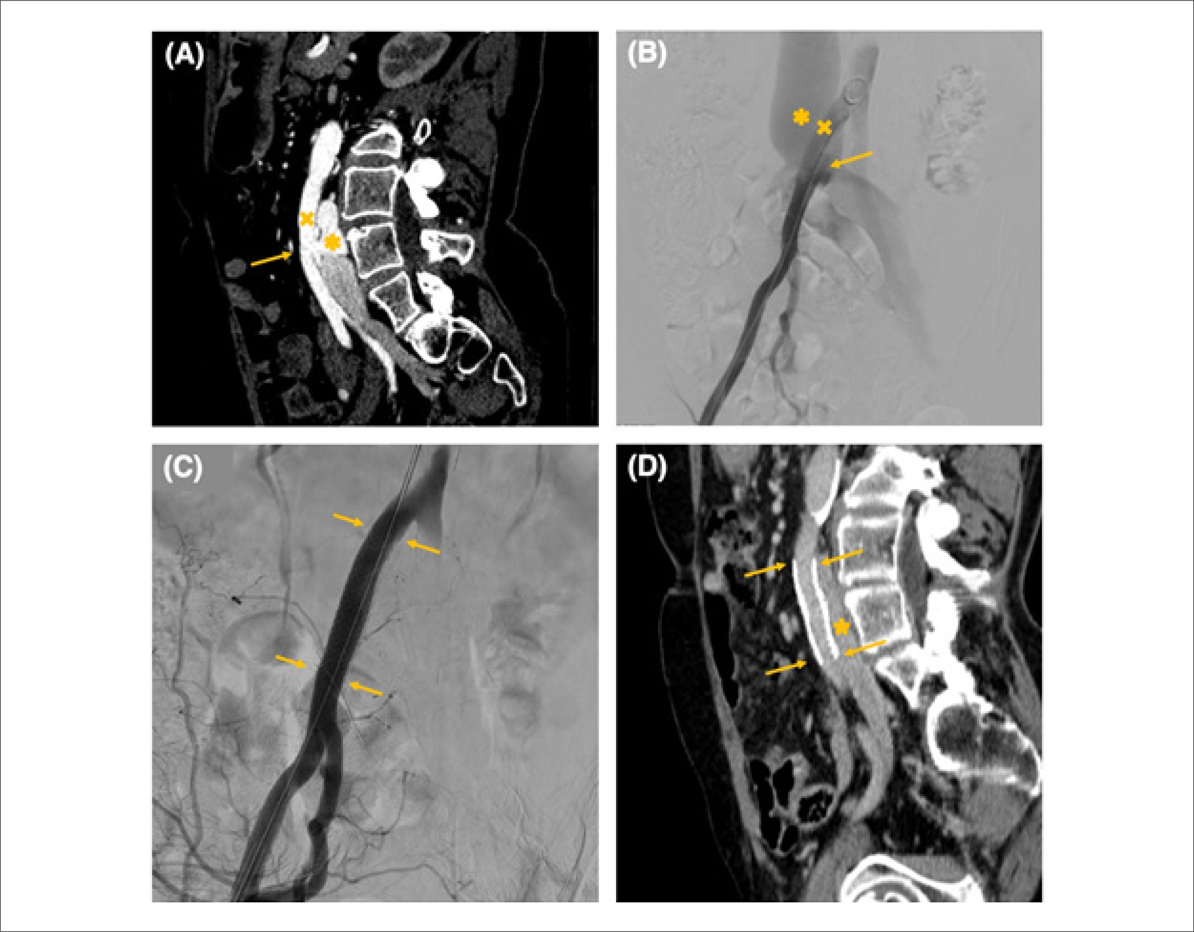
Full-body CTA revealed marked dilation of the inferior vena cava and iliac veins, as well as an arteriovenous fistula (AVF) between the posterior wall of the right common iliac artery and the confluence of both common iliac veins, at the level of L3-L4 vertebrae (Figure 1A). This finding was confirmed by magnetic resonance venous angiography.
(A) Pre-procedural CT angiogram showing the fistulous communication (arrow) between the right common iliac artery and the inferior vena cava at the L3L4 vertebral level. The right common iliac artery is marked (x), and early contrast opacification of the inferior vena cava through the fistulous tract is noted during the arterial phase (asterisk). (B) Pre-intervention angiography demonstrating the arteriovenous fistula (arrow), the right common iliac artery (x), and a markedly dilated inferior vena cava (asterisk). (C) Post-intervention angiography showing the covered metal stent in place (arrows). (D) Post-procedural CT angiogram confirming appropriate stent positioning (arrows) and normal opacification of the inferior vena cava, with no evidence of residual fistulous flow.
Three months later, the patient underwent successful endovascular closure of the AVF with a balloon-expandable covered metal stent (GORE® VIABAHN® VBX 11x59mm) implanted in the right common iliac artery. Post-procedure angiography confirmed proper stent positioning and patency (Figures 1B and 1C).
At the 30-day follow-up, the patient reported complete resolution of symptoms and near normalization of NTproBNP levels (2036>207 pg/mL). Repeat echocardiogram demonstrated normalization of right heart chamber dimensions and resolution of PH (trace tricuspid regurgitation). Follow-up CTA revealed normal opacification of the inferior vena cava and right iliac common artery, without evidence of AVF (Figure 1D).
Discussion
PH is a hemodynamic disorder characterized by a mean pulmonary arterial pressure (mPAP) greater than 20 mmHg at rest, as measured by RHC.1 Though uncommon, with an estimated global prevalence of approximately 1%,1 PH can arise from a broad spectrum of etiologies. The most recent classification system divides PH into five groups based on clinical presentation, hemodynamic profiles, and treatment strategies.1 While PH is most often associated with left heart disease and pulmonary disorders, rarer causes, such as hyperdynamic states, warrant special attention.
PH with unclear and/or multifactorial mechanisms (Group 5) encompass a group of diseases which include, but are not limited to, high CO states, such as left-to-right shunts, cirrhosis, hyperthyroidism, and anemia.2 Contrary to PH, in which intrinsic vasculopathy is the primary driver of disease, PH related to high-output states arises from a sustained increase in blood flow, which induces persistent shear stress in the pulmonary circulation. In this matter, several mechanisms have been proposed to explain the hyperdynamic circulatory state, such as the reduction in systemic vascular resistance in systemic to pulmonary shunts, or the activation of neurohormonal pathways.2–4 In patients with end-stage kidney disease on dialysis via arteriovenous access, several explanations justify the development of PH: increased cardiac output, anemia, fluid overload, and hormonal and metabolic derangement with increased pulmonary vascular tone (e.g., impaired nitric oxide production, higher levels of endothelin-1).5 Other causes included in Clinical Group 5 PH, such as sickle cell disease, have explored other potential mechanisms for the development of PH, such as chronic hemolysis, leading to inhibition of nitric oxide production; microthrombotic embolization (with chronic thromboembolic PH as a potential complication), and chronic hypoxia, with altered gene regulation favoring adverse vascular remodeling.6
This rare case highlights an unusual cause of PH resulting from an extrathoracic AVF, which induced a significant left-to-right shunt and was fully reversible following closure of the shunt.
Importantly, despite the pre-capillary hemodynamic phenotype observed in this patient, there was a significant chance that in less experienced centers, such a presentation could be misclassified as pulmonary arterial hypertension. This could lead to the inappropriate use of PH specific vasodilator therapies, which would be highly deleterious in this context, exacerbating symptoms and worsening the patient's prognosis.
The distinction between PH, an intrinsic disease of the pulmonary vasculature, and PH driven by chronic hyperflux — often accompanied by pre-capillary and/or capillary vascular remodeling that, although ultimately detrimental to right heart chambers, is theoretically adaptive as a protective mechanism against chronic hyperflux into the left heart chambers — is critical to ensure accurate management and to avoid therapeutic missteps.
This highlights the complexity of interpreting hemodynamic profiles in patients with PH due to hyperdynamic circulatory states and emphasizes the importance of specialized centers in managing such challenging cases.
Additionally, on physical examination, one might expect to detect palpable thrills or auscultatory bruits in a patient with such a large AVF. However, in this case, these findings were absent, likely due to the patient's abdominal adiposity and the considerable size of the shunt orifice, which may have allowed blood to flow through with less turbulence than expected.
Several cases of AVF-induced PH have been documented in the literature.7–10 In one case, an acquired AVF between the common iliac artery and vein was reported as a iatrogenic complication of lumbar spinal surgery,9 a likely etiology in our patient. Of note, anterior longitudinal ligament perforation with subsequent major vessel injury has been described as a rare, but potentially life-threatening complication in patients who are submitted to laminectomy, with several cases reported in the last decades.11–13
As demonstrated in other cases, prompt identification and correction of the AVF led to rapid clinical and hemodynamic improvement. Timely diagnosis is essential to prevent irreversible structural heart disease and to reduce morbidity and mortality associated with untreated PH.
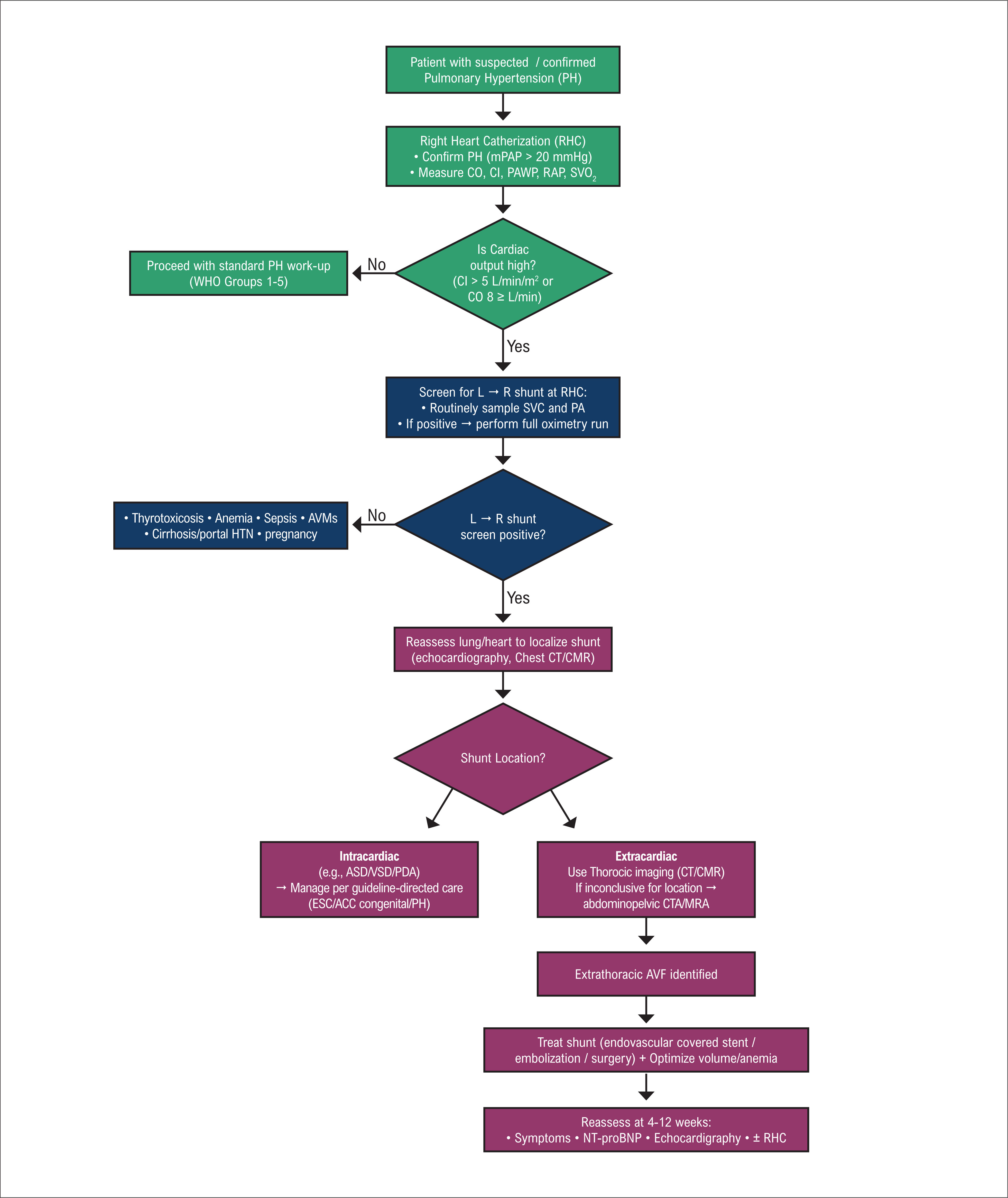
This case emphasizes the importance of thorough clinical investigation in patients with PH, encouraging clinicians to consider less common but potentially reversible causes in order to optimize patient outcomes. We propose a diagnostic algorithm for patients with suspected/confirmed PH and high cardiac output (Figure 2).
Diagnostic Algorithm – Suspected High-Output PH / AVF. Diagnostic algorithm for suspected high-output pulmonary hypertension with possible AVF. High-output state defined as CI > 5 L/min/m² or CO ≥ 8 L/min. L→R shunt screening at RHC is considered positive if ΔO2 (SVC – PA) ≥ 8% or if PA O2 saturation ≥ 80%; when positive, a full oximetry run is recommended. If an extracardiac shunt is suspected and thoracic imaging (CT/CMR) is inconclusive for localization, abdominopelvic CTA/MRA may be used. Intracardiac shunts should be managed according to guideline-directed care (ESC/ACC). AVF: arteriovenous fistula; AVM: arteriovenous malformation; CI: cardiac index; CO: cardiac output; CMR: cardiovascular magnetic resonance; CTA: computed tomographic angiography; mPAP: mean pulmonary artery pressure; PA: pulmonary artery; PAWP: pulmonary artery wedge pressure; PH: pulmonary hypertension; RAP: right atrial pressure; RHC: right heart catheterization; SvO2: mixed venous oxygen saturation; SVC: superior vena cava; HTN: hypertension.
In conclusion, PH secondary to an extrathoracic AVF is a rare but reversible cause of PH. Early detection and intervention are crucial in preventing the development of severe structural heart disease. This case underscores the importance of comprehensive clinical evaluation in PH patients and reinforces the need for vigilance in identifying less common etiologies of hyperdynamic circulatory states.
Sources of funding
Study association
Ethics approval and consent to participate
Use of Artificial Intelligence
Data Availability Statement
The underlying content of the research text is contained within the manuscript.
References
-
1 Humbert M, Kovacs G, Hoeper MM, Badagliacca R, Berger RMF, Brida M, et al. 2022 ESC/ERS Guidelines for the Diagnosis and Treatment of Pulmonary Hypertension. Eur Heart J. 2022;43(38):3618-731. doi: 10.1093/eurheartj/ehac237.
» https://doi.org/10.1093/eurheartj/ehac237 -
2 Qaiser KN, Sahay S, Tonelli AR. Pulmonary Hypertension Due to High Cardiac Output. Respir Med. 2023;206:107034. doi: 10.1016/j.rmed.2022.107034.
» https://doi.org/10.1016/j.rmed.2022.107034 -
3 Anand IS, Florea VG. High Output Cardiac Failure. Curr Treat Options Cardiovasc Med. 2001;3(2):151-9. doi: 10.1007/s11936-001-0070-1.
» https://doi.org/10.1007/s11936-001-0070-1 -
4 Narechania S, Tonelli AR. Hemodynamic Consequences of a Surgical Arteriovenous Fistula. Ann Am Thorac Soc. 2016;13(2):288-91. doi: 10.1513/AnnalsATS.201509-636CC.
» https://doi.org/10.1513/AnnalsATS.201509-636CC -
5 Abassi Z, Nakhoul F, Khankin E, Reisner SA, Yigla M. Pulmonary Hypertension in Chronic Dialysis Patients with Arteriovenous Fistula: Pathogenesis and Therapeutic Prospective. Curr Opin Nephrol Hypertens. 2006;15(4):353-60. doi: 10.1097/01.mnh.0000232874.27846.37.
» https://doi.org/10.1097/01.mnh.0000232874.27846.37 -
6 Gordeuk VR, Castro OL, Machado RF. Pathophysiology and Treatment of Pulmonary Hypertension in Sickle Cell Disease. Blood. 2016;127(7):820-8. doi: 10.1182/blood-2015-08-618561.
» https://doi.org/10.1182/blood-2015-08-618561 -
7 Bhatia S, Morrison JF, Bower TC, McGoon MD. Pulmonary Hypertension in the Setting of Acquired Systemic Arteriovenous Fistulas. Mayo Clin Proc. 2003;78(7):908-12. doi: 10.4065/78.7.908.
» https://doi.org/10.4065/78.7.908 -
8 Kilic S. Pulmonary Hypertension Caused by Arteriovenous Fistula. J Am Coll Cardiol. 2018;71(11):A2470. doi:10.1016/S0735-1097(18)33011-0.
» https://doi.org/10.1016/S0735-1097(18)33011-0 -
9 Başarıcı İ, Onaç M. Unusual Case of Pulmonary Hypertension due to Acquired Arteriovenous Fistula. Turk Kardiyol Dern Ars. 2023;51(7):512-5. doi: 10.5543/tkda.2023.67362.
» https://doi.org/10.5543/tkda.2023.67362 -
10 Pupiales-Dávila KA, Sánchez-Amaya DJ, Zebadúa-Torres R, López-Reyes JC, Zayas-Hernández NG. Pulmonary Hypertension Secondary to Arteriovenous Fistula: A Case Report. Br J Cardiol. 2024;31(1):004. doi: 10.5837/bjc.2024.004.
» https://doi.org/10.5837/bjc.2024.004 -
11 Goodkin R, Laska LL. Vascular and Visceral Injuries Associated with Lumbar Disc Surgery: Medicolegal Implications. Surg Neurol. 1998;49(4):358-70. doi: 10.1016/s0090-3019(97)00372-8.
» https://doi.org/10.1016/s0090-3019(97)00372-8 -
12 Inamasu J, Guiot BH. Vascular Injury and Complication in Neurosurgical Spine Surgery. Acta Neurochir. 2006;148(4):375-87. doi: 10.1007/s00701-005-0669-1.
» https://doi.org/10.1007/s00701-005-0669-1 - 13 Torun F, Tuna H, Deda H. Abdominal Vascular Injury during Lumbar Disc Surgery: Report of Three Cases. Ulus Travma Acil Cerrahi Derg. 2007;13(2):165-7.