Artificial Intelligence-Enhanced Electrocardiography Analysis for Diagnosing Chagas Disease
Abstract
Background: Chagas disease affects millions worldwide and remains a leading cause of cardiomyopathy in Latin America. Early diagnosis remains challenging in endemic regions. Artificial intelligence (AI)-based electrocardiography (ECG) analysis may offer a low-cost strategy for large-scale screening in resource-limited settings.
Objectives: To evaluate the performance of an AI-ECG algorithm combined with clinical data for detecting Chagas disease in a community-based screening program conducted in a highly endemic region in Northeastern Brazil.
Methods: In August 2024, 1,115 adults underwent standardized 12-lead ECG acquisition during a field campaign in Feira de Santana, Bahia, Northeast Brazil. A previously trained AI model analyzed ECG tracings and incorporated three clinical variables: i) prior residence in triatomine-infested areas, ii) poor housing conditions, and iii) family history of Chagas disease. Individuals flagged as AI-positive were classified as suspected cases. Suspected cases and matched controls (2:1) underwent point-of-care serological testing.
Results: The algorithm flagged 121 individuals (10.9%), corresponding to an estimated AI-based prevalence of Chagas disease of 7.8% (95%CI: 6.2-9.6). Among the 112 individuals who completed serological testing, 13 tested positive, all within the AI-positive suspected group; no seropositive cases were identified among controls. Sensitivity was 100% (95%CI: 69-100), specificity 40% (95%CI: 30-50), negative predictive value 100% (95%CI: 91-100), and positive predictive value 12% (95%CI: 11-14), with a diagnostic odds ratio of 6.6.
Conclusions: An AI-ECG algorithm combined with simple clinical variables demonstrated excellent sensitivity for the detection of Chagas disease and may represent a valuable triage tool in endemic, resource-constrained settings.
Keywords
Chagas Disease; Artificial Intelligence; Electrocardiography; Mass Screening
Resumo
Fundamento: A doença de Chagas afeta milhões de pessoas em todo o mundo e permanece como uma das principais causas de cardiomiopatia na América Latina. O diagnóstico precoce ainda representa um desafio em regiões endêmicas. A análise de eletrocardiografia (ECG) aprimorada por inteligência artificial (IA) pode oferecer uma estratégia de baixo custo para rastreamento em larga escala em contextos com recursos limitados.
Objetivos: Avaliar o desempenho de um algoritmo de ECG aprimorada por IA combinado com dados clínicos para a detecção da doença de Chagas em um programa de rastreamento comunitário realizado em uma região altamente endêmica do Nordeste do Brasil.
Métodos: Em agosto de 2024, 1.115 adultos foram submetidos à aquisição padronizada de ECG de 12 derivações durante uma campanha de campo em Feira de Santana, Bahia, nordeste do Brasil. Um modelo de IA previamente treinado analisou os traçados de ECG e incorporou três variáveis clínicas: i) residência prévia em áreas infestadas por triatomíneos, ii) condições habitacionais precárias e iii) histórico familiar de doença de Chagas. Indivíduos identificados como positivos pela IA foram classificados como casos suspeitos. Casos suspeitos e controles pareados (2:1) foram submetidos a testes sorológicos no ponto de cuidado.
Resultados: O algoritmo identificou 121 indivíduos (10,9%), correspondendo a uma prevalência estimada de doença de Chagas baseada em IA de 7,8% (intervalo de confiança de 95% [IC95%]: 6,2-9,6). Entre os 112 indivíduos que completaram o teste sorológico, 13 apresentaram resultado positivo, todos pertencentes ao grupo suspeito positivo pela IA; nenhum caso soropositivo foi identificado entre os controles. A sensibilidade foi de 100% (IC95%: 69-100), a especificidade de 40% (IC95%: 30-50), o valor preditivo negativo de 100% (IC95%: 91-100) e o valor preditivo positivo de 12% (IC95%: 11-14), com uma razão de chances diagnóstica de 6,6.
Conclusões: Um algoritmo de ECG aprimorada por IA combinado com variáveis clínicas simples demonstrou excelente sensibilidade para a detecção da doença de Chagas e pode representar uma ferramenta valiosa de triagem em contextos endêmicos com recursos limitados.
Palavras-chave:
Doença de Chagas; Inteligência Artificial; Eletrocardiografia; Programas de Rastreamento
Introduction
Chagas disease, which is caused by the protozoan parasite Trypanosoma cruzi, is one of the most prevalent neglected tropical diseases. It affects approximately 6-7 million people worldwide, primarily in Latin America.1–3 Although transmission through vectors (notably triatomine insects) and blood transfusions has declined substantially in recent decades due to socioeconomic improvements, better housing conditions, and public health initiatives, chronic Chagas cardiomyopathy remains a major cause of morbidity and mortality. It is the most common form of organ involvement, affecting more than 30% of seropositive individuals, and often progresses silently until advanced stages.4
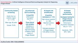
AI: artificial intelligence; CNN: convolutional neural network; ECG: electrocardiography; NPV: negative predictive value.
The gold standard for etiological diagnosis of Chagas disease is serological testing for T. cruzi antibodies, whereas echocardiography remains the cornerstone for detecting advanced heart involvement, particularly ventricular dysfunction, and for guiding risk stratification and prognosis.1 However, access to these diagnostic modalities remains restricted in many endemic, resource-constrained settings.
In contrast, electrocardiography (ECG) is a widely accessible, cost-effective tool capable of identifying early signs of heart involvement in chronic Chagas cardiomyopathy, including right bundle branch block (RBBB), left anterior fascicular block (LAFB), premature ventricular contractions, and fragmented QRS complexes, which are well-established markers of heart involvement in Chagas disease.5 Despite its clinical utility and recommendation for annual assessment, conventional ECG interpretation has limited sensitivity and specificity, particularly for detecting subclinical disease. Moreover, ECG has the potential to be incorporated into population-based screening strategies as a point-of-care tool to estimate the likelihood of Chagas disease, especially in underserved settings where diagnostic resources are scarce.
In recent years, deep learning models based on artificial intelligence (AI) have demonstrated high diagnostic performance in ECG interpretation. Notably, prior studies have shown that AI-ECGs can identify T. cruzi seropositivity6 and predict left ventricular systolic dysfunction in individuals with chronic Chagas cardiomyopathy, with area under the curve (AUC) values of up to 0.84.7 These findings underscore the potential of AI-ECG algorithms as affordable and scalable tools for early detection and risk stratification in Chagas disease, even before structural heart changes become evident.
This study evaluated the diagnostic performance of an AI-ECG algorithm for detecting T. cruzi seropositivity during a community-based screening campaign in individuals without clinical suspicion of Chagas disease, aiming to assess its potential as a low-cost population-based triage strategy in endemic regions. The main findings are summarized in Central Illustration.
Methods
Study design and population
Upon reasonable request to the corresponding author, study data and analytical methods may be made available to other researchers for the purpose of reproducing the results or replicating the procedures of this study. The AI-ECG model evaluated herein is currently a proprietary research tool and has not been publicly released; access to the code or deployment interface is subject to institutional agreements, data governance requirements, and future regulatory considerations.
This was a cross-sectional external validation study in which a pre-existing AI-enhanced, point-based algorithm was applied to participants enrolled in a community health campaign conducted in August 2024 in a Chagas disease-endemic region in Feira de Santana and surrounding regions in Bahia, Northeast Brazil. The cardiovascular screening effort was part of a multicenter initiative named PROVAR+ (Programa de Rastreamento da Valvopatia Reumática), conducted through an international collaboration between academic institutions between Hospital das Clínicas, Universidade Federal de Minas Gerais (UFMG), Brazil, and Children's National Hospital, Washington, DC, United States. The study protocol was approved by the local Institutional Review Board of UFMG under CAAE number 37228120.9.0000.5149 and by regional health authorities, in accordance with ethical standards for research involving human participants.
The study cohort consisted of adults (≥ 18 years) recruited through the primary care network, dedicated health campaigns, and social media outreach, without a prior diagnosis or clinical suspicion of Chagas disease. Eligible participants were randomly identified by local health authorities and underwent a standardized screening protocol that included a brief clinical questionnaire, a 12-lead ECG, and transthoracic portable echocardiography, irrespective of symptom status or previous indication for cardiovascular evaluation. Individuals with a known history of Chagas disease were excluded. All participants provided written informed consent prior to any study-related procedures. A detailed flowchart of the study population and exclusions is presented in Figure 1.

Electrocardiography acquisition and artificial intelligence algorithm
All participants underwent a resting 12-lead ECG, acquired using commercially available devices and transmitted to the Telehealth Center at UFMG. ECGs were initially interpreted using an automated system developed by an external academic institution (version 28.5, January 2014), which generated real-time clinical reports.
Additionally, for study standardization, all ECG tracings were subsequently reviewed by a trained cardiologist and classified according to the standardized Minnesota Code.8 The Minnesota Code is the most widely used ECG classification system worldwide, developed in the 1950s by Dr. Henry Blackburn, and is based on predefined measurement criteria to assign specific numerical codes according to the severity of findings.9 In cases of discrepancy between automated reports and cardiologist interpretation, examinations were adjudicated to establish a final classification. Although modified versions of the Minnesota Code adapted for Chagas disease have been widely used in disease-specific cohorts, ECG coding in this study was used exclusively for descriptive standardization and did not serve as an input to the AI model or as a reference standard for the diagnosis of Chagas disease.
Prior to ECG acquisition, a brief structured interview was conducted to collect demographic data, clinical indications for testing, and risk factors for Chagas disease. Based on these data, an AI-enhanced algorithm was applied, integrating ECG signals with three clinical variables derived from national guidelines for Chagas disease10: i) history of residence in areas with triatomine vector presence; ii) residence in housing conditions associated with vector exposure; and iii) family history of confirmed Chagas disease. Participants flagged by the combined algorithm were classified as AI-positive for screening purposes.
The proprietary AI-ECG algorithm for Chagas disease was originally developed using two large datasets SaMi-Trop and CODE derived from a heterogeneous population, including individuals with Chagas disease and a smaller group of healthy controls. One dataset comprised a cohort study conducted in the SaMi-Trop cohort, whereas the other consisted of a large tele-ECG database collected from a broad population through the CODE tele-ECG dataset.
The algorithm has been previously validated in the REDS-II and ELSA-Brasil datasets, including individuals with different serological profiles and a longitudinal cohort of adults.6,7
The deep learning model was based on a residual network architecture adapted for one-dimensional signals. It included convolutional layers both preceding and within residual blocks, consistent with architectures previously used to classify multiple ECG abnormalities, with modification of the final output layer to generate a binary classification output for Chagas disease. Model parameters were optimized by minimizing the binary cross-entropy loss function, with data processed in batches of size 32.
Regularization strategies included a dropout rate of 0.5 and weight decay of 0.001. An ensemble approach was implemented by training the model 15 times using different random seeds, with final predictions obtained by averaging the outputs. The resulting values ranged from 0 to 1 and can be interpreted as the predicted probability of Chagas disease based on reference serological testing.6
The predicted probability was subsequently integrated with the aforementioned clinical and demographic variables to generate a final point-based probability of Chagas disease, independent of conventional ECG abnormalities or echocardiographic findings.
Two-phase serological verification strategy
Because serological testing was not performed in the entire screened population, a two-phase verification design was implemented. All participants classified as AI-positive were invited for serological confirmation, whereas AI-negative participants were selected as matched controls through age- and sex-matched sampling in a 2:1 ratio. Controls were randomly selected using proprietary trigger software based on computer-generated random numbers.
Serological testing was performed using a point-of-care rapid diagnostic test based on chimeric antigens (IBMP), designed to detect T. cruzi antibodies. This assay has demonstrated high sensitivity and specificity and was considered the diagnostic reference standard in this study.11 This sampling strategy was adopted to ensure feasibility within a field-based screening campaign.
Echocardiography
A simplified echocardiographic protocol, based on recommendations from the American Society of Echocardiography,12 was performed by physicians using portable ultrasound systems on the same day as the clinical evaluation and ECG triage. However, echocardiographic findings were not available at the time of AI-based classification and were not used in the development of the algorithm or in point-of-care decision-making. Such data were used solely for ancillary post hoc analyses.
Heart structural abnormalities were defined according to predefined criteria described in PROVAR+ studies and ASE-REWARD studies.10,13 These included moderate to severe valvular disease (regurgitation or stenosis), ventricular dysfunction or hypertrophy, congenital heart disease, pericardial effusion, and regional wall-motion abnormalities. Both objective measurements and qualitative assessments were recorded. Only participants with clinically significant abnormalities were referred for specialized care.
Statistical analysis
Because of the descriptive nature of the analysis, all computations were performed using Microsoft Excel 2019 (Microsoft Corporation, Redmond, WA, USA). As this was an exploratory study, no a priori sample size calculation was performed; instead, the total number of participants enrolled during the 4-day screening period was analyzed.
Continuous variables are presented as mean ± standard deviation or median and interquartile range as appropriate, based on distribution assessed using the Shapiro-Wilk test. Categorical variables are presented as absolute frequencies and percentages. Baseline characteristics of participants classified as AI-positive and AI-negative were summarized descriptively.
When applicable, comparisons between groups were performed using the unpaired Student's t test or the Mann-Whitney U test for continuous variables, and the chi-square test or Fisher's exact test for categorical variables, as appropriate. A p < 0.05 was considered statistically significant.
Diagnostic performance metrics of the AI algorithm, including sensitivity, specificity, positive predictive value (PPV), negative predictive value (NPV), and diagnostic odds ratio (DOR), were calculated using point-of-care seropositivity as the reference standard. 95%CIs were calculated for all estimates.
Since serological testing was not performed in all screened participants but followed a two-phase verification strategy, diagnostic performance measures should be interpreted as exploratory estimates within the tested sample. A detailed flowchart describing the study design and participant selection is provided in Figure 1.
Results
A total of 1,115 individuals were screened during the community-based health campaign. Of these, 1,031 (92.5%) successfully completed the 12-lead ECG protocol. The mean age of participants was 56 years, and most were women (78.4%). According to the Minnesota Code classification, 402 individuals (39.0%) exhibited ECG abnormalities, including 284 (27.5%) with minor alterations and 118 (11.4%) with major abnormalities.
The AI-ECG algorithm generated 121 alerts for point-of-care serological testing. Of these, 80 corresponded to individuals classified as having suspected Chagas disease, and 41 to participants not initially considered at high risk. After excluding nine individuals who did not undergo testing due to loss to follow-up or logistical constraints, 112 participants completed serological evaluation. These included 73 individuals from the suspected Chagas disease group and 39 from the non-high-risk group. Detailed baseline characteristics are provided in Table 1.
Of the 112 individuals tested, 13 (all from the suspected Chagas disease group) had positive serology confirming Chagas disease. No seropositive cases were identified among participants not initially considered at high risk. These findings yielded a sensitivity of 100% (95%CI: 69-100), specificity of 40% (95%CI: 30-50), NPV of 100% (95%CI: 91-100), and PPV of 12% (95%CI: 11-14), with a DOR of 6.6 for the combined AI-based algorithm.
Among the 99 participants classified as AI-positive but with negative serology, 46 (46.4%) exhibited ECG abnormalities, including 13 with major abnormalities and 33 with minor alterations.
In the subgroup of 13 individuals who were both AI-positive and seropositive, ECG findings showed that five had major abnormalities (four with RBBB associated with LAFB and one with isolated RBBB) while two had minor abnormalities and six had normal ECGs.
Although echocardiography was not incorporated into the algorithm or used for diagnostic classification, echocardiographic data were available for exploratory analysis in this subgroup. Seven participants had normal echocardiograms, three had mild findings (including isolated tricuspid regurgitation or right ventricular dilation), and three had moderate valvular abnormalities, including two with mitral regurgitation and one with combined mitral and tricuspid regurgitation suggestive of a rheumatic pattern. None of the seropositive individuals showed evidence of left ventricular systolic dysfunction.
Discussion
The present study demonstrates that the integration of an AI-ECG algorithm with simple clinical variables can accurately identify seropositive cases of Chagas disease in a community-based screening campaign involving a non-selected population at high sociodemographic risk. The combined algorithm achieved a DOR of 6.6, with 100% sensitivity and NPV, supporting its potential utility as a rule-out tool in resource-limited settings. These findings indicate strong applicability as a triage strategy, particularly in contexts where prioritization of confirmatory testing is required. The ability of the algorithm to identify all seropositive individuals, even in the absence of overt structural heart disease, reinforces its clinical value in endemic regions with limited access to specialized diagnostic resources. Although specificity remained modest (40%), this trade-off may be acceptable in early screening strategies where minimizing false-negative results is a priority. These findings are particularly relevant in the context of neglected tropical diseases, in which underdiagnosis is common and early detection may reduce disease progression.
ECG remains the most accessible cardiovascular diagnostic tool in low-resource settings and has long been used in the evaluation of Chagas disease, as a core component of routine clinical assessment to identify heart involvement. Classical abnormalities (e.g., RBBB, LAFB, and premature ventricular contractions) are well-established markers of disease severity and progression,14 correlating with ventricular involvement, risk of malignant ventricular arrhythmias, and mortality.15 However, evidence indicates that a normal ECG, particularly with a narrow QRS complex, does not exclude early heart involvement in Chagas disease.16 These observations support the use of advanced computational approaches, such as AI, which may detect subclinical electrical patterns not identifiable through conventional visual interpretation.
Recent deep learning models have demonstrated promising results in detecting Chagas disease-related abnormalities from ECG data. Jidling et al. trained a neural network using the CODE and SaMi-Trop cohorts to identify T. cruzi seropositivity, achieving an AUC of 0.80 in internal validation and 0.68 in external validation.6 Similarly, prior work has shown that AI-ECG analysis can predict left ventricular systolic dysfunction in Chagas disease with AUC values of up to 0.84.7 The present study extends these findings by applying this approach in a real-world, prospective, community-based setting, involving individuals without prior clinical suspicion of Chagas disease.
An important methodological aspect of this study is the integration of AI-ECG with targeted clinical variables, which is consistent with evidence that ECG data alone may be insufficient for early-stage detection. In addition, a substantial proportion of individuals classified as AI-positive but with negative serology presented ECG abnormalities, suggesting the presence of early or alternative cardiovascular conditions detectable through ECG screening. This finding warrants further investigation, particularly regarding its prognostic implications.
Notably, a subgroup of individuals in this study presented normal conventional ECG findings but were classified as AI-positive and had confirmed seropositivity. This observation suggests AI-ECG analysis may detect subtle electrical patterns beyond the limits of expert visual interpretation, potentially reflecting very early myocardial involvement. This concept is supported by previous studies using the Selvester QRS score, which have demonstrated that myocardial fibrosis, as identified by cardiac magnetic resonance imaging, may be present even in the absence of overt ECG abnormalities.17,18 These findings reinforce the potential role of advanced ECG analysis, including AI-enhanced approaches, in the detection of early heart involvement in Chagas disease.
The clinical variables incorporated into the algorithm (i.e., residence in endemic areas, housing conditions associated with triatomine infestation, and family history of Chagas disease) were selected based on national clinical guidelines.10 These factors likely improved diagnostic yield by capturing key sociodemographic determinants of the disease. The AI-ECG model was originally trained in a general population rather than exclusively in individuals with Chagas disease, highlighting the importance of contextual adaptation when applied to high-risk populations.6 The integration of environmental and epidemiological variables enhanced its performance in this setting. However, prospective multicenter validation and implementation studies are necessary before broader clinical adoption of this triage strategy.
Finally, the distribution of seropositive cases observed in this community-based sample is consistent with previously reported epidemiological patterns in high-risk populations in Latin America, particularly in endemic rural and semi-urban regions. For example, Cucunubá et al. reported higher seropositivity rates among older adults in rural endemic areas.4 Although the present study was not designed to estimate disease prevalence, the identification of confirmed cases reinforces the need for scalable and accessible screening strategies in underserved populations as well as for appropriate prioritization of diagnostic and therapeutic resources.
Limitations of study
This study has some limitations. First, the cross-sectional design and the use of an artificial intelligence model previously trained on general population datasets may limit generalizability to other endemic or clinical settings and to specific high-risk subpopulations.
Second, although the AI-ECG algorithm demonstrated excellent sensitivity, the observed value of 100% should be interpreted with caution given the limited number of individuals who underwent serological testing, particularly among those classified as AI-negative. In small samples, estimates of perfect sensitivity are prone to overestimation, especially when no seropositive cases are identified in the AI-negative group.
Additionally, the relatively small number of AI-negative individuals who underwent serological testing, selected through age- and sex-matched sampling, may have introduced selection bias and reduced statistical power to detect false-negative cases. This limitation also affects the precision of specificity and PPV estimates, increasing uncertainty around the observed diagnostic performance metrics.
Furthermore, using selective serological verification may have introduced partial verification bias. As a result, the reported diagnostic metrics may not fully reflect the performance of the screening strategy in the entire study population. Additional studies are warranted to validate the model in broader and more representative cohorts.
Moreover, although the inclusion of basic clinical variables likely contributed to improved model performance, unmeasured confounders may have influenced the results. Finally, the selected clinical variables reflect traditional risk factors associated with vector-borne transmission. In regions with different transmission dynamics, such as the Amazon basin, where oral transmission predominates, the model may not adequately capture infection risk and may require adaptation to local epidemiological contexts.
Future prospective studies involving larger, more diverse populations, as well as comprehensive follow-up including echocardiographic correlation, are needed to further validate and calibrate the algorithm.
Conclusion
This study highlights the high sensitivity and operational feasibility of an AI-ECG approach for detecting T. cruzi seropositivity in underserved settings. By integrating clinical variables with a neural network trained on large-scale ECG data from one of the largest telemedicine networks in Latin America, this strategy represents a promising and scalable solution for early detection and triage in the context of neglected diseases. Further research should focus on improving specificity and evaluating the integration of this approach into broader health system strategies for the control of Chagas disease.
-
Sources of Funding
This study was partially funded by Edwards Lifesciences Foundation, Every HEartbeat Matters Program 2025, CNPq grants 465518/2014-1, 310790/2021-2, 409604/2022-4, 445011/2023-8 and 310749/2022-0, and FAPEMIG grants RED 00192-23 and APQ-03291-18.
-
Study Association
This study is not associated with any thesis or dissertation work.
-
Ethics Approval and Consent to Participate
This study was approved by the Ethics Committee of the Universidade Federal de Minas Gerais under the protocol number CAAE 37228120.9.0000.5149. All the procedures in this study were in accordance with the 1975 Helsinki Declaration, updated in 2013. Informed consent was obtained from all participants included in the study.
-
Use of Artificial Intelligence
During the preparation of this work, the author(s) used ChatGPT for improve grammar, language clarify and manuscript organization. After using this tool/service, the author(s) reviewed and edited the content as needed and take full responsibility for the content of the published article.
Availability of Research Data
All datasets supporting the results of this study are available upon request from the corresponding author.
References
-
1 Marin-Neto JA, Rassi A Jr, Oliveira GMM, Correia LCL, Ramos NA Jr, Luquetti AO, et al. SBC Guideline on the Diagnosis and Treatment of Patients with Cardiomyopathy of Chagas Disease - 2023. Arq Bras Cardiol. 2023;120(6):e20230269. doi: 10.36660/abc.20230269.
» https://doi.org/10.36660/abc.20230269 -
2 Andrade GN, Bosch-Nicolau P, Nascimento BR, Martins-Melo FR, Perel P, Geissbühler Y, et al. Prevalence of Chagas Disease among Latin American Immigrants in Non-Endemic Countries: An Updated Systematic Review and Meta-Analysis. Lancet Reg Health Eur. 2024;46:101040. doi: 10.1016/j.lanepe.2024.101040.
» https://doi.org/10.1016/j.lanepe.2024.101040 -
3 GBD 2023 Chagas Disease and RAISE Study Collaborators. Global, Regional, and National Burden of Chagas Disease, 1990-2023: A Systematic Analysis for the Global Burden of Disease Study 2023. Lancet Infect Dis. 2026;26(3):284-301. doi: 10.1016/S1473-3099(25)00562-6.
» https://doi.org/10.1016/S1473-3099(25)00562-6 -
4 Cucunubá ZM, Gutiérrez-Romero SA, Ramírez JD, Velásquez-Ortiz N, Ceccarelli S, Parra-Henao G, et al. The Epidemiology of Chagas Disease in the Americas. Lancet Reg Health Am. 2024;37:100881. doi: 10.1016/j.lana.2024.100881.
» https://doi.org/10.1016/j.lana.2024.100881 -
5 Nascimento BR, Araújo CG, Rocha MO, Domingues JD, Rodrigues AB, Barros MV, et al. The Prognostic Significance of Electrocardiographic Changes in Chagas Disease. J Electrocardiol. 2012;45(1):43-8. doi: 10.1016/j.jelectrocard.2011.04.011.
» https://doi.org/10.1016/j.jelectrocard.2011.04.011 -
6 Jidling C, Gedon D, Schön TB, Oliveira CDL, Cardoso CS, Ferreira AM, et al. Screening for Chagas Disease from the Electrocardiogram Using a Deep Neural Network. PLoS Negl Trop Dis. 2023;17(7):e0011118. doi: 10.1371/journal.pntd.0011118.
» https://doi.org/10.1371/journal.pntd.0011118 -
7 Brito BOF, Attia ZI, Martins LNA, Perel P, Nunes MCP, Sabino EC, et al. Left Ventricular Systolic Dysfunction Predicted by Artificial Intelligence Using the Electrocardiogram in Chagas Disease Patients-The SaMi-Trop Cohort. PLoS Negl Trop Dis. 2021;15(12):e0009974. doi: 10.1371/journal.pntd.0009974.
» https://doi.org/10.1371/journal.pntd.0009974 -
8 Prineas RJ, Crow RS, Zhang ZM. The MINNESOTA Code Manual of Electrocardiographic Findings. London: Springer; 2010. doi:10.1007/978-1-84882-778-3.
» https://doi.org/10.1007/978-1-84882-778-3 -
9 Ribeiro AL, Marcolino MS, Prineas RJ, Lima-Costa MF. Electrocardiographic Abnormalities in Elderly Chagas Disease Patients: 10-Year Follow-Up of the Bambui Cohort Study of Aging. J Am Heart Assoc. 2014;3(1):e000632. doi: 10.1161/JAHA.113.000632.
» https://doi.org/10.1161/JAHA.113.000632 - 10 Brasil. Ministério da Saúde. Protocolo Clínico e Diretrizes Terapêuticas Doença de Chagas. Brasília: Ministério da Saúde; 2023.
-
11 Santos FLN, Campos ACP, Amorim LDAF, Silva ED, Zanchin NIT, Celedon PAF, et al. Highly Accurate Chimeric Proteins for the Serological Diagnosis of Chronic Chagas Disease: A Latent Class Analysis. Am J Trop Med Hyg. 2018;99(5):1174-9. doi: 10.4269/ajtmh.17-0727.
» https://doi.org/10.4269/ajtmh.17-0727 -
12 Mitchell C, Rahko PS, Blauwet LA, Canaday B, Finstuen JA, Foster MC, et al. Guidelines for Performing a Comprehensive Transthoracic Echocardiographic Examination in Adults: Recommendations from the American Society of Echocardiography. J Am Soc Echocardiogr. 2019;32(1):1-64. doi: 10.1016/j.echo.2018.06.004.
» https://doi.org/10.1016/j.echo.2018.06.004 -
13 Singh S, Bansal M, Maheshwari P, Adams D, Sengupta SP, Price R, et al. American Society of Echocardiography: Remote Echocardiography with Web-Based Assessments for Referrals at a Distance (ASE-REWARD) Study. J Am Soc Echocardiogr. 2013;26(3):221-33. doi: 10.1016/j.echo.2012.12.012.
» https://doi.org/10.1016/j.echo.2012.12.012 -
14 Brito BOF, Ribeiro ALP. Electrocardiogram in Chagas Disease. Rev Soc Bras Med Trop. 2018;51(5):570-7. doi: 10.1590/0037-8682-0184-2018.
» https://doi.org/10.1590/0037-8682-0184-2018 -
15 Rassi A Jr, Rassi A, Little WC, Xavier SS, Rassi SG, Rassi AG, et al. Development and Validation of a Risk Score for Predicting Death in Chagas’ Heart Disease. N Engl J Med. 2006;355(8):799-808. doi: 10.1056/NEJMoa053241.
» https://doi.org/10.1056/NEJMoa053241 -
16 Ribeiro AL, Rocha MO, Barros MV, Rodrigues AR, Machado FS. A Narrow QRS does Not Predict a Normal Left Ventricular Function in Chagas’ Disease. Pacing Clin Electrophysiol. 2000;23(11 Pt 2):2014-7. doi: 10.1111/j.1540-8159.2000.tb07076.x.
» https://doi.org/10.1111/j.1540-8159.2000.tb07076.x -
17 Couceiro KN, Ortiz JV, Correia MN, Silva e Silva MRH, Brandão AR, Silva PRL, et al. The Selvester QRS Score as an Estimative of Myocardial Injury in Acute Chagasic Patients from the Brazilian Amazon. BMC Infect Dis. 2021;21(1):396. doi: 10.1186/s12879-021-06083-x.
» https://doi.org/10.1186/s12879-021-06083-x -
18 Sivri F, Icen YK, Koca H, Coşkun M, Ardınç M, Deniz O, et al. Selvester QRS Score is a Predictor of Mortality in Heart Failure with Preserved Ejection Fraction. Arq Bras Cardiol. 2023;120(9):e20230235. doi: 10.36660/abc.20230235.
» https://doi.org/10.36660/abc.20230235
Edited by
-
Editor responsible for the review:
Gláucia Maria Moraes de Oliveira