 Stem adaptation in Heteranthera zosterifolia Mart. (Pontederiaceae) across distinct water depths
Abstract
A significant correlation between water level fluctuations and morphological traits in macrophytes is well-documented, especially in shallow water species. However, predicting morpho-anatomical characteristics in submerged plants across water depths remains challenging. We investigated morpho-anatomical variations in Heteranthera zosterifolia stems from a deep flooded doline at three depths: surface, 2.5m, and 5m. The anatomical survey followed standard protocols, and variance analysis was used based on the measurements obtained. Our study revealed significant differences in the stem and aerenchyma area, number of air channels, cortex thickness, stele-cortex ratio, and average vessel diameter between surface specimens and those from greater depths. The anatomical survey highlighted a greater investment in the cortex and aerenchyma area in water surface specimens, potentially related to structural stability and buoyancy. This challenges the conventional belief that deeper water increases the stem’s area and number of air channels, questioning the traditional association of aerenchyma solely with oxygen transport. Moreover, a balance in vessel diameter between surface and deeper specimens suggests considerations for transport efficiency, protection against water stress, and buoyancy needs. The morpho-anatomical adaptation of the H. zosterifolia stem yields significant implications for the strategies employed by the species for survival and growth in deep aquatic environments.
Keywords:
Aerenchyma; aquatic macrophytes; flooded doline; hydrophytes; Poales; Pontederiaceae; stem anatomy
Introduction
The aquatic habit is reported in about 80 families of flowering plants (Chambers et al., 2008), some of which include species that can occur in aquatic or palustrine environments (Alves et al., 2015; Govaerts et al., 2019). In contrast, several families are exclusively aquatic, such as Alismataceae, Cabombaceae, Hydrocharitaceae, Nymphaeaceae, Pontederiaceae, and Potamogetonaceae (Les & Tippery, 2013; Les, 2020). Among these exclusively aquatic families, Pontederiaceae stands out with six genera and 40 species, with more than half of the species occurring in Brazilian wetlands (Sousa, 2023). A significant morphological variation of vegetative and reproductive structures within the family is related to the fluctuations in water levels (Barrett & Grahan, 1997; Sousa et al., 2016; Leandro et al., 2021) that results in a variety of hydrophytic life forms, including emergent, rooted-floating, free-floating, and submerged herbs (Cook, 1998).
The role of water level fluctuations as a modifying factor in morphological traits has been previously documented as morphological and anatomical patterns such as an increase in leaf blade size and a decrease in cuticle thickness, aimed at enhancing water contact and improving gas exchange efficiency (Armstrong et al., 1994; Engloner & Papp, 2006; Santos et al., 2015; Pan et al., 2021). Increases in the size and number of aerenchyma air channels as an adaptation to enhance oxygen availability underwater are recurrently reported in aquatic plants (Justin & Armstrong, 1991; Armstrong et al., 1994; Coops et al., 1996; Jackson & Armstrong, 1999). Additionally, a reduction is observed in the number of metaxylem as an adaptation to less water transport demand into the plant due to the direct contact with the aquatic environment (Armstrong et al., 1994; Engloner & Papp, 2006; Santos et al., 2015; Pan et al., 2021). These morphological and anatomical traits are primarily associated with the submerged life form which interaction between the individual plant and the aquatic environment is most pronounced.
While most of the trait studies on aquatic plants focus on species in temporary pools, riverbanks, and stream margins with shallow water depths (i.e. Scheffer et al., 1993; Maltchik et al., 1999; Riis & Hawes, 2002; Schutten et al., 2005; Sousa et al., 2016), morphological and anatomical predictability is low when considering submerged life forms across a significant water depth gradient. For example, although an increase in aerenchyma tissue with water depth has been reported (Armstrong et al., 1994; Coops et al., 1996; Jackson & Armstrong, 1999), changes may not occur within a specific range of water depth gradients (Engloner & Papp, 2006). Hence, increased evidence of how aquatic plants' structural traits respond to depth gradients is important. This is especially relevant in scenarios of climate change, where extreme peaks of flooding and drought are more recurrent. These changes are driven by an acceleration of the water cycle (Kerr, 2012), which is more relevant in natural wetlands (e.g., Alho & Silva, 2012; Marengo et al., 2016).
In this matter, deep lotic water bodies are suitable models for better understanding the morphological traits related to water depth. This is the case of the study site, regionally called “Lagoa Misteriosa”. Despite being known as a lake, the “Lagoa Misteriosa” is a flooded doline encircled by an extensive karstic wall with a depth exceeding 220 meters (Scheffler et al., 2019), making it an ideal environment for aquatic organisms across various depth gradients (Zelnik et al., 2012; Castello et al., 2021). The relevance of studying organisms in flooded doline is due to the particular features of this environment, such as the calcarean nature of its sediment, the carbonate, transparent and lotic water, as well as the stable water temperature all over the year (Scheffler et al., 2019). Moreover, the “Lagoa Misteriosa” exhibits a low diversity of herbivores, with two species of fish (Scheffler et al., 2019) with a reported diet based on the consumption of macrophytes fragments and filamentous algae (Caldatto, 2023), which makes this environment relevant to understanding the adaptation and role of aquatic plants. Notably, the occurrence of Heteranthera zosterifolia is remarkable in the studied area, as it dominates the water surface margins and spreads to distinct water depths within the doline walls, reaching depth rates up to seven meters (A. Giaretta pers. obs.), which is not common among macrophytes in general, mainly occurring up to four meters (Bornette & Puijalon, 2011). In addition to their ecological role as a food source for aquatic fauna, it has been reported that the deposition and/or dissolution of calcium carbonate in karst spring-fed streams is controlled by aquatic plants (Spiro & Pentecost, 1991; Liu et al., 2006; 2008). Daytime deposition of calcium carbonate is primarily associated with photosynthesis, while nighttime dissolution is linked to the respiration of these plants (Liu et al., 2008). Given that flooded doline ecosystems in Brazil are poorly studied (Scheffler et al., 2019; Kuerten et al., 2022), there is a clear need for research to understand these ecosystems and their key biotic component strategies, such as macrophytes.
Therefore, we aim to conduct an anatomical and morphological survey of H. zosterifolia across distinct water depths (surface, 2.5- and 5-meter depths) to address the following questions: 1) Are there significant changes in the external morphology at different water depths? 2) Are there significant changes in the anatomical structures? These will be assessed by morphological biometry and anatomical sections of the stem to analyze the number and length of stem internodes, stem area, aerenchyma, cortex, and vascular tissue traits. The results of this study will enhance our understanding of the role of deep aquatic environments in shaping the stability and adaptability of morphological and anatomical features across varying water depth environments.
Material and methods
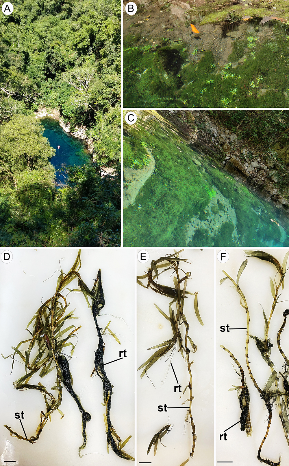
Study Area Characterization - The study area is known as "Lagoa Misteriosa," located in the municipality of Jardim, southeastern Mato Grosso do Sul (21°27’30” S, 56°27’12” W) (Fig. 1 A -C). "Cavidade Natural Lagoa Misteriosa" is a water body formed by a depressed calcareous doline, surrounded by an extensive karstic wall with a depth of 75 meters to the water level, a surface area of approximately 1,650 m², and a water column reaching at least 220 meters (Scheffler et al., 2019). The calcareous nature of its sediment results in carbonate-rich, transparent water with visibility up to 45 meters. The site has an extensive depth and stable water temperature ranging from 23°C to 26°C throughout the year, along with lotic water conditions (Scheffler et al., 2019).
Plant Material - For the morphological and anatomical survey, mature individuals of Heteranthera zosterifolia Mart. (Fig. 1 D -F) were collected, with vouchers deposited in the herbarium DDMS at the Federal University of Grande Dourados (Vouchers: 7825 and 7829). Collections occurred at three distinct water depths: 1) at the water surface, 2) at a 2.5-meter depth, and 3) at a 5-meter depth. The latter represents the species deepest occurrence and although it can occur at depths of up to seven meters, their presence at these levels is sparse. For each depth, five samples were processed for morphological and anatomical surveys. Samples were individualized by identifying the primary stem, each isolated individual was labeled and documented by its depth of origin, ensuring accurate morphological and anatomical analysis despite the species' tendency to form dense, interconnected growths.
Anatomical Assessment - Samples of mature individuals of Heteranthera zosterifolia (Fig. 1 D -F) were fixed in FAA50 (formaldehyde, glacial acetic acid, 50% ethanol, 1:1:18 v/v) (Johansen, 1940) and stored in 70% ethanol for morphological and anatomical studies. The samples were dissected under a Leica EZ4 stereomicroscope (Leica Microsystems, Wetzlar, Germany). For the anatomical survey, free-hand cross-sections of the median regions of the stem were obtained using a razor blade (Johansen, 1940). Sections were clarified with 20% sodium hypochlorite, rinsed in distilled water, and stained with safrablau (5 mL of an aqueous solution of 1% safranin, 95 mL of an aqueous solution of 1% Astra blue, and two drops of glacial acetic acid) (Bukatsch, 1972, modified). Semi-permanent glass slides were mounted with Glycerol Gelatine (Johansen, 1940). Light microscope micrographs were taken using a Leica DFC 320 camera (Leica Microsystems, Wetzlar, Germany) coupled to a Leica DMLB microscope (Leica Microsystems, Wetzlar, Germany), using Scan System Images (IM50). For starch detection in the cortical cells of the stem, Lugol’s reagent test was performed (Johansen, 1940). The aerenchyma pattern was described based on Jung et al. (2008). For the average vessel diameter and vessel lumen area, only the metaxylem was investigated. Specifically, ten metaxylem vessels were measured per individual. To assess variations in stem morphology and anatomy across distinct water depths, measurements were conducted as described in Table 1. The quantitative measurements were performed using ImageJ software (Image J, 1.54d, National Institutes of Health, Bethesda, MD, USA).
Morphological and anatomical traits analyzed in Heteranthera zosterifolia individuals from three water depths: surface, 2.5- and 5-meter, along with their respective descriptions.
Statistical Analysis - Comparative analyses using variance analysis (ANOVA) followed by the Tukey test were conducted based on the obtained measurements. Statistical significance was considered at p < 0.05. All statistical analyses were carried out using PAST software version 2.17c. Given our measurements from three different depths, ANOVA was chosen to compare more than two groups simultaneously. It provides a comprehensive analysis of how water depth affects the plant's morphology and anatomy, identifying variations that could be missed with pairwise comparisons. The subsequent Tukey test helps pinpoint specific group differences.
Results
The analyses of stem biometry and anatomy highlight significant variations in the stem cross-sectional area, aerenchyma area, the number of air channels within the aerenchyma, cortex thickness, stele-cortex ratio, and average vessel diameter between H. zosterifolia specimens from the surface and those from 2.5- and 5-meter depths (Figs. 2 and 3; Table 2). Additionally, a more pronounced development of adventitious roots was observed in surface specimens of H. zosterifolia compared to those occurring at depths of 2.5- and 5-meter (Fig. 1 D -F). All the analyzed traits and their respective values are listed in Table 2.
Collection site and morphology of Heteranthera zosterifolia. Aerial view of the “Lagoa Misteriosa” flooded doline (A). Heteranthera zosterifolia growing in the flooded doline karstic wall (B-C). Specimens from the water surface; note the more pronounced root development (D). Specimens from 2.5 meters (E). Specimens from 5 meters (F). "rt: root; st: stem" Scale bars = 1mm (D-F).
Mean values followed by standard deviation (±) of morphological and anatomical traits analyzed in Heteranthera zosterifolia individuals from three water depths: surface, 2.5- and 5 meters. Values of p < 0.05 represent statistical significance between depths depths (asterisk), with values indicating significant differences being subscripted.
Stem biometry
Significant differences in stem cross-sectional area were observed among specimens collected from various water depths, specifically between those from the surface and deeper water (p < 0.0001; Fig. 2 A ; Table 2). However, no substantial variations were found in the number and length of stem internodes and the relative area of the stem and aerenchyma.
Anatomy of the aerenchyma
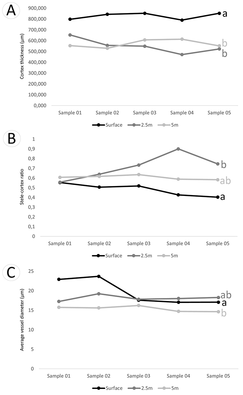
The anatomy of the stem among specimens reveals a significant difference in total aerenchyma area between surface specimens and those from deeper waters (p = 0.001; Fig. 2 C ; Table 2). Regarding the number of air channels within the aerenchyma, surface specimens exhibit a significantly higher count compared to samples from depths of 2.5 and 5-meter (p = 0.0005; Fig. 2 A ; Table 2). Conversely, both the number of air channels and the aerenchyma area show no significant differences between specimens from depths of 2.5 and 5-meter (Fig. 2 A , C). Additionally, a significant contrast is observed in the cortex thickness between surface specimens and those from deeper waters (p < 0.0001; Fig. 3 A ; Table 2), as well as in the vascular traits (Fig. 3 B , C), such as the stele-cortex ratio between surface and 2.5-meter specimens (p = 0.003; Fig. 3 B , Table 2).

Graphics illustrating significant variations in morphological and anatomical features of the stem of Heteranthera zosterifolia. Number of air channels in the aerenchyma (A). Steam area (mm2) (B). Aerenchyma area (μm2) (C). In each graphic, black lines and dots correspond to specimens from the water surface, dark gray lines and dots represent specimens from 2.5 meters, and light gray lines and dots depict specimens from 5 meters. Small letters (a, b, ab) indicate significant differences (p<0.05) among specimens from different water depths.

Graphics illustrating significant variations in anatomical features of the stem of Heteranthera zosterifolia. Cortex thickness (μm) (A). Stele-cortex ratio (B). Average vessel diameter (μm) (C). In each graphic, black lines and dots correspond to specimens from the water surface, dark gray lines and dots represent specimens from 2.5 meters, and light gray lines and dots depict specimens from 5 meters. Small letters (a, b, ab) indicate significant differences (p<0.05) among specimens from different water depths.
The stem displays a uniform anatomical structure among specimens (Fig. 4 A -C) with specific variations in the distribution of amyloplasts and idioblasts. The epidermis is single-layered with thin walls, positioned just above a uniseriate hypodermis, and all specimens exhibit a distinctly thick and uniform cuticle (Fig. 4 D -F). The cortex is primarily composed of aerenchyma with large air channels (Fig. 4 G -I), and the absence of remnants of internal tissues indicates the schizogenous origin of air channels (Fig. 4 D -F). The aerenchyma pattern is characterized by a honeycomb structure. Within the aerenchyma, we observed amyloplasts and idioblasts containing phenolic compounds and druses (Fig. 5 A -H), with their distribution varying considerably along the depth variation.
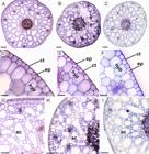
Anatomical features of the stem of Heteranthera zosterifolia, shown in cross-section. General view of specimens from the water surface (A), 2.5 meters (B), and 5 meters (C). Detailed view of the epidermis (ep) and hypodermis (hyp) of specimens from the water surface (D), 2.5 meters (E), and 5 meters (F). Detailed view of the aerenchyma pattern and air channels (ac) of specimens from the water surface (G), 2.5 meters (H), and 5 meters (I). ac: air channels; ct: cuticle; ep: epidermis; hyp: hypodermis. Scale bars = 200 μm (A-C), 20 μm (D-F), 100 μm (G-I).
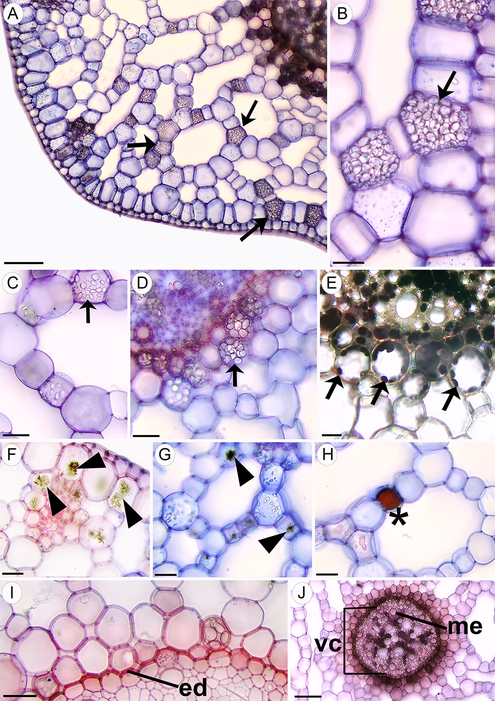
The presence of amyloplasts in the aerenchyma was more pronounced and widespread throughout the cortex in surface specimens (Fig. 5 A , B - arrows). In specimens from 2.5-meter depth, amyloplasts were more prominent near the vascular cylinder (Fig. 5 C - arrow), while in specimens from 5-meter depth, they were restricted to the central region around the vascular cylinder within the cortex (Fig. 5 D - arrow). The presence of starch in amyloplasts was confirmed by a positive Lugol’s reagent test (Fig. 5 E - arrow). Idioblasts with druses were absent in surface specimens but were abundant in specimens from 2.5-meter depth, predominantly found in the cortex’s first layer (Fig. 5 F - arrowhead). In specimens from a 5-meter depth, these druses were rare and scattered throughout the cortex (Fig. 5 G - arrowhead). Idioblasts containing phenolic compounds were only observed in specimens from 5-meter depth distributed throughout the cortex (Fig. 5 H - asterisk).
Anatomy of the vascular tissue
The number of vascular bundles distributed throughout the cortex showed no significant differences among specimens from different water depths (Table 2). In all samples, the endodermis displayed distinct Casparian strips (Fig. 5 I ), and the vascular cylinder consisted of a vascular plexus without distinct bundles, with metaxylem scattered within parenchymatous tissue (Fig. 5 J ). The average vessel diameter in the vascular cylinder showed a significant difference between surface specimens and those from 5-meter depths (p = 0.01; Fig. 3 C ; Table 2). Notably, the number of metaxylems in the vascular cylinder and the stele diameter exhibited no significant differences among specimens from different water depths.

Anatomical features of the stem of Heteranthera zosterifolia, shown in cross-section. Detailed view of the aerenchyma of the specimen from the water surface, highlighting the presence of amyloplasts (arrow) spread in the cortex (A). Detailed view of the amyloplasts (arrow) and starch grains specimen from the water surface (B), 2.5 meters (C), and 5 meters (D), with the latter surrounding the vascular plexus. Detail of the histochemical analysis positive for starch (E). Detailed view of idioblast with druses (arrowhead) in specimen from 5 meters (F) and 2.5 meters (G). Detailed view of idioblasts containing phenolic compounds (asterisks) in the specimen from 5 meters (H). Detailed view of the endodermis (ed) displaying distinct Casparian strips (I). Vascular cylinder (vc) without distinct metaxylem (me), spread within a parenchymatous pith and enclosed by the pericycle (J). ed: endodermis; me: metaxylem; vc: vascular cylinder. Scale bars = 20 μm (B-I), 100 μm (A-J).
Discussion
In this study, we investigated the stem anatomy and morphology of H. zosterifolia specimens across varying water depths to address a significant research gap in our understanding of aquatic plant adaptation to deeper aquatic environmental conditions. Our findings reveal notable differences in the stem area, aerenchyma features, cortex thickness, stele-cortex ratio, and average vessel diameter among specimens from different water depths. Additionally, we observed variations in the distribution of amyloplasts across the cortex, which is discussed in detail below.
Characteristics of the stem and aerenchyma vary across different water depths
The results indicate that surface specimens of H. zosterifolia exhibit a significant expansion in stem area, total aerenchyma area, an increase in the number of air channels, and a thicker cortex compared to specimens from greater water depths. This finding contrasts with previous reports suggesting a trend toward increased area and number of air channels in specimens inhabiting deeper waters (Blom et al., 1990; Blom & Voesenek, 1996; Braendle & Crawford, 1999). The conventional association between deeper water depths and increased dimension and quantity of aerenchyma, driven by hypoxic conditions and lower oxygen diffusion (Justin & Armstrong, 1991; Armstrong et al., 1994; Coops et al., 1996; Jackson & Armstrong, 1999), is contradicted by our findings.
Therefore, our findings lead to the hypothesis of a distinctive strategy employed by surface specimens of H. zosterifolia. In shallow water depths, where macrophytes face intensified susceptibility to abiotic factors and undergo alternating periods of submergence and emersion, surface specimens seem to invest in a larger stem and aerenchyma area. This investment may be linked with enhanced structural stability and buoyancy, enabling these plants to withstand the dynamic forces of their environment (Björn et al., 2022). While aerenchyma tissue has traditionally been associated with facilitating oxygen transport in response to hypoxic or anaerobic conditions (Justin & Armstrong, 1991; Armstrong et al., 1994; Coops et al., 1996; Jackson & Armstrong, 1999), our research underscores that aerenchyma serves a role beyond oxygen diffusion-it significantly contributes to the structural stability of the flexible stems by storing gases, providing buoyancy with relatively small biomass investment (Björn et al., 2022).
However, the effectiveness of the buoyancy provided by aerenchyma is only up to a certain depth and is determined by the pressure exerted by the water. Once the water pressure becomes too high, it can compress the stems, affecting the distribution of these macrophytes in aquatic environments (Björn et al., 2022). In this context, our results report a decrease in aerenchyma area with depth. This decrease might be related to the fact that in deeper water depths, where hydrostatic pressure is higher, plants might reduce the thickness of the cortex and the volume of aerenchyma to minimize compression effects and maintain structural integrity-a strategy to balance buoyancy requirements with the limitations imposed by increased pressure.
Regarding the pattern of aerenchyma, the studied species exhibits a honeycomb aerenchyma formed via schizogeny, which is the most common pattern occurring in aquatic plant shoots (Jung et al., 2008). Schizogenous aerenchyma is characterized by the presence of large air spaces formed by the separation and expansion of cell walls during development, followed by chemical changes in the lamella media, leading to the development of air channels and spaces (Seago et al., 2005; Jung et al., 2008). Besides the air channels, the parenchymatic cells in the aerenchyma show variation in starch storage, possibly linked to resource availability, light intensity, and depth-related trade-offs in resource allocation (Hao et al., 2021). Surface specimens of H. zosterifolia, with amyloplasts spread throughout the parenchyma cells of the cortex, likely have a more efficient storage system due to pronounced adventitious root development. This may represent a strategy for better fixation in a more unstable water zone compared with specimens at 2.5- and 5-meter. In deeper water depths, starch storage is restricted around the vascular bundles, probably related to reduced light penetration and limited photosynthetic rates. This concentrates amyloplasts near the vascular bundles to facilitate resource exchanges (Brock et al., 1987).
Variation in the average vessel diameter and the stele-cortex ratio across different water depths
The investigation of H. zosterifolia specimens from various water depths revealed no significant differences in the number of vascular bundles throughout the cortex. In the central region, the vascular cylinder is characterized by a vascular plexus composed of several metaxylem and a parenchymatous pith, a common feature in Pontederiaceae (Sousa et al., 2016). Additionally, the number of metaxylem in the vascular cylinder of H. zosterifolia exhibited no significant variations. Previous reports have indicated that variations in vascular bundle area and number are affected by water fluctuation (Engloner & Papp, 2006), often resulting in an increase in the pith cavity at deeper depths, which serves as a pathway for oxygen transport. However, for the studied species, no variation was observed in the pith cavities. This suggests that transport efficiency within the stem remains relatively consistent and that oxygen flow possibly mainly occurs in the aerenchyma across the depth gradient.
A significant difference in average vessel diameter was observed between surface specimens and those from 5-meter depths, with surface specimens having significantly larger vessel diameters. This difference might be related to the environmental variations that surface specimens are subjected to; therefore, larger vessels could be an adaptation to maximize transport efficiency under conditions where water availability is less predictable or more necessary to compensate for loss through evapotranspiration (Tyree & Ewers, 1991; Hacker et al., 2006). Conversely, the smaller vessel diameter of 5-meter specimens is possibly linked to the increased hydrostatic pressure at greater depths, which can affect vessel structure and function. Smaller vessels may be an adaptation to better resist the additional pressure, preventing collapse and maintaining internal transport integrity (Sperry & Tyree, 1988; Tyree & Ewers, 1991; Hacker et al., 2006).
Notably, there is a significant difference in the stele-cortex ratio between surface and 2.5-meter specimens. Specimens from a depth of 2.5 meters reported an increase in the stele-to-cortex ratio compared to surface specimens. This demonstrates that surface specimens have a cortex proportionally thicker than the stele compared to those from 2.5 meters deep. The stele-to-cortex ratio has been previously studied mainly in absorptive roots to evaluate the balance between resource absorption via the cortex and transportation via the stele (Kong et al., 2017; Wang et al., 2018; Liu et al., 2023). For H. zosterifolia stems, the significant differences may be related to the balance between transportation and buoyancy in these specimens, with surface specimens investing in a greater cortex for better buoyancy and structural stability. It is notable that the stele-to-cortex ratio was not shown to be linear with depth and can be influenced by different environmental or physiological factors as depth increases.
Therefore, the anatomical features of the stem of H. zosterifolia at distinct water depths reveal potential strategies of the species to adapt to varying water levels. The increased investment in the cortex, aerenchyma area, number of air channels, and vessel diameter in surface specimens suggest adaptations for maximizing transport efficiency and structural stability in fluctuating water levels, which is less crucial in deeper zones due to higher hydrostatic pressure. In contrast, deeper specimens have smaller vessel diameters, likely an adaptation to resist higher hydrostatic pressure, and the concentration of amyloplasts near the vascular plexus indicates a strategy for better resource allocation. Future ecophysiological research on Heteranthera zosterifolia in doline ecosystems would significantly enhance our understanding of the species' ecological roles and adaptation, as well as the strategies employed by macrophytes to thrive in deep lotic aquatic habitats.
Acknowledgments
The authors thank CAPES (Coordenação de Aperfeiçoamento de Pessoal de Nível Superior) for the Post-doctorate grant for MM. This study was financed by the Coordenação de Aperfeiçoamento de Pessoal de Nível Superior - Brasil (CAPES grant #001). We thank the Recanto Ecológico Rio da Prata staff for facilities and professional divers for the collection of Heteranthera zosterifolia samples. We also thank Dr Lucas C. M. Lopes for his support with the statistical analyses.
References
-
Alho CJ, Silva JS. 2012. Effects of severe floods and droughts on wildlife of the Pantanal wetland (Brazil) - a review. Animals 2: 591-610. doi: 10.3390/ani2040591.
» https://doi.org/10.3390/ani2040591. -
Alves M, Trovó M, Forzza RC, Viana P. 2015. Overview of the systematics and diversity of Poales in the Neotropics with emphasis on the Brazilian flora. Rodriguésia 66: 305-328. doi: 10.1590/2175-7860201566203.
» https://doi.org/10.1590/2175-7860201566203 -
Armstrong W, Brändle R, Jackson MB. 1994. Mechanisms of flood tolerance in plants. Acta Botanica Neerlandica 43: 307-358. doi: 10.1111/j.1438-8677.1994.tb00756.x.
» https://doi.org/10.1111/j.1438-8677.1994.tb00756.x. - Barrett SCH, Graham SW. 1997. Adaptive radiation in the aquatic plant family Pontederiaceae: Insights from phylogenetic analysis. In: Givnish TJ, Sytsma KJ (eds.). Molecular evolution and adaptive radiation. Cambridge University Press, Cambridge. p. 225-258.
-
Björn LO, Middleton BA, Germ M, Gaberščik A. 2022. Ventilation systems in wetland plant species. Diversity 14: 1-21. doi: 10.3390/d14070517.
» https://doi.org/10.3390/d14070517. -
Blom CWPM, Bogemann GM, Laan P, van der Sman AJM, van der Steeg HM, Voesenek LACJ. 1990. Adaptations to flooding in plants from river areas. Aquatic Botany 38: 29-47. doi: 10.1016/0304-3770(90)90097-5.
» https://doi.org/10.1016/0304-3770(90)90097-5. -
Blom CWPM, Voesenek LACJ. 1996. Flooding: The survival strategies of plants. Trends in Ecology and Evolution 11: 290-295. doi: 10.1016/0169-5347(96)10034-3.
» https://doi.org/10.1016/0169-5347(96)10034-3. -
Bornette G, Puijalon S. 2011. Response of aquatic plants to abiotic factors: A review. Aquatic Sciences 73: 1-14. doi: 10.1007/s00027-010-0162-7.
» https://doi.org/10.1007/s00027-010-0162-7. -
Braendle R, Crawford RMM. 1999. Plants as amphibians. Perspectives in Plant Ecology, Evolution and Systematics 2: 56-78. doi: 10.1078/1433-8319-00065.
» https://doi.org/10.1078/1433-8319-00065. - Brock TCM, Van der Velde G, Van de Steeg HM. 1987. The effects of extreme water level fluctuations on the wetland vegetation of a Nymphaeid-dominated oxbow lake in the Netherlands. Archives für Hydrobiologie 27: 57-73.
- Bukatsch F. 1972. Bemerkungen zur Doppelfärbung Astrablau-Safranin. Mikrokosmos 61: 255.
- Caldatto AM. 2023. Influência temporal e ontogenética na dieta de um caracídeo na dolina cárstica Lagoa Misteriosa - MS, Brasil. MSc Thesis, Universidade Federal da Grande Dourados, Brasil.
-
Castello M, Poldini L, Altobelli A. 2021. The aquatic and wetland vegetation of Lake Doberdò: An analysis for conservation value assessment of a disappearing lake of the Classical Karst (Northeast Italy). Plant Sociology 58: 75-106. doi: 10.3897/pls2020581/05.
» https://doi.org/10.3897/pls2020581/05. -
Chambers PA, Lacoul P, Murphy KJ, Thomaz SM. 2008. Global diversity of aquatic macrophytes in freshwater. Hydrobiologia 595: 9-26. doi: 10.1007/s10750-007-9154-6.
» https://doi.org/10.1007/s10750-007-9154-6. -
Cook CDK. 1998. Pontederiaceae. In: Kubitzki K (ed.). Flowering Plants: Monocotyledons. The Families and Genera of Vascular Plants. Springer, Berlin. p. 395-403. doi: 10.1007/978-3-662-03531-3_37.
» https://doi.org/10.1007/978-3-662-03531-3_37. -
Coops H, van den Brink FW, Van Der Velde G. 1996. Growth and morphological responses of four helophyte species in an experimental water-depth gradient. Aquatic Botany 54: 11-24. doi: 10.1016/0304-3770(96)01025-X.
» https://doi.org/10.1016/0304-3770(96)01025-X. -
Engloner AI, Papp M. 2006. Vertical differences in Phragmites australis culm anatomy along a water depth gradient. Aquatic Botany 85: 137-146. doi: 10.1016/j.aquabot.2006.03.007.
» https://doi.org/10.1016/j.aquabot.2006.03.007. -
Govaerts R, Simpson DA, Goetghebeur P, Wilson KL, Egorova T, Bruhl J. 2019. World checklist of Cyperaceae. The board of trustees of the Royal Botanic Gardens. https://wcsp.science.kew.org/13 Feb. 2024.
» https://wcsp.science.kew.org/ -
Hacker UG, Sperry JS, Wheeler JK, Castro L. 2006. Scaling of angiosperm xylem structure with safety and efficiency. Tree Physiology 26: 689-701. doi: 10.1093/treephys/26.6.689.
» https://doi.org/10.1093/treephys/26.6.689. -
Hao S, Su W, Li QQ. 2021. Adaptive roots of mangrove Avicennia marina: Structure and gene expressions analyses of pneumatophores. Science of The Total Environment 757: 1-10. doi: 10.1016/j.scitotenv.2020.143994.
» https://doi.org/10.1016/j.scitotenv.2020.143994. -
Jackson MB, Armstrong W. 1999. Formation of aerenchyma and the processes of plant ventilation in relation to soil flooding and submergence. Plant Biology: 274-287. doi: 10.1111/j.1438-8677.1999.tb00253.x.
» https://doi.org/10.1111/j.1438-8677.1999.tb00253.x. - Johansen DA. 1940. Plant Micro technique. New York, McGraw-Hill Book Company.
-
Jung J, Lee SC, Choi HK. 2008. Anatomical patterns of aerenchyma in aquatic and wetland plants. Journal of Plant Biology 51: 428-439. doi: 10.1007/BF03036065.
» https://doi.org/10.1007/BF03036065. -
Justin SHFW, Armstrong W. 1991. Evidence for the involvement of ethene in aerenchyma formation in adventitious roots of rice (Oryza sativa L.). New Phytologist 118: 49-62. doi: 10.1111/j.1469-8137.1991.tb00564.x.
» https://doi.org/10.1111/j.1469-8137.1991.tb00564.x. -
Kerr RA. 2012. The Greenhouse Is Making the Water-Poor Even Poorer. Science 336: 405. doi: 10.1126/science.336.6080.405.
» https://doi.org/10.1126/science.336.6080.405. -
Kong DL, Wang JJ, Zeng H, Liu MZ, Miao Y, Wu HF. 2017. The nutrient absorption-transportation hypothesis: optimizing structural traits in absorptive roots. New Phytologist 213: 1569-1572. doi: 10.1111/nph.14344.
» https://doi.org/10.1111/nph.14344. -
Kuerten S, McGlue MM, Rasbold GG et al 2022. Doline pond sediments reveal Late Holocene hydro-geomorphological changes in the highlands of the Pantanal, western Brazil. Journal of South American Earth Sciences 118: 103945. doi: 10.1016/j.jsames.2022.103945.
» https://doi.org/10.1016/j.jsames.2022.103945. -
Leandro TD, Holsback ZDR, Scremin-Dias E. 2021. The aquatic species Pontederia azurea and P. crassipes (Pontederiaceae) in the Pantanal, Brazil: Evidence of how plant structure can simultaneously reflect phylogeny and ecology. Acta Botanica Brasilica 35: 79-91. doi: 10.1590/0102-33062020abb0278.
» https://doi.org/10.1590/0102-33062020abb0278. - Les DH. 2020. Aquatic Monocotyledons of North America: Ecology, Life History, and Systematics. Boca Raton, CRC Press.
-
Les DH, Tippery NP. 2013. In time and with water... the systematics of alismatid monocotyledons. In: Wilkin P, Mayo SJ (eds.). Early events in monocot evolution. Cambridge, Cambridge University Press. p. 118-164. doi: 10.1017/CBO9781139002950.007.
» https://doi.org/10.1017/CBO9781139002950.007. -
Liu Z, Li Q, Sun H et al 2006. Diurnal Variations of Hydrochemistry in a Travertine-depositing Stream at Baishuitai, Yunnan, SW China. Aquatic Geochemistry 12: 103-121. doi: 10.1007/s10498-005-2962-2.
» https://doi.org/10.1007/s10498-005-2962-2. -
Liu Z, Liu X, Liao C. 2008. Daytime deposition and nighttime dissolution of calcium carbonate controlled by submerged plants in a karst spring-fed pool: Insights from high time-resolution monitoring of physico-chemistry of water. Environmental Geology 55: 1159-1168. doi: 10.1007/s00254-007-1062-6.
» https://doi.org/10.1007/s00254-007-1062-6. -
Liu X, Du Y, Ren Y et al 2023. New insights into the cortex-to-stele ratio show it to effectively indicate inter- and intraspecific function in the absorptive roots of temperate trees. Frontiers in Plant Science 14: 1061503. doi: 10.3389/fpls.2023.1061503.
» https://doi.org/10.3389/fpls.2023.1061503. - Maltchik L, Costa MAJ, Duarte MCD. 1999. Inventory of Brazilian semiarid temporary shallow lakes. Anais da Academia Brasileira de Ciências 71: 801-808.
-
Marengo JA, Oliveira GS, Alves LM. 2016. Climate change scenarios in the Pantanal. In: Bergier I, Assine M (eds.). Dynamics of the Pantanal Wetland in South America. Berlin, Springer Cham. p. 227-238. doi: 10.1007/978-3-319-18735-8.
» https://doi.org/10.1007/978-3-319-18735-8. -
Pan Y, Yuan D, Wu Q et al 2021. Effect of water exchange rate on interspecies competition between submerged macrophytes: Functional trait hierarchy drives competition. Plant Soil 466: 631-647. doi: 10.1007/s11104-021-05081-x.
» https://doi.org/10.1007/s11104-021-05081-x. -
Riis T, Hawes I. 2002. Relationships between water level fluctuations and vegetation diversity in shallow water of New Zealand lakes. Aquatic Botany 74: 133-148. doi: 10.1016/S0304-3770(02)00074-8.
» https://doi.org/10.1016/S0304-3770(02)00074-8. -
Santos KR, Pereira MP, Ferreira ACG et al 2015. Typha domingensis Pers. growth responses to leaf anatomy and photosynthesis as influenced by phosphorus. Aquatic Botany 122: 47-53. doi: 10.1016/j.aquabot.2015.01.007.
» https://doi.org/10.1016/j.aquabot.2015.01.007. -
Scheffer M, Hosper SH, Meijer ML, Moss B, Jeppesen E. 1993. Alternative equilibria in shallow lakes. Trends in Ecology & Evolution 8: 175-279. doi: 10.1016/0169-5347(93)90254-M.
» https://doi.org/10.1016/0169-5347(93)90254-M. - Scheffler SM, Silva WM, Pellin A, Duleba S, Pellin A. 2019. Floração de algas na Cavidade Natural Lagoa Misteriosa (MS 043), Jardim, MS, Brasil: uma análise integrada de dados físico-químicos, biológicos e climatológicos. Terra Plural 13: 400-420.
-
Schutten J, Dainty J, Davy AJ. 2005. Root anchorage and its significance for submerged plants in shallow lakes. Journal of Ecology 93: 556-571. doi: 10.1111/j.1365-2745.2005.00980.x.
» https://doi.org/10.1111/j.1365-2745.2005.00980.x. -
Seago JL, Marsh LC, Stevens KJ, Soukup A, Votrubová O, Enstone DE. 2005. A re-examination of the root cortex in wetland flowering plants with respect to aerenchyma. Annals of Botany 96: 565-79. doi: 10.1093/aob/mci211.
» https://doi.org/10.1093/aob/mci211. -
Sousa DJL. 2023. Pontederiaceae in Flora e Funga do Brasil 2022. Instituto de Pesquisas Jardim Botânico do Rio de Janeiro. https://floradobrasil.jbrj.gov.br/reflora/floradobrasil/FB197 13 Feb. 2024.
» https://floradobrasil.jbrj.gov.br/reflora/floradobrasil/FB197 -
Sousa DJL, Scatena VL, Giulietti AM, Oriani A. 2016. Morphological and anatomical patterns in Pontederiaceae (Commelinales) and their evolutionary implications. Aquatic Botany 129: 19-30. doi: 10.1016/j.aquabot.2015.11.003.
» https://doi.org/10.1016/j.aquabot.2015.11.003. -
Sperry JS, Tyree MT. 1988. Mechanism of water stress-induced xylem embolism. Plant Physiology 88: 581-587. doi: 10.1104/pp.88.3.581.
» https://doi.org/10.1104/pp.88.3.581. - Spiro B, Pentecost A. 1991. One day in the life of a stream - a diurnal inorganic carbon mass balance for a travertine-depositing stream (Waterfall Beck, Yorkshire). Geomicrobiology Journal 9: 1-11.
-
Tyree MT, Ewers FW. 1991 The hydraulic architecture of trees and other woody plants. New Phytologist 119: 345-360. doi: 10.1111/j.1469-8137.1991.tb00035.x.
» https://doi.org/10.1111/j.1469-8137.1991.tb00035.x. -
Wang WN, Wang Y, Hoch G, Wang ZQ, Gu JC. 2018. Linkage of root morphology to anatomy with increasing nitrogen availability in six temperate tree species. Plant Soil 425: 189-200. doi: 10.1007/s11104-018-3563-3.
» https://doi.org/10.1007/s11104-018-3563-3. - Zelnik I, Potisek M, Gaberščik A. 2012. Environmental Conditions and Macrophytes of Karst Ponds. Polish Journal of Environmental Studies 21: 1911-1920.