 What about the striated phyllaries in Calea (Asteraceae: Neurolaeneae)? Anatomical analyses and the implications for the genus taxonomy
Abstract
Calea L. (Asteraceae Bercht. & J.Presl) has 166 species that can be characterized by striated phyllaries, modified leaves that surround the flowers forming an involucre to protect these structures, yellow florets, and pappus scales. Despite being one of the diagnostic characters for the genus, this is the first in-depth paper about the striated phyllaries of Calea. We analyzed 166 different Calea species to verify if there are differences in the width of these striations and their taxonomic value. Additionally, we examined 16 Calea species for anatomical characterization and investigated the nature of the phyllary striations. Each stria is characterized by a pair of secretory ducts that run side by side with a vascular bundle surrounded by fibers. The lumen of the secretory ducts was characterized according to its width: narrow striae (46.5-80 μm) and wide striae (100-265.4 μm). The characterization of the type of striae has great informative taxonomic potential for Calea. We present images with anatomical sections of the phyllaries of the species analyzed. We also provide the classification of all Calea species analyzed macroscopically regarding the type of striae on the inner phyllaries.
Keywords:
Anatomy; Capitula; Compositae; Heliantheae Alliance; Microcharacter
Introduction
Neurolaeneae Rydb. is one of the 50 tribes of Asteraceae Bercht. & J.Presl (Susanna et al., 2020), and it is included in the Heliantheae Alliance (Susanna et al., 2020). Members of this tribe are commonly shrubby, having opposite leaves, cymose capitulescences, heterogamous capitula, pistillate ray florets, and pappus with scales, rarely absent, bristles or coroniform (Bueno et al., 2021; Bueno, 2023). Neurolaeneae comprises 187 species (Bueno, 2023; Pruski, 2023) of neotropical and pantropical distribution (in the latter, only Enydra Lour.) (Bueno et al., 2021; Bueno, 2023). All the species are classified in six genera: Calea L., Enydra, Greenmaniella W.M.Sharp, Heptanthus Griseb., Neurolaena R.Br., and Staurochlamys Baker (Bueno, 2023).
Calea is the most species-rich genus, with 166 species (Bueno, 2023; Pruski, 2023; Bueno et al., 2025; Melquíades et al., 2025; Silva et al., 2025), occurring from Mexico to Argentina, as well as Jamaica and Trinidad and Tobago (Bueno, 2023). Calea species are morphologically diagnosed by their shrubby habit, opposite leaves, dichasial cymose or umbelliform capitulescences or solitary capitula, heterogamous capitula, striated phyllaries, yellow to brown anthers, and cypselae with pappus scales (Bueno et al., 2021; Bueno, 2023). Pruski (2023) proposed the segregation of the genus into seven genera based on morphological characters. However, Bueno (2023) provided the first phylogeny for Calea and showed its monophyly; these analyses took into account molecular, morphological, and geographic characters.
Phyllaries are modified leaves grouped, enclosing, or closely subtending an inflorescence in Asteraceae (Roque et al., 2009). The phyllaries differ from the leaves in terms of the position associated with the inflorescence, as well as in color, shape, and texture (Roque et al., 2009). In the capitulum, the phyllaries act functionally as sepals, protecting the inflorescence during development, and can be attractive for insects (Roque et al., 2009).
The application of microcharacters in the taxonomy of Asteraceae has been performed since Cassini (1821) until today (Robinson, 2009; Martínez-Quezada et al., 2023). These studies used microscopy techniques to study taxonomically informative characters at an anatomical level, such as trichomes and ducts (Martínez-Quezada et al., 2023), and morphologically, such as receptacles, corollas, anthers, styles, and pappi (Robinson, 2009). Examples of microcharacter applications have even been reported throughout the history of Calea: anatomical studies of the cypsela of Calea species provided subsidies to justify the transfer of some species of Calea to Alloispermum Willd. (Robinson, 2009).
Several studies on Calea (Pruski, 1984; Pruski & Urbatsch, 1983; 1988; Roque & Carvalho, 2011; Reis-Silva & Nakajima, 2020; 2021; Bueno & Heiden, 2021; 2022a;b; Bueno, 2023; Bueno et al., 2024a) have cited the striated phyllaries as diagnostic characters for the genus taxonomy. Other studies (Pruski & Urbatsch, 1988; Pruski, 1997; 2011; Bueno & Heiden, 2022a) have used phyllaries as a source of important taxonomic information for species comparison, but only their external morphology, such as shape, dimensions, indument, and number of series. Although Robinson (1975) superficially suggested that the striae are resinous ducts, he does not indicate which species he analyzed or which methodology he used, and he does not discuss it. Therefore, to date, no paper has investigated what the Calea phyllary striations are and whether these structures are taxonomically informative.
Furthermore, all the Calea literature that cites the striated phyllaries refers to the striae without mentioning differences in these striations (Robinson, 1975; Pruski, 1984; Pruski & Urbatsch, 1983; 1988; Roque & Carvalho, 2011; Reis-Silva & Nakajima, 2020; 2021; Bueno & Heiden, 2021; 2022a;b; Bueno, 2023; Bueno et al., 2024a).
In Calea, there are species with two types of phyllaries or only one (Calea in Flora e Funga do Brasil, 2025). The foliaceous phyllaries are the outermost series in several species, while the innermost ones are always scarious. In this way, through anatomical analyses on the inner phyllaries of Calea species, we intend to investigate the nature of the striae in phyllaries. Additionally, we morphologically examined whether there are differences in the width of these striations and their taxonomic value.
Materials and Methods
Macromorphological analyses were performed on inner phyllaries of at least one specimen from each of the 166 species of Calea, using stereoscopic microscopes (Leica M205C, Leica M60) and Leica Application Suite X 3.7.5. The list of the examined materials is provided in Supplementary material. These specimens are from 53 herbaria: ALCB, BHCB, BHZB, BLA, BM, BRIT, CEN, CESJ, DIAM, ECT, EFC, ESAL, FLOR, FUEL, FURB, G, HAS, HBR, HDJF, HEPH, HRB, HUCS, HUEFS, HUFSJ, HUFU, HURB, IBGE, ICN, K, MBM, MO, NYBG, P, PACA, PAMG, PEL, R, RB, RFA, SJRP, SMDB, SP, SPF, SPSF, TEPB, UB, UEC, UFG, UPCB, US, VIC, and VIES (Thiers, 2025). We analyzed all available Calea specimens in these herbaria. Some species are only known from the type specimen, whereas others have a large number of specimens available, all of which were analyzed. The analyses were performed on the inner phyllaries, which are always scarious and are present in all Calea species (Bueno, 2023).
The striae of the 166 species of Calea were previously classified based on macroscopic and qualitative analyses of the herbarium specimens using the Leica Application Suite X 3.7.5. Based on qualitative macroscopic analyses of the striae width in herbarium specimens, two distinct groups of striae were identified: narrow and wide. This classification, which divided the Calea species into two groups, is easily observed using a stereoscopic microscope.
To confirm this macroscopic classification, we performed anatomical analyses on a subset of the material from 16 species (Table 1), representing six subgenera (60 % of Calea) (Bueno, 2023). For anatomical analyses and diaphanization, we used 16 species that had at least three specimens available, except for Calea triantha (Vell.) Pruski, for which only two specimens were found (Table 1).
Diaphanization was performed according to a modified protocol from Handro (1964). The samples were placed in sodium hydroxide (2 %) for two hours, washed in distilled water (four successive times), clarified in sodium hydroxide solution (11 %), washed again in distilled water (four times sequentially), stained with safranin, and finally washed with 50 % ethanol to remove excess safranin (Kraus & Arduin, 1997). The material was then mounted on a slide with glycerin (Kraus & Arduin, 1997) and analyzed under a light microscope (Olympus CX 41).
For anatomical analyses, the herborized samples were rehydrated (Smith & Smith 1942), dehydrated in an ethyl series (10-70 %), and stored in 70 % ethanol. The samples were subsequently dehydrated in an ethyl series (85 %-95 %) and embedded in Historesin (Leica Biosystems, Heidelberg, Germany). Cross-sections and longitudinal sections (5 µm thick) were obtained with a rotary microtome (RM 2155, Leica Microsystems, Heidelberg, Germany). The samples were stained with toluidine blue (O’Brien et al., 1964). The slides were mounted with Permount synthetic resin (Fisher Scientific, New Jersey, USA) and analyzed under an Olympus CX 41 microscope. Analyses and photographic documentation were carried out with a Nikon photomicroscope (Eclipse E200) equipped with a digital camera.
The width of the lumen of the secretory ducts was measured in cross-sections images using the ImageJ software (Schneider et al., 2012). Each measurement was made in triplicate, with three replicates in each sample and three replicates per species whenever possible. The measurements taken are available in the supplementary material. To test the measurements and determine whether the two morphological groups of stretch marks were significantly different, we performed a t-test to compare the measurements taken for the two groups. The statistical analyses were performed using R (version 4.5.1; R Development Core Team, 2025; www.r-project.org/).
Results
Anatomically, in species classified as narrowly striate based on macroscopic analysis, the lumen of the secretory ducts ranged from 46.5 to 80 μm in width, whereas in widely striate species, it ranged from 100 to 265.4 μm (Supplementary Material). Statistical analysis of the data objectively reinforced the distinction between narrow and wide striae, confirming what was already observed macroscopically under a stereoscopic microscope (Figure 1A-B). Welch's t-test revealed a highly significant difference between the two groups (p < 0.00000000002), with averages of approximately 64 µm for the narrow striae and 155 µm for the wide striae. Furthermore, the confidence intervals did not overlap, reinforcing the robustness of the separation between the categories.
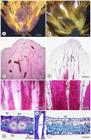
External morphology and anatomy of inner phyllaries of Calea graminifolia (A, C, E, G, and H) and C. pinnatifida (B, D, F, and I). Two types of striae in the inner phyllaries of Calea: Widely striate - 100-180.7 μm (A) and Narrowly striate - 46.5-86 μm (B). Frontal view of inner phyllaries in light microscopy - diafanization (C-F). General viewer of inner phyllaries (C and D). Detail of vascularization and fibers of inner phyllaries (E and F). Anatomy of inner phyllaries in cross (G and I) and longitudinal (H) section - light microscopy. Ad: adaxial surface epidermis; Ab: abaxial surface epidermis; Fi: Fibers; Vb: Vascular bundles; St: Stomata; Sd: Secretory duct; White arrows: striae inner phyllaries. Scales: A and B = 1 mm; C and D = 500 μm; E and F = 200 μm; G and I = 45 μm; H = 150 μm.
We propose here the characterization of all Calea species regarding the type of striae on the inner phyllaries based on macroscopic classification (Table 2). It was seen that there is no variation within each species. As can be seen in Fig. 1A-B, in addition to the width being visibly different in the stereoscopic images, the narrow striae (Fig. 1B) are clearly less prominent, less delimited and less conspicuous than the wide striae (Fig. 1A), this is uniform for all 166 species classified here.
As observed in the diaphanized samples (Fig. 1C-F), the striae are the unstained regions between the fibers. In cross-section, the striae are characterized by a pair of secretory ducts that run side by side with a vascular bundle surrounded by fibers (Fig. 1G-H). In some cases, the analyzed inner phyllaries region may present a continuous layer of fibers on the abaxial side, below the secretory ducts (Fig. 1I). The secretory ducts are arranged longitudinally in the phyllaries of all the species evaluated (Figs. 1-2).
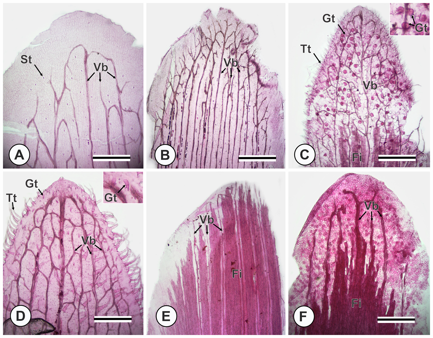
In the diaphanized samples, we observed the presence of fibers, vascular bundles, and stomata in all species evaluated (Fig. 1C-F; Fig. 2A-F). In some species, the inner phyllaries' surface is glabrous when viewed from the median frontal region, such as in C. graminifolia, C. pinnatifida (Fig. 1C-F), C. candolleana, C. hymenolepis, C. lemmatioides, and C. sickii (Fig. 2A-B and 2E-F). However, in other species, such as C. mediterranea (Fig. 2C) and C. acaulis (Fig. 2D), the inner phyllaries' surface presents both tector and glandular trichomes. Furthermore, it was possible to observe variations in the organization of vascular bundles (venation patterns) and distribution of fibers in the species evaluated in the diaphanized material. Some species have fibers that extend to the apex of the phyllaries, such as C. lemmatioides and C. sickii (Fig. 2 E-F); others have less elongated fibers restricted to the base of the phyllaries, such as C. graminifolia, C. pinnatifida (Fig. 1 C-F), C. candolleana, C. hymenolepis, C. mediterranea, and C. acaulis (Fig. 2 A-D).

Frontal view of inner phyllaries of Calea in light microscopy - diafanization (A-F). Widely striate (A, B, C, and D) and narrowly striate (E and F). Calea candolleana (A); C. hymenolepis (B); C. mediterranea (C); C. acaulis (D); C. lemmatioides (E), and C. sickii (F). St: Stomata; Vb: Vascular bundles; Fi: Fibers; Tt: Tector trichome; Gt: Glandular trichome. Scales: A-F = 600 μm.
In anatomical cross-sections, the inner phyllaries of all the species evaluated have a unstratified epidermis with stomata that can occur on both the adaxial and abaxial surfaces (Fig. 1G, 1I; Fig. 3). The mesophyll of all the species evaluated was composed of parenchymatic cells and fibers (Fig. 1G-I; Fig. 3). The secretory ducts (with longitudinally elongated internal space delimited by a secretory epithelium; Fig. 1H) are associated with the vascular bundles (Fig. 1G-I; Fig. 3A-N). In some species, such as Calea acaulis, C. candolleana, C. cymosa, and C. mediterranea, there are tector and glandular trichomes on the surface of the epidermis (Fig. 3G-J).

Anatomy of inner phyllaries of Calea in cross section - light microscopy (A-N). Widely striate (B, C, D, F, G, H, I, J, and M) and narrowly striate (A, E, K, L, and N). C. quadrifolia (A); C. lutea (B); C. hymenolepis (C); C. teucriifolia (D); C. lemmatioides (E); C. clematidea (F); C. mediterranea (G); C. cymose (H); C. candolleana (I); C. acaulis (J); C. sickii (K); C. lantanoides (L); C. clausseniana (M) and C. triantha (N). Ad: adaxial surface epidermis; Ab: abaxial surface epidermis; Fi: Fibers; Vb: Vascular bundles; St: Stomata; Sd: Secretory duct; Gt: Glandular trichome; Tt: tector trichome; asterisk: secretory duct. Scales: A-N = 45 μm.
Discussion
Our data demonstrated that the striae in Calea phyllaries are secretory ducts arranged longitudinally, associated with vascular bundles, and may be surrounded by fibers, as demonstrated in the anatomical analyses. Among all the Calea literature concerning the striae in the phyllaries, only Robinson (1975) makes a superficial mention of structures that could anatomically correspond to striae in the inner phyllaries. We confirm that Robinson's hypothesis (1975) is correct: the Calea inner phyllaries' striae are longitudinal ducts associated with the vascular bundle. As pointed out by Robinson (1975), this is one of the few characteristics that are shared by all species of Calea. Although this author originally used the term 'resinous duct', no chemical analyses of the secretion were performed, so we prefer to refer to them as 'secretory ducts'.
The secretory ducts we describe in Calea phyllaries exhibit anatomical similarities to those in other Asteraceae species (Fahn, 1979; Martínez-Quezada et al., 2023). In Asteraceae, it is reported that the predominant internal secretory structures are ducts, also described as secretory canals and resinous ducts, and they are widely distributed in vegetative organs and floral units (Robinson, 1981; Metcalfe & Chalk, 1983; Mauseth, 1988; Martínez-Quezada et al., 2023). Secretory ducts, as we verified, are characterized by the presence of a secretory epithelium that delimits a lumen, an elongated internal space in which secretion accumulates and is generally associated with vascular bundles (Fahn, 1979; Prado & Demarco, 2018). The composition of the duct secretion can vary, but it is predominantly resinous in some families in which it occurs, including Asteraceae, and has taxonomic value (Metcalfe & Chalk, 1983; Prado & Demarco, 2018; Martínez-Quezada et al., 2023). The presence of ducts is common in Asteraceae, especially in Asteroideae Lindl., Mutisioideae Lind. and Carduoideae Cass. ex Sweet (Robinson, 1981; Metcalfe & Chalk, 1983; Martínez-Quezada et al., 2023).
Even though the striations have never been studied in depth in Calea, there are records of ducts in the inner phyllaries of other groups. Anderberg (2009) reported a unique duct in the inner phyllaries of Iphionopsis Anderb. (Inuleae Cass.); Funk et al. (2009) reported the presence of a single duct in inner phyllaries of Feddea Urb. (Feddeae Urb.); Robinson (1981) (as Coreopsidineae Lindl.) and Crawford et al. (2009) noted that there are few or many ducts in the inner phyllaries of Coreopsideae Lindl.; Pruski et al. (2015) also found ducts in the inner phyllaries in Electranthera Mesfin, D.J.Crawford & Pruski (Coreopsideae); and Lizarazu & Freire (2019) documented these ducts in the phyllaries for Heterosperma Cav. (Coreopsideae). Except for Iphionopsis (Inuleae), all the other groups mentioned are included in the Heliantheae Alliance (Susanna et al., 2020). Interestingly, although the presence of secretory ducts in these species has been noted, their anatomical structure remains unexplored. Anatomical data on these structures in Calea are notably scarce in the literature. In this context, our study is pioneering, as it provides a detailed description of the anatomy of secretory ducts in the phyllaries of Calea, thus serving as a foundation for future anatomical and taxonomic studies.
As for Electranthera and Narvalina (Pruski et al., 2015) and Heterosperma (Lizarazu & Freire, 2019), we propose that all Calea species have ducts. This is supported by our anatomical analyses, which found ducts within the striae that are macromorphologically visible in all Calea species. Robinson (1975) or any of the other authors who mention Calea's striae (Pruski, 1984; Pruski & Urbatsch, 1983; 1988; Roque & Carvalho, 2011; Reis-Silva & Nakajima, 2020; 2021; Bueno & Heiden 2021; 2022a; 2022b; Bueno, 2023; Bueno et al., 2024a) do not discuss the informative taxonomic potential of these striae within Calea. All genera of Neurolaeneae have striate phyllaries (Bueno, 2023). We demonstrated that herbarium specimens can be used in anatomical studies of phyllaries and that this approach can be extended to other genera within Neurolaeneae.
No Calea species presents both types of striae, which shows that this is a stable character for each species. Because the striae types are easily distinguishable macroscopically, their characterization has several taxonomic applications. This information can help to differentiate infrageneric groups and species, as seen in recently published subgroups: Calea subgen. Teucriifoliae V.R. Bueno, Gostel & G.Heiden (Bueno et al., 2024a) and C. ser. Candolleanae V.R. Bueno, Gostel & G. Heiden (Bueno et al., 2024b) are both characterized by widely striate phyllaries, whereas Calea subg. Meyeria can be characterized by narrowly striate phyllaries (Bueno et al. 2025).
Bueno & Heiden (2022b) compared C. arachnoidea G.A. Reis-Silva & J.N.Nakaj., C. sessilifolia V.R.Bueno & G.Heiden, C. heteropappa Pruski, and C. semirii Pruski & D.J.N.Hind due to the similar pappus. According to Table 2, our results could contribute to the key, since the presence of widely striate phyllaries only exists in C. semirii, while the other three species have narrowly striate phyllaries.
Furthermore, Bueno (2023) discussed the morphological relationships of the taxonomy of the Nana clade of Calea that includes five species: Calea coridifolia Pruski, Calea linearifolia Maguire & Wurdack, Calea nana Maguire, Calea saxatilis Cuatrec., and Calea spiralis V.R.Bueno, Gostel & G.Heiden. With our results, we can see that Calea nana presents widely striate phyllaries, while the remaining species have narrow striate phyllaries. The type of striate phyllaries is also informative for Calea sect. Haplocalea (Less.) Pruski, which has eight species (Pruski, 1998) that present similar morphology regarding their capitulescence. However, three have widely striate phyllaries (Calea acaulis, C. cymosa, and C. mediterranea) and five have narrowly striate phyllaries (C. crenata Chodat, C. hassleriana Chodat, C. reticulata Gardner, and C. rhombifolia S.F.Blake).
In taxonomic studies of Calea with geographic scope, the differentiation of the types of striate phyllaries could also be very informative. Silva & Teles (2018) listed 30 species for Goiás state, Brazil, and produced a key for these species. Our results contribute directly to the differentiation of these species, since seven of these species have narrowly striate phyllaries and the other 23 have widely striate phyllaries. Pruski (1997) listed the Calea species for the Flora of the Venezuelan Guayana; 26 species have already been published, of which 14 are widely striate phyllary species and 12 are narrowly striate. Therefore, this character could be quite useful in the taxonomy of these species.
Our results contribute novel insights that fill gaps in the understanding of the anatomy of Calea phyllaries. These insights may enhance the characterization of the genus and support comparative studies across related taxa. Additionally, although our study was directed towards the analysis of inner phyllaries striae, anatomical analyses also revealed that only some species of Calea species present tector and glandular trichomes on the surface of their inner phyllaries. Thus, we suggest that in addition to striae, other anatomical characters of inner phyllaries, such as the shape of epidermal cells, venation patterns, and distribution of fibers, may also be potentially useful in taxonomic studies of Calea. However, this study provides a foundation for future research, and additional anatomical studies are needed to better understand the chemical nature of secretory duct secretion and the usefulness of other anatomical structures for taxonomic purposes.
This study is the first to identify anatomically what these striations are and their significant potential for generating taxonomic information within the genus. Anatomical analyses solved the great mystery surrounding the striated inner phyllaries of Calea by elucidating that they are secretory ducts associated with vascular bundles. We propose that these striae can be classified morphologically into two types: narrowly and widely striate. This characteristic is present in all Calea species analyzed, and our analyses demonstrate that it is highly informative for the genus, with the potential to characterize groups and to be applied in taxonomic keys. Further studies could elucidate whether the inner phyllaries of other genera of Neurolaeneae (Bueno et al., 2021; Bueno, 2023) show structural differences when compared with Calea and among themselves. Furthermore, other anatomical characters of inner phyllaries, such as vascularization and distribution of fibers, may be potentially useful in these investigations.
Supplementary Material
The following online material is available for this article
Supplementary Material 1.
Supplementary Material 2.
Acknowledgments
The authors acknowledge and are grateful to the staff of all the herbaria visited for the availability, receptivity, and assistance: CEN, ECT, EPAMIG, HALP, HEPH, HUFU, ICN, and MBM. In addition, the authors also thank all the institutions and their staff in which activities were developed to prepare this paper: UFRGS, UFU, UFV (acronyms for Federal Universities of Brazil), the National Museum of Natural History, the Missouri Botanical Garden, and the New York Botanical Garden. We thank the Histotechnical Laboratory of the Federal University of Viçosa - Campus Rio Paranaíba for the analyses carried out.
References
- Anderberg AA. 2009. Inuleae. In: Funk VA, Susanna A, Stuessy TF, Bayer RJ (eds.). Systematics, evolution, and biogeography of the Compositae. Vienna, IAPT. p. 667-680.
- Bueno VR. 2023. Neurolaeneae Systematics. PhD Thesis, Universidade Federal do Rio Grande do Sul, Brasil.
-
Bueno VR, Bentes MS, Ramos R, Zavatin DA, Prata-Silveira A. 2025. Three new species of Calea (Asteraceae, Neurolaeneae) from the sky islands of the Espinhaço Range. Phytotaxa 689: 185-203. doi: 10.11646/phytotaxa.689.2.2
» https://doi.org/10.11646/phytotaxa.689.2.2 -
Bueno VR, Gostel MR, Heiden G. 2021. An overview of Neurolaeneae (Compositae). Capitulum 1: 36-43. doi: 10.53875/capitulum.01.1.03
» https://doi.org/10.53875/capitulum.01.1.03 -
Bueno VR, Heiden G. 2021. Calea funkiana (Compositae, Neurolaeneae), A New Species Endemic From Serra do Cipó, Minas Gerais, Brazil. Systematic Botany 46: 470-475. doi: 10.1600/036364421X16431782047497.
» https://doi.org/10.1600/036364421X16431782047497 -
Bueno VR, Heiden G. 2022a. Novelties in Calea sect. Meyeria (Asteraceae, Neurolaeneae) from Brazil. Systematic Botany 47: 575-585. doi: 10.1600/036364422X16512564801652
» https://doi.org/10.1600/036364422X16512564801652 -
Bueno VR, Heiden G. 2022b. Calea sessilifolia (Asteraceae, Neurolaeneae), a new species from the Diamantina Plateau, Minas Gerais, Brazil. Systematic Botany 47: 586-592. doi: 10.1600/036364422X16512564801687
» https://doi.org/10.1600/036364422X16512564801687 -
Bueno VR, Silva GHL, Reis-Silva GA et al 2024a. Synopsis of Calea subgen. Teucriifoliae (Asteraceae: Neurolaeneae), a new subgenus endemic to Cerrado. Phytotaxa 637: 62-72. doi: 10.11646/phytotaxa.637.1.4
» https://doi.org/10.11646/phytotaxa.637.1.4 -
Bueno VR, Silva GHL, Reis-Silva GA et al 2024b. Nomenclatural synopsis of the new Calea ser. Candolleanae (Asteraceae: Neurolaeneae), a clade endemic to Eastern and Central Brazil. Phytotaxa 660: 46-54. doi: 10.11646/phytotaxa.660.1.4
» https://doi.org/10.11646/phytotaxa.660.1.4 -
Bueno VR, Reis-Silva GA, Nakajima JN, Gostel MR, Heiden G. 2025. A new narrower circumscription and a nomenclatural synopsis for Calea subg. Meyeria (Asteraceae: Neurolaeneae). Phytotaxa 683: 273-280. doi: 10.11646/phytotaxa.683.3.8
» https://doi.org/10.11646/phytotaxa.683.3.8 -
Flora e Funga do Brasil. 2025. Calea in Flora e Funga do Brasil. https://floradobrasil.jbrj.gov.br/FB103751 26 Mar. 2025.
» https://floradobrasil.jbrj.gov.br/FB103751 - Cassini H. 1821. Helianthées. In: Cuvier G (ed.). Dictionnaire des sciences naturelles. Paris, Le Normant. vol. XX, p. 260-261.
- Crawford DJ, Tadesse M, Mort ME, Kimball RT, Randle CP. 2009. Coreopsideae In: Funk VA, Susanna A, Stuessy TF, Bayer RJ (eds.). Systematics, evolution, and biogeography of the Compositae. Vienna, IAPT . p. 713-730.
- Fahn A. 1979. Secretory Tissues in Plants. London, Academic Press.
- Funk VA, Susanna A, Stuessy TF, Robinson H. 2009. Classification of Compositae, In: Funk VA, Susanna A, Stuessy TF, Bayer RJ (eds.). Systematics, evolution, and biogeography of the Compositae. Vienna, IAPT. p. 171-176.
- Handro W. 1964. Contribuição ao estudo da venação e anatomia foliar das Amarantáceas dos cerrados. Anais da Academia Brasileira de Ciências 36: 479-499.
- Kraus JE, Arduin M. 1997. Manual básico de métodos em morfologia vegetal. Seropédica, EDUR.
-
Lizarazu MA, Freire SE. 2019. A taxonomic revision of Heterosperma (Asteraceae: Coreopsideae). Annals of the Missouri Botanical Garden 104: 633-663. doi: 10.3417/2019374
» https://doi.org/10.3417/2019374 -
Martínez-Quezada DM, Rivera P, Rojas-Leal A, Villaseñor JL, Terrazas T. 2023. Leaf secretory structures in Asteraceae: A synthesis of their diversity and evolution. The Botanical Review 89: 59-90. doi: 10.1007/s12229-022-09276-4
» https://doi.org/10.1007/s12229-022-09276-4 - Mauseth JD. 1988. Plant anatomy. Menlo Park, Benjamin/Cummings Publishing Company.
-
Melquíades IM, Bueno VR, Teles AM, Koch AK. 2025. A new species and an geographic distribution update of Calea (Asteraceae, Neurolaeneae) from the Mato Grosso state, Brazil. Nordic Journal of Botany 2025: e04684. doi: 10.1111/njb.04684
» https://doi.org/10.1111/njb.04684 - Metcalfe CR, Chalk L. 1983. Anatomy of the Dicotyledons: Wood Structure and Conclusion of the General Introduction. Oxford, Clarendon Press. vol. II.
- O’Brien TP, Feder N, McCully ME. 1964. Polychromatic staining of plant cell walls by toluidine blue O. Protoplasma 59: 367-373.
-
Prado E, Demarco D. 2018. Laticifers and Secretory Ducts: Similarities and Differences. In: Hufnagel E (ed.). Ecosystem Services and Global Ecology. Rijeka, IntechOpen. doi: 10.5772/intechopen.75705
» https://doi.org/10.5772/intechopen.75705 - Pruski JF, Tadesse M, Crawford DJ. 2015. Studies of Neotropical Compositae-XI. The new generic name Electranthera (Coreopsideae). Phytoneuron 68: 1-17.
-
Pruski JF, Urbatsch LE. 1983. Calea bucaramangensis (Asteraceae), a new species from the Colombian Andes. Systematic Botany 8: 93-95. doi: 10.2307/2418567
» https://doi.org/10.2307/2418567 -
Pruski JF, Urbatsch LE. 1988. Five new species of Calea (Compositae: Heliantheae) from Planaltine Brazil. Brittonia 40: 341-56. doi: 10.2307/2807644
» https://doi.org/10.2307/2807644 -
Pruski JF. 1984. Calea brittoniana and Calea kristiniae: Two new compositae from Brazil. Brittonia 36: 98-103. doi: 10.2307/2806617
» https://doi.org/10.2307/2806617 - Pruski JF. 1997. Calea L. In: Steyermark JA, Berry PE, Holst BK (eds.). Flora of the Venezuelan Guayana. St. Louis, Missouri Botanical Garden. vol. III. p. 221-236.
-
Pruski JF. 1998. Novelties in Calea (Compositae: Heliantheae) from South America. Kew Bulletin 53: 683-693. doi: 10.2307/4110487
» https://doi.org/10.2307/4110487 - Pruski JF. 2011. Compositae of the Guayana Highland, XIV. Four new species of Calea (Neurolaeneae) from Tepui summits in Venezuela. Phytoneuron 52: 1-9.
- Pruski JF. 2023. The new genera Laceanthos, Podocalea, Tepuipappus, the resurrected genera Lemmatium, Meyeria, Tonalanthus and other novelties in the Calea Alliance I. Phytoneuron 42: 1-171.
-
R Core Team. 2025. R: A language and environment for statistical computing. https://www.R-project.org/ 26 Mar. 2025.
» https://www.R-project.org/ -
Reis-Silva GA, Nakajima JN. 2020. A new species of Calea (Neurolaeneae, Asteraceae) from the Espinhaço Range, Minas Gerais, Brazil. Phytotaxa 432: 199-205. doi: 10.11646/phytotaxa.432.2.9
» https://doi.org/10.11646/phytotaxa.432.2.9 -
Reis-Silva GA, Nakajima JN. 2021. A new species of Calea (Neurolaeneae, Asteraceae) from the Atlantic Forest, Minas Gerais, southeastern Brazil. Phytotaxa 490: 129-136. doi: 10.11646/phytotaxa.494.1.9
» https://doi.org/10.11646/phytotaxa.494.1.9 - Robinson HE. 1975. Studies in the Heliantheae (Asteraceae). VI. Additions to the genus Calea Phytologia 32: 426-430.
- Robinson, HE. 1981. A Revision of the Tribal and Subtribal Limits of the Heliantheae (Asteraceae). Washington, Smithsonian Institution Press.
- Robinson HE. 2009. An introduction to micro-characters of Compositae. In: Funk VA, Susanna A, Stuessy TF, Bayer RJ (eds.). Systematics, evolution, and biogeography of the Compositae. Vienna, IAPT. p. 89-100.
-
Roque N, Carvalho VC. 2011. Estudos taxonômicos do gênero Calea (Asteraceae, Neurolaeneae) no estado da Bahia, Brasil. Rodriguésia 62: 547-561. doi: 10.1640/2175-7860201164308
» https://doi.org/10.1640/2175-7860201164308 - Roque N, Keil DJ, Susanna A. 2009. Illustrated glossary of Compositae. In: Funk VA, Susanna A, Stuessy TF, Bayer RJ (eds.). Systematics, evolution, and biogeography of the Compositae. Vienna, IAPT. p. 781-806.
-
Schneider CA, Rasband WS, Eliceiri KW. 2012. NIH Image to ImageJ: 25 years of image analysis. Nature Methods 9: 671-675. doi: 10.1038/nmeth.2089
» https://doi.org/10.1038/nmeth.2089 -
Silva GHL, Teles AM. 2018. Calea (Asteraceae, Neurolaeneae) no estado de Goiás, Brasil. Rodriguésia 69: 1851-1875. doi: 10.1640/2175-7860201869422
» https://doi.org/10.1640/2175-7860201869422 -
Silva MS, Bueno VR, Scatigna AV, Marinho LC. 2025. Calea breviflora (Asteraceae): A new species endemic to white sand ecosystems of easternmost Amazon, Maranhão, Brazil. Phytotaxa 682: 91-100. doi: 10.11646/phytotaxa.682.1.6
» https://doi.org/10.11646/phytotaxa.682.1.6 -
Smith FH, Smith EC. 1942. Anatomy or the inferior ovary of Darbya American Journal of Botany 29: 464-471. doi: 10.1002/j.1537-2197.1942.tb10236.x
» https://doi.org/10.1002/j.1537-2197.1942.tb10236.x -
Susanna A, Baldwin BG, Bayer BG et al 2020. The classification of the Compositae: A tribute to Vicki Ann Funk (1947-2019). Taxon 69: 807-814. doi: 10.1002/tax.12235
» https://doi.org/10.1002/tax.12235 -
Thiers B. 2025 [continuously updated]. Index Herbariorum: A global directory of public herbaria and associated staff. http://sweetgum.nybg.org/science/ih/ 17 Jan. 2025.
» http://sweetgum.nybg.org/science/ih/
-
Data Availability
No new data were created or analyzed in this study.
-
Funding Information
This work was supported by FAPEMIG (Fundação de Amparo à Pesquisa do Estado de Minas Gerais), grant number [APQ-01846-24, 2022] and the CNPq (Centro Nacional de Desenvolvimento Científico e Tecnológico) [141645/2018; 151676/2024-0], CAPES/PROAP for research support to VRB, the Department of Botany at the National Museum of Natural History for funding the 2020 Harold E. Robinson and Vicki A. Funk Award to VRB, the Society of Systematic Biologists for funding the 2021 Mini-ARTS Award to VRB, and the International Association for Plant Taxonomy Research Grants to VRB in 2021.
No new data were created or analyzed in this study.