
 Eukaryotic diversity associated with the phycosphere of the seaweed Ulvaria obscura (Kützing) Gayral (Chlorophyta, Ulvophyceae) in the Svalbard Archipelago, Arctic region assessed using DNA metabarcoding
Abstract
Microorganisms often occur in association with macroalgae, with the term “phycosphere” referring to the seaweed surface where they may be present. Phycosphere represents a poorly explored niche of marine diversity, especially in the polar regions. DNA metabarcoding provides a new and accessible method for the detection of DNA from different organisms, especially applicable for poorly known groups where taxonomic expertise is limited or unavailable. In this study we used DNA metabarcoding to provide an initial survey of eukaryotic communities associated with marine macroalgae obtained from the shores of Svalbard. Samples of Ulvaria obscura were sequenced and the DNA reads found were assigned to 75 taxa of six Kingdoms and 17 phyla: Chromista (Ciliophora, Haptophyta, Ochrophyta, Oomycota and Chrysophyta), Fungi (Ascomycota, Basidiomycota, Chytridiomycota, Mortierellomycota and Rozellomycota), Holozoa (Ichthyosporia), Metazoa (Cnidaria), Protozoa (Cercozoa, Discosea and Heterolobosea) and Viridiplantae (Bryophyta and Chlorophyta). The most abundant group was Viridiplantae, followed by Fungi. Our environmental DNA study confirmed that the phycosphere of U. obscura shelters a rich and complex microbiome, suggesting that Arctic macroalgae provide a hotspot of currently undescribed polar biodiversity. Additionally, our results were obtained during the first official Brazilian Arctic expedition, representing a historic step for the Brazilian Antarctic Program (PROANTAR).
Key words
Ecology; environmental DNA; macroalgae; polar; taxonomy
INTRODUCTION
Located in the High Arctic, the Norwegian archipelago of Svalbard (74°-81° N; 10°-34° E) is surrounded by the Arctic Ocean to the north, Barents Sea to the east and Greenland Sea to the west. The archipelago is considered a global warming hotspot with parts warming by at least 2°C per decade since the mid to late Twentieth Century, a trend predicted to continue in the current century (Hanssen-Bauer et al. 2019, van Pelt et al. 2019, Pedersen et al. 2022).
The drastic levels of climate change in the High Arctic are expected to lead to wide but still poorly documented consequences for the local biota (Hanssen-Bauer et al. 2019, Pedersen et al. 2022). In the European Arctic, the best-studied macroalgal communities are those present on Svalbard, where they are important primary producers and creators of biogenic habitat in fjords ecosystems, producing up to 50% of the organic carbon available for the benthos (Renaud et al. 2015). In total, 197 species of macroalgae have been recorded from Svalbard, with 84 species recorded from Kongsfjorden alone (Fredriksen et al. 2019). Among Arctic macroalgal groups, green seaweeds generally have lower richness compared to red and brown species (Pellizzari et al. 2025).
The green algal (Chlorophyta) genus Ulvaria Ruprecht, 1850, is a foliaceous genus of monostromatic marine chlorophytes. Its taxonomy is complex, forming a group together with the genera Monostroma, Protomonostroma and Gayralia, and the presence of cryptic diversity. Guiry & Guiry (2025) list six described species within Ulvaria of which only three are currently considered valid taxonomic names (U. obscura, U. blyttii and U. shepherdii; type localities France, Norway and Australia, respectively), even then with U. blyttii in an uncertain position. Members of the genus are commonly found on the shores of Svalbard (Gulliksen et al. 1999) and the White Sea (Gobi 1878). The genus was originally described from the Aleutian Islands (Ruprecht 1850) and is widely distributed in marine cold waters throughout the Northern Hemisphere. Ulvaria obscura (Kützing) Gayral ex Bliding is broadly distributed in temperate and sub-Arctic regions of the Northern Hemisphere, occurring in the shallow subtidal and intertidal zones, occurring on stones, shells, larger brown algae, and other hard (natural or artificial) substrates. U. obscura is reported to the Arctic region including Svalbard (Gulliksen et al. 1999, Malavenda 2021).
Microorganisms often occur in association with macroalgae. They may participate in true symbioses or occur commensally, with the term “phycosphere” referring to the seaweed surface in an analogous fashion to the rhizosphere in soils around terrestrial plant roots, where microbial growth can be stimulated by extracellular products of the algae (Bell & Mitchell 1972). Microalgae, ciliates and fungi, amongst other microbial groups may be present within the phycosphere. However, with the exception of bacteria, little is known about these microorganisms and the phycosphere represents a poorly explored niche of marine diversity, especially in the polar regions.
Recent advances in molecular biology have provided new tools and analytical approaches for the assessment of diversity in environmental samples obtained from various ecosystems. DNA metabarcoding using high-throughput sequencing (HTS) provides a new and accessible method for the detection of DNA from different organisms, and is especially applicable for poorly-known groups where taxonomic expertise is limited or unavailable or where resting stages are overlooked when using traditional taxonomic methods based on morphology alone (Fernandes et al. 2021, Campello-Nunes et al. 2024). Câmara et al. (2021a, b) used this approach to compare periphytic diversity between two lakes in the South Shetland Islands (Maritime Antarctic), but it has yet to be used widely in the assessment of marine macroalgal periphytic biodiversity. In this study, we used DNA metabarcoding to provide an initial survey of eukaryotic communities associated with marine macroalgae obtained from the shores of Svalbard, Arctic region.
MATERIALS AND METHODS
Macroalgal sampling and taxonomy
Five specimens of Ulvaria obscura were sampled from the intertidal zone at Longyearbyen, Svalbard (78°13’24.02” N; 15°38’48.8” E) in July 2023 (Figure 1). The samples were washed using fresh water, stored between sheets of blotting paper and cold dried. The specimens were labelled and transported to the plant molecular biology laboratory at University of Brasilia, Brazil for further analysis. In the laboratory, samples for morphological analysis were re-hydrated to enable histological procedures to be carried out before examination using a stereoscope (Motic SMZ-161) and optical microscope with phase contrast (Olympus CX31) and coupled image capture (UC90). Analyses focused on the morpho-anatomical features of the vegetative and reproductive regions. Histological sections were used to assess cell size, number, position and shape of chloroplasts, shape and size of uni- and multi-cellular fertile structures, type, size and shape of the apical cells, and exclusive morphological structures. Taxonomic identification was performed by comparing the specimens obtained with the available specialized bibliography of seaweeds from the Arctic (Fredriksen et al. 2019). Nomenclature follows Guiry & Guiry (2025). Specimens have been deposited in the collection of the Museu Botânico Municipal (MBM) Herbarium (Curitiba Botanical Garden, Paraná, Brazil).

Satellite images of the study region from which Arctic macroalgae samples were obtained. a) Arctic region, b) Longyearbyen, Svalbard Archipelago, c) sampling location (red triangle) between Longyearbyen Airport and downtown. Satellite images obtained from Google Earth Pro, 2019 (https://earth.google.com).
DNA sampling, extraction and sequencing
From each of the five collected samples, a subsample (ca. 2cm2) was immediately placed in a sterile plastic tube, frozen immediately at -20°C and stored until further analyses in Brazil. From one subsample, the total DNA was extracted using the FastDNA Spin Kit for Soil (MPBIO, Ohio, USA), following the manufacturer’s instructions. DNA quality was analyzed using agarose gel electrophoresis (1% agarose in 1 x Trisborate-EDTA) and then quantified using the Quanti-iT ™ Pico Green dsDNA Assay (Invitrogen). Negative controls did not render any detectable DNA. We used the internal transcribed spacer 2 (ITS2) region of the nuclear ribosomal DNA (Chen et al. 2010, Richardson et al. 2015, Câmara et al. 2021a, b, c, 2022) as a barcode, which is widely applied to identify a diverse range of eukaryote organisms including fungi, animals, protozoans, chromists and plants (Ruppert et al. 2019), and has proved effective in recent studies of Antarctic diversity using environmental samples (Câmara et al. 2022, Rosa et al. 2020, Ogaki et al. 2021, Carvalho-Silva et al. 2021). Library construction and DNA amplification were performed using the Library kit Herculase II Fusion DNA Polymerase Nextera XT Index Kit V2, following the Illumina 16S Metagenomic Sequencing Library Preparation Part #15,044,223 Rev. B protocol. Paired-end sequencing (2 × 300 bp) was performed on a MiSeq System (Illumina) by Macrogen Inc. (South Korea).
Data analyses and taxonomic assignment
Quality analysis was carried out using BBDuk v. 38.87 in BBmap software (BBMap - Bushnell B.; sourceforge.net/projects/bbmap) with the following parameters: Illumina adapters were removed (Illumina artefacts and the PhiX Control v3 Library); ktrim = l; k = 23; mink = 11; hdist = 1; minlen = 50; tpe; tbo; qtrim = rl; trimq = 20; ftm = 5; maq = 20. The remaining sequences were imported to QIIME2-amplicon version 2023.9 (https:// qiime2.org/) for bioinformatics analyses (Bolyen et al. 2019). The qiime2-dada2 plugin was used for filtering, dereplication, turn paired-end fastq files into merged, and remove chimeras, using default parameters (Callahan et al. 2016). Taxonomic assignments of ASVs (amplicon sequence variants) were determined using the qiime2-feature-classifier (Bokulich et al. 2018) classify-sklearn against different databases, using a sequence similarity threshold of 97%. First, ASVs were classified against the PLANiTS2 database (Banchi et al. 2020). After this step, ASVs that remained unclassified were filtered and classify-sklearn classified against the UNITE Eukaryotes ITS database version 8.3 (Abarenkov et al. 2020). Finally, remaining unclassified ASVs were filtered and aligned against the filtered NCBI non-redundant nucleotide sequences (nt) database (May 2024) using BLASTn (Camacho et al. 2009) with default parameters; the nt database was filtered using the following keywords: “ITS1”, “ITS2”, “Internal transcribed spacer” and “internal transcribed spacer”. Taxonomic assignments were performed using MEGAN6 (Hudson et al. 2016). For simplicity we henceforth refer to the assigned ASVs as “taxa”. For comparative purposes, we consider reads as a proxy for relative abundance (Giner et al. 2016). Taxonomic profiles were plotted using the Krona (Ondov et al. 2011).
Diversity and ecology
Those taxa with >1,000 DNA reads were considered abundant. The number of DNA reads were used to assess taxon diversity, richness and dominance, using the following indices: (i) Fisher’s α, (ii) Margalef’s and (iii) Simpson’s, respectively. Species accumulation curves were obtained using the Mao Tao index (based on a presence-absence matrix). All results were obtained with 95% confidence, and bootstrap values were calculated from 1,000 replicates using the PAST program v. 1.90 (Hammer et al. 2001).
RESULTS
Macroalgal taxonomy
The macroalgal specimens sampled (up to 20 cm long) had dark green monostromatic foliose thalli when submerged, and brownish color on drying (Figure 2a and b). Rhizoids were present only at the basal cell. The cells in frontal view varied in form along the length of the blade, and were quadratic, rounded to polygonal, sometimes arranged in a regular pattern, or in groups of four cells (Figure 2d). In the basal region of the blade the cells were longer and narrower than in the median region, approximately 20-23.5 µm × 30-37.5 µm, with a cellular lumen of 3-5 µm. A parietal chloroplast and single pyrenoid were present (Figure 2e). In cross-section (Figure 2f), only one layer of cells was present. The cells in the marginal and median region were, on average (measured 10 times), 30 µm deep, with a cell lumen of 4 µm, and were wider than longer. According to the macro- and micromorphological characteristics the macroalgae was identified as Ulvaria obscura (Kützing) Gayral (Chlorophyta, Ulvophyceae).

Macro- and micromorphology of Ulvaria obscura sampled in the intertidal zone at Longyearbyen, Svalbard. a) specimen in natural habitat; b) and c) wet and dried herbarium specimens; d) cells in frontal view; e) transverse histological section; f) basal cells in frontal view.
Eukaryotic DNA taxonomy and diversity
A total of 1,106,858 paired-end DNA reads were generated in the sequencing run of which 45,139 reads remained after quality filtering and the removal of the host macroalgal DNA. These reads were assigned to 75 ASVs and included representatives of six Kingdoms and 17 phyla: Chromista (Ciliophora, Haptophyta, Ochrophyta, Oomycota and Chrysophyta), Fungi (Ascomycota, Basidiomycota, Chytridiomycota, Mortierellomycota and Rozellomycota), Holozoa (Ichthyosporia), Metazoa (Cnidaria), Protozoa (Cercozoa, Discosea and Heterolobosea) and Viridiplantae (Bryophyta and Chlorophyta). Some sequences could only be assigned at higher taxonomic level (family, order or division) and the calculated rarefaction curves indicated that the DNA reads gave an accurate representation of the local diversity in the sample (Figure 3).
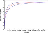
Rarefaction curves, with 95% confidence limits, of amplicon sequence variants (ASVs) obtained from the phycosphere of the Arctic macroalga Ulvaria obscura.
The most abundant group was Viridiplantae, followed by Fungi (Figure 4; Table I), with the dominant taxa being Ulothrix sp., Chlamydomonas sp., Kornmannia leptoderma, Acrosiphonia sp., Chlamydomonas raudensi, Pseudothrix groenlandica, Pseudendoclonium sp. (Chlorophyta, Viridiplantae) and Rhizophydiales sp. (Chytridiomycota, Fungi). The most diverse and rich (Fisher α and Margalef indices, respectively) groups were Fungi, Metazoa and Viridiplantae, in rank; however, Viridiplantae displayed the highest dominance (Simpson’s index) (Table II).
Assigned amplicon sequence variants associated with phycosphere of Ulvaria obscura. *Taxa not previously recorded from Svalbard. Taxa in gray were ranked as abundant.
Diversity indices of amplicon sequence variants (ASVs) associated with phycosphere of Ulvaria obscura.

Krona chart showing the abundances of different amplicon sequence variant (ASV) taxonomic levels associated with the phycosphere of Ulvaria obscura, assessed using PlantITS, UNITE and GenBank databases.
DISCUSSION
The host macroalgal species Ulvaria obscura
The algal flora of Svalbard generally comprises species that are also present in other regions of the North Atlantic and Arctic, as is the case for U. obscura (Weslawski et al. 1997). Fredriksen et al. (2019) listed a total of 197 macroalgal species recorded from Svalbard, representing 51 green, 76 brown and 70 red macroalgae. A total of 84 species are recorded from Kongsfjorden, comprising 19 green, 36 brown and 29 red algae are representing 42% of the total number of species know from Svalbard (refs needed). Malavenda (2021), in a study of seaweeds at the mouth of a Spitsbergen fjord (Svalbard) considered that most species of the 63 species present had high-boreal or Arctic-boreal distributions, suggesting that richness remained underestimated.
Eukaryotic diversity associated with the Artic macroalgae Ulvaria obscura
The assignment of a sequence obtained from eDNA does not confirm the presence of a living organism or a viable propagule in the sample and is also limited by the quality and completeness of available databases. This is particularly the case in the polar regions, where much diversity has yet to be sequenced. There is also no universal DNA barcode capable of covering all groups of organisms, while the use of different markers targeting certain sets of taxonomic groups inevitably generates different results for specific groups. Taxa assigned as ‘unknown’ or only assigned at higher taxonomic levels are likely to be absent from the consulted databases but could also represent currently undescribed taxa.
Viridiplantae
The most abundant Viridiplantae ASVs detected in Arctic U. obscura thalli were Ulothrix sp., Chlamydomonas sp., Kornmannia leptoderma, Acrosiphonia sp., Chlamydomonas raudensis, Pseudothrix groenlandica and Pseudendoclonium sp. (Table I). Chlamydomonas is a poorly known genus with more than 500 species, mostly reported from freshwater habitats but also with marine representatives. Chlamydomonas nivalis (F.A. Bauer) Wille is one of the most common algae present in snow communities and been reported from Svalbard (Matula et al. 2007). Its presence here is likely due to the proximity of the intertidal zone to adjacent terrestrial and now habitats. The genus Raphidonema includes 12 species of which R. nivale Lagerheim is recorded from Svalbard (Kvíderová 2012), similarly being common in snow algal communities. Kornmannia leptoderma (Kjellman) Bliding is also a monostromatic marine green alga that is widespread in temperate and sub-polar zones of the Northern Hemisphere and known from Svalbard (Fredriksen et al. 2019). Acrosiphonia is a diverse genus containing 45 described species, including A. arcta (Dillwyn) Gain, A. flagellata Kjellman, A. incurva Kjellman and A. sonderi (Kützing) Kornmann recorded from Svalbard (Gulliksen et al. 1999, Fredriksen et al. 2019). Pseudothrix groenlandica (J.Agardh) Hanic & S.C.Lindstrom, is a widespread marine species also reported from Svalbard (Fredriksen et al. 2019), while Pseudendoclonium is a genus containing nine described of which Pseudendoclonium submarinum Wille is present in Svalbard (Gulliksen et al. 1999, Fredriksen et al. 2019).
Amongst the other taxa assigned, the genus Hazenia includes five species, none of which have been reported from Svalbard, although representatives occur in Europe and Antarctica (Guiry & Guiry 2025). The assignment of Monostroma grevillei (Thuret) Wittrock represents the first record from Svalbard, although the species has been recorded from the adjacent Barents Sea (Malavenda 2018) and Arctic Canada (Taylor 1957). It has also been recorded from locations in Europe and the subArctic, and recently by Pellizzari et al. (2023) from Deception Island in the Maritime Antarctica. Its congener, M. lubricum Kjellman (the homotypic synonym of Monostroma grevillei var. lubricum (Kjellman) Collins), has been reported from Svalbard (Fredriksen et al. 2019). Planophila is, similarly, a genus containing around eight species which has not previously been reported from Svalbard but is present in the Baltic Sea (Nielsen et al. 1995) and Russian Arctic (Patova et al. 2015), and has also been reported in Antarctica in an eDNA study (Câmara et al. 2021b). Pseudochlorella pyrenoidosa (Zeitler) J.W.G. Lund is a European species, while other members of the genus have been reported from Russia and Antarctica (Guiry & Guiry 2025). The genus Ulvaria includes seven species worldwide, with two being found on Svalbard (U. splendens (Ruprecht) Vinogradova, U. obscura (Kützing) P. Gayral) (Gulliksen et al. 1999, Fredriksen et al. 2019). We excluded the DNA reads assigned to the host species sampled here, suggesting that these reads are from another source. Ulvella is another diverse but poorly known genus containing 65 species of which two are recorded from Svalbard (U. lens P.L. Crouan & H.M. Crouan, U. scutata (Reinke) R. Nielsen, C.J.O’ Kelly & B. Wysor). The genus Urospora includes 10 species of which three are present in Svalbard (U. elongata (Rosenvinge) Hagem, U. penicilliformis (Roth) Areschoug, U. wormskioldii (Mertens ex Hornemann) Rosenvinge) (Fredriksen et al. 2019). The two assigned bryophyte taxa found are widespread in the Northern Hemisphere and are likely sourced from the adjacent terrestrial environment.
Fungi
Fungal diversity associated with Antarctic macroalgae have, until recently, only been assessed using culturing approaches (Rosa et al. 2019). Davey et al. (2019) used a metabarcoding approach to determine the species composition of the microbial community in snow microalgal blooms in Antarctica, reporting a eukaryotic community dominated by unknown fungi. Câmara et al. (2024) also used metabarcoding to survey eukaryotic communities associated with the Antarctic macroalgae Desmarestia menziesii, Monostroma hariotii, and Ulothrix sp. (Chlorophyta), Desmarestia sarcopeltis and Iridaea sp. (Rhodophyta), and Adenocystis utricularis, and Ascoseira mirabilis (Phaeophyta), detecting 18 fungal ASVs representing the phyla Ascomycota and Basidiomycota.
The most abundant fungal ASV detected in Arctic U. obscura thalli was the chytrid taxon Rhizophydiales (Chytridiomycota). The order Rhizophydiales includes zoosporic species often present in wet, cold habitats and widely reported in association with algae (Schmidt et al. 2012, Naff et al. 2013, Seto & Degawa 2018). In addition, Rhizophydiales includes parasitic taxa able to affect different organisms, including algae, and may provide ecological control of aquatic populations (Powell 1993, Ibelings et al. 2004, Christaki et al. 2017). Davey et al. (2019) also reported Rhizophydiales as one of the dominant taxa present in snow algal blooms, and Ilicic et al. (2022) detected Betamyces (Rhizophydiales) in benthic diatoms in coastal zones of Antarctic environments.
Chromista
The most abundant chromist ASVs detected in Arctic U. obscura thalli represented the phylum Ciliophora. This is a poorly investigated and reported group. Knowledge of marine ciliates from the polar regions, to date, has concentrated on pelagic forms and, to a lesser extent on benthic periphytic communities (Song & Wilbert 2002). Only two ciliate ASVs were assigned to species level in the present study, the scuticociliate Mesanophrys carcini Small & Lynn in Aescht, 2001 and the dysteriid Planilamina ovata Ma et al. (2006), Mesanophrys carcini was originally described as Paranophrys carcini, extracted from the haemolymph of the crab Cancer pagurus Linnaeus, 1758 (Grolière & Léglise 1977), but also occurring in the free-living form (Pan et al. 2016). Planilamina ovata was described from blowhole mucus of the Atlantic bottlenose dolphin Tursiops truncatus Montagu, 1821 and the false killer whale Pseudorca crassidens Owen, 1846 (Ma et al. 2006).
Little can be inferred from ASVs identified at genus level or above, because their referenced taxa all have widespread geographic distributions. The genera Cyrtohymena and Holosticha are both hypotrichs, a group for which taxonomy is currently fluid and dependent on combined observations of live and protargol-stained specimens, and molecular data (Berger 1999, Paiva et al. 2014). Cyrtohymena sensu Berger (2018) contains six species, of which only C. marina (Kahl 1932) Foissner, 1989 occurs in marine environments, originally being reported from the North Sea and apparently restricted to the Northern Hemisphere (Berger 2018, Li et al. 2023). Holosticha was historically considered to be a large genus of hypotrichs containing ca. 100 species occurring in various habitats. It was subsequently disassembled until Berger (2006) redefined it, recognizing only eight species, all marine. Presently, about 10 species are accepted, depending whether Uncinata is accepted as a sub-genus of Holosticha (Paiva 2020, Chen et al. 2023). The genus Homalogastra includes three known species – H. setosa (Kahl 1926), H. parasetosa (Liu et al. 2020a), and H. binucleata (Liu et al. 2020b) - all known mostly from soil (sometimes saline) and brackish environments (Liu et al. 2020a, b).
Pseudovorticella is a peritrich genus containing ca. 60 species. Most species of Pseudovorticella were transferred from Vorticella, which still includes more than 50 species, many with questionable genus assignment. Available molecular databases have poor coverage of these genera (Sun et al. 2013, Jiang et al. 2019) and, as with other peritrichs, detailed taxonomic reassessment is required (Liao et al. 2021). Similarly, Prorodon is a problematic prostomatid genus due to overly simplistic descriptions and taxonomic errors (Foissner 2021).
The widespread crysophyte genus Paraphysomonas includes 46 accepted species, some of which are recorded from the Baltic Sea (Hällfors 2004) and Scandinavia (Karlson al. 2018). Representatives have been reported from Svalbard in molecular studies (van den Brink et al. 2021, Sørensen et al. 2012). The haptophyte order Pavlolales contains only four marine genera (Exanthemachrysis with four species, Diacronema with six species, Pavlova with about 12 species and Rebecca with three species), none of which have previously been recorded from Svalbard. However, Rebecca salina (N.Carter) J.C.Green has been reported from Scandinavia (Karlson et al. 2018) and Diacronema lutheri (Droop) Bendif & Véron from the Baltic Sea (Hällfors 2004).
The ochrophyte genus Navicula (Diatom) includes more than 1,600 accepted species, with at least seven species reported from Svalbard: N. kongsfjordensis Stachura, N. moskalii Metzeltin, Witkowski & Lange-Bertalot, N. bipustulata Van der Werff & Huls, N. directa (W.Smith) Ralfs, N. gregaria Donkin, N. kariana Grunow in Cleve & Grunow, N. transitans Cleve, N. trigonocephala Cleve and N. perminuta Grunow (Fredriksen et al. 2019, Guiry & Guiry 2025, Schaub et al. 2017). The oomycote genus Phytium is a widespread parasitic genus, mostly plant pathogens, causing diseases in crops but also in humans (Vanittanakom et al. 2004).
Holozoa, Metazoa and Protozoa
The ichthyophonid genus Sphaeroforma includes four species that are reported as saprotrophs from animal tissue, with records from the Bering Sea (Hasset et al. 2015). However, ITS2 markers as used in this study are not very useful in studies of metazoans, where COX1 is generally used (Folmer et al. 1994). Consequently, only two species assignments were made here, both to marine Cnidaria. Catablema vesicarium is an Atlantic species confined to Arctic cold waters (Schuchert 2007) and Obelia dichotoma is a widely distributed species occurring from the Arctic to the tropics, including Svalbard (Orejas et al. 2013). All three Protist phyla assigned in this study are widely distributed groups occurring in marine, terrestrial and freshwater habitats and including both free living and parasitic forms (Thompson et al. 2019).
CONCLUSIONS
Despite studying only one Arctic macroalgae species once, our environmental DNA study confirmed that the phycosphere of Ulvaria obscura (Ulvophyceae) shelters a rich and complex microbiome, indicating that Arctic macroalgae provide a hotspot of currently undescribed polar biodiversity. The sequence assignments made included diverse taxa with cosmopolitan, polar and restricted distributions, performing different ecological functions and services. Our data reinforce the need for further detailed studies of Arctic macroalgae, using larger sample sizes, a wide range of study locations, and multiple markers in order to increase knowledge of the diversity and composition of the phycosphere communities of Arctic macroalgae. Finally, our results were obtained during the first official Brazilian Arctic expedition, representing a historic step for the Brazilian Antarctic Program (PROANTAR – Programa Antártico Brasileiro).
Acknowledgements
This study received financial support from Conselho Nacional de Desenvolvimento Cientifico e Tecnológico (CNPq), Programa Antártico Brasileiro (PROANTAR), and Coordenação de Pessoal de Ensino Superior (CAPES). We are grateful to the Brazilian Navy, Brazilian Ministries of Foreign Affairs, Science Technology and Innovations and CNPq funding agency. The Government of Svalbard, the Norwegian Embassy in Brazil. Thiago Paiva is also grateful for CNPq Universal Grant 421766/2021-2. Peter Convey is supported by NERC core funding to the British Antarctic Survey’s ‘Biodiversity, Evolution and Adaptation’ Team.
References
- ABARENKOV ET AL. 2020. UNITE QIIME Release for Eukaryotes 2. Version 4.
- BANCHI ET AL. 2020. PLANiTS: a curated sequence reference dataset for plant ITS DNA metabarcoding. Database: baz155.
- BELL W & MITCHELL R. 1972. Chemotactic and Growth Responses of Marine Bacteria to Algal extracellular products. The Biol Bull 143: 265-277.
- BERGER H. 1999. Monograph of the Oxytrichidae (Ciliophora, Hypotrichia). Monogr Biol 78: 1-180.
- BERGER H. 2006. Monograph of the Urostyloidea (Ciliophora, Hypotricha). Monogr Biol 85: 1-1304.
- BERGER H. 2018. Six mainly little-known Cyrtohymena species (Ciliophora, Hypotricha): update 1.0 on monographic treatment. - Series Monographiae Ciliophorae. Helmut Berger Salzburg 2: 1-24.
-
BOKULICH ET AL. 2018. Optimizing taxonomic classification of marker-gene amplicon sequences with QIIME 2’s q2-feature-classifier plugin. Microbiome 6: 1-17. https://doi.org/10.1186/s40168-018-0470-z.
» https://doi.org/10.1186/s40168-018-0470-z -
BOLYEN ET AL. 2019. Reproducible, interactive, scalable and extensible microbiome data science using QIIME 2”. Nature Biotechnol 37: 852-857. https://doi.org/10.1038/s41587-019-0209-9.
» https://doi.org/10.1038/s41587-019-0209-9 -
CALLAHAN ET AL. 2016. DADA2: High-Resolution Sample Inference from Illumina Amplicon Data. Nature Methods 13: 581-583. https://doi.org/10.1038/nmeth.3869.
» https://doi.org/10.1038/nmeth.3869 -
CAMACHO ET AL. 2009. BLAST+: architecture and applications. BMC Bioinformatics 10: 1-9. https://doi.org/10.1186/1471-2105-10-421.
» https://doi.org/10.1186/1471-2105-10-421 -
CÂMARA ET AL. 2021a. Periphyton diversity in two different Antarctic lakes assessed using metabarcoding. Antarctic Sci 33: 596-604. https://doi.org/10.1017/S0954102021000316.
» https://doi.org/10.1017/S0954102021000316 -
CÂMARA ET AL. 2021b. Diversity and ecology of Chlorophyta (Viridiplantae) assemblages in protected and non-protected Sites in Deception Island (Antarctica, South Shetland Islands) assessed using an NGS approach. Microb Ecol 81: 323-334. https://doi.org/10.1007/s00248-020-01584-9.
» https://doi.org/10.1007/s00248-020-01584-9 -
CÂMARA ET AL. 2021c. The largest moss carpet transplant in Antarctica and its bryosphere cryptic biodiversity. Extremophiles 25: 369-384. https://doi.org/10.1007/s00792-021-01235-y.
» https://doi.org/10.1007/s00792-021-01235-y -
CÂMARA ET AL. 2022. Diversity of Viridiplantae DNA present on rock surfaces in the Ellsworth Mountains, continental Antarctica. Polar Biol 45: 637-646. https://doi.org/10.1007/s00300-022-03021-8
» https://doi.org/10.1007/s00300-022-03021-8 - CÂMARA ET AL. 2024. DNA metabarcoding reveal hidden diversity of periphytic eukaryotes on marine Antarctic macroalgae. An Acad Bras Cienc 96: e20240570. DOI 10.1590/0001-3765202420240570.
-
CAMPELLO-NUNES ET AL. 2024. Ciliate diversity in rodrigo de freitas lagoon (Rio de Janeiro, Brazil) from an integrative standpoint. Braz J Microbiol 55: 1489-1505. https://doi.org/10.1007/s42770-024-01291-4.
» https://doi.org/10.1007/s42770-024-01291-4 -
CARVALHO-SILVA ET AL. 2021. Exploring the plant environmental DNA diversity in soil from two sites on Deception Island (Antarctica, South Shetland Islands) using metabarcoding. Antarctic Scie 33: 469-478. https://doi.org/10.1017/S0954102021000274
» https://doi.org/10.1017/S0954102021000274 -
CHEN ET AL. 2010. Validation of the ITS2 region as a novel DNA barcode of identifying medicinal plant species. PLoS ONE 5: e8613. https://doi.org/10.1371/journal.pone.0008613.
» https://doi.org/10.1371/journal.pone.0008613 -
CHEN ET AL. 2023. Refining the “melting pot” genus Holosticha s. l. (Protozoa, Ciliophora, Hypotrichia) based on multigene datasets with establishment of a new species Caudikeronopsis monilata sp. nov. J Zoological Syst Evolut Res 2023: 3411188. https://doi.org/10.1155/2023/3411188.
» https://doi.org/10.1155/2023/3411188 - CHRISTAKI U ET AL. 2017. Parasitic eukaryotes in a meso-eutrophic coastal system with marked Phaeocystis globosa blooms. Front Mar Sci 4: 416.
- DAVEY MP ET AL. 2019. Snow algae communities in Antarctica: metabolic and taxonomic composition. New Phytol 222: 1242-1255.
-
FERNANDES ET AL. 2021. Ciliate diversity from aquatic environments in the Brazilian Atlantic Forest as revealed by high-throughput DNA sequencing. Microb Ecol 81: 630-643. https://doi.org/10.1007/s00248-020-01612-8.
» https://doi.org/10.1007/s00248-020-01612-8 -
FOISSNER W. 2021. A detailed description of a Brazilian Holophrya teres (Ehrenberg, 1834) and nomenclatural revision of the genus Holophrya (Ciliophora, Prostomatida). Eur J Protistol 80: 125662. https://doi.org/10.1016/j.ejop.2019.125662.
» https://doi.org/10.1016/j.ejop.2019.125662 - FOLMER O ET AL. 1994. DNA primers for amplification of mitochondrial cytochrome c oxidase subunit I from diverse metazoan invertebrates. Mol Mar Biol Biotechnol 3: 294-299.
- FREDRIKSEN ET AL. 2019. Chapter 9 Biodiversity of benthic macro- and microalgae from Svalbard with special focus on Kongsfjorden. In: Ecosystem of Kongsfjorden, Svalbard, Advances in Polar Ecology 2, Switzerland: Springer Nature, p. 331-371.
- GINER CR ET AL. 2016. Environmental sequencing provides reasonable estimates of the relative abundance of specific picoeukaryotes. Appl Environ Microbiol 82: 4757-4766.
- GOBI C. 1878. Die Algenflora des Weissen Meeres und der demselben zunächstliegenden Theile des nördlichen Eismeeres. Mémoires de l’Académie Impériale des Sciences de St. Petersburg, VIIé série 26: 1-92.
- GROLIÈRE CA & LÉGLISE M. 1977. Paranophrys carcini n. sp., Cilié Philasterina récolté dans l’hémolymphe du crabe Cancer pagurus Linné. Protistologica 13: 503-507.
-
GUIRY MD & GUIRY GM. 2025. AlgaeBase World-wide electronic publication, University of Galway. Available at: https://www.algaebase.org Accessed on March 11, 2025.
» https://www.algaebase.org - GULLIKSEN B ET AL. 1999. Distribution of marine benthic macro-organisms at Svalbard (including Bear Island) and Jan Mayen. Research Report for DN 1999-4: 1-148. Trondheim: Directorate for Nature Management.
- HÄLLFORS G. 2004. Checklist of Baltic Sea phytoplankton species (including some heterotrophic protistan groups). Baltic Sea Environ Proc 95: 1-208.
- HAMMER Ø ET AL. 2001. PAST: paleontological statistics software package for education and data analysis. Palaeontol Electron 4: 9.
- HANSSEN-BAUER I ET AL. 2019. Climate in Svalbard 2100 - a knowldge base for climate adaptation, NCCS report 1/2019, 105 p.
- HASSETT B, LÓPEZ JA & GRADINGER R. 2015. Two new species of marine saprophytic Sphaeroformids in the Mesomycetozoea isolated from the Sub-Arctic Bering Sea. Protist 166: 310-322. 10.1016/j.protis.2015.04.004.
-
HUDSON DH ET AL. 2016. MEGAN community edition-interactive exploration and analysis of large-scale microbiome sequencing data. PLoS Comp Biol 12: e1004957. https://doi.org/10.1371/journal.pcbi.1004957.
» https://doi.org/10.1371/journal.pcbi.1004957 - IBELINGS BW ET AL. 2004. Host parasite interactions between freshwater phytoplankton and chytrid fungi (Chytridiomycota). J Phycol 40: 437-453.
- ILICIC D ET AL. 2022. Antarctic glacial meltwater impacts the diversity of fungal parasites associated with benthic diatoms in shallow coastal zones. Front Microbiol 13: 805694.
- JIANG M ET AL. 2019. Morphology and phylogeny of three Pseudovorticella species (Ciliophora: Peritrichia) from brackish waters of China. J Eukaryot Microbiol 66: 869-881. DOI 10.1111/jeu.12738.
-
KARLSON B ET AL. 2018. Nordic Microalgae. World-wide electronic publication. Norrköping: Swedish Meteorological and Hydrological Institute. Available at: http://nordicmicroalgae.org
» http://nordicmicroalgae.org - KVÍDEROVÁ J. 2012. Research on cryosestic communities in Svalbard: the snow algae of temporary snowfields in Petuniabukta, Central Svalbard. Czech Polar Rep 2: 8-19.
-
LI X ET AL. 2023. Biodiversity of marine interstitial ciliates in the intertidal zone of the White Sea: a dataset from the Chernaya River estuary, Kandalaksha Gulf. Diversity 15: 873. https://doi.org/10.3390/d15070873.
» https://doi.org/10.3390/d15070873 -
LIAO W ET AL. 2021. Incorporating mitogenome sequencing into integrative taxonomy: the multidisciplinary redescription of the ciliate Thuricola similis (Peritrichia, Vaginicolidae) provides new insights into the evolutionary relationships among Oligohymenophorea subclasses. Mol Phyl Evol 158: 107089. https://doi.org/10.1016/j.ympev.2021.107089.
» https://doi.org/10.1016/j.ympev.2021.107089 -
LIU M ET AL. 2020a. Two new scuticociliates from southern China: Uronema apomarinum sp. nov. and Homalogastra parasetosa sp. nov., with improved diagnoses of the genus Homalogastra and its type species Homalogastra setosa (Ciliophora, Oligohymenophorea). Int J Syst Evol Microbiol 70: 2405-2419. https://doi.org/10.1099/ijsem.0.004046.
» https://doi.org/10.1099/ijsem.0.004046 - LIU M ET AL. 2020b. Taxonomy and molecular phylogeny of three species of Scuticociliates from China: Citrithrix smalli gen. nov., sp. nov., Homalogastra binucleate sp. nov. and Uronema orientalis Pan et al., 2015 (Protozoa, Ciliophora, Oligohymenophorea), with the proposal of a new family, Citrithrixidae fam. nov. Front Mar Sci 7: 604704. DOI 10.3389/fmars.2020.604704.
-
MA H ET AL. 2006. Two new species of symbiotic ciliates from the respiratory tract of cetaceans with establishment of the new genus Planilamina n. gen. (Dysteriida, Kyaroikeidae). J Eukar Microbiol 53: 407-419. https://doi.org/10.1111/j.1550-7408.2006.00124.x.
» https://doi.org/10.1111/j.1550-7408.2006.00124.x - MALAVENDA SV. 2018. Flora vodorosley makrofitov Kol’skogo zaliva (Barentsevo more) [Flora of macrophyte algae Kola Bay (the Barents Sea)]. Bull Moscow State Techn Univ 21: 245.
- MALAVENDA SV. 2021. Species diversity of macroalgae in Grønfjorden, Spitsbergen, Svalbard. Polar Res 40: 3682. DOI 10.33265/polar.v40.3682.
- MATULA J ET AL. 2007. Cyanoprokaryota and algae of Arctic terrestrial ecosystems in the Hornsund area, Spitsbergen. Polish Polar Res 28: 283-315.
- NAFF CS ET AL. 2013. Phylogeny and biogeography of an uncultured clade of snow chytrids. Environ Microbiol 15: 2672-2680.
- NIELSEN R, KRISTIANSEN A, MATHIESEN L & MATHIESEN H. 1995. Distributional index of the benthic marine macroalgae of the Baltic Sea area. Acta Bot Fen 155: 1-70.
- OGAKI MB ET AL. 2021. Diversity of fungal DNA in lake sediments on Vega Island, north-east Antarctic Peninsula assessed using DNA metabarcoding. Extremophiles 25: 257-265. 10.1007/s00792-021-01226-z.
- ONDOV BD ET AL. 2011. Interactive metagenomic visualization in a web browser. BMC Bioinform 12: 385.
- OREJAS C ET AL. 2013. Feeding ecology and trophic impact of the hydroid Obelia dichotoma in the Kongsfjorden (Spitsbergen, Arctic). Polar Biol 36: 61-72. DOI 10.1007/s00300-012-1239-7.
-
PAIVA TS. 2020. Systematic redefinition of the Hypotricha (Alveolata, Ciliophora) based on combined analyses of morphological and molecular characters. Protist 171: 125755. https://doi.org/10.1016/j.protis.2020.125755.
» https://doi.org/10.1016/j.protis.2020.125755 -
PAIVA TS ET AL. 2014. Description and phylogeny of Tetrakeronopsis silvanetoi gen. nov., sp. nov. (Hypotricha, Pseudokeronopsidae), a new benthic marine ciliate from Brazil. PLoS ONE 9: e88954. https://doi.org/10.1371/journal.pone.0088954.
» https://doi.org/10.1371/journal.pone.0088954 -
PAN X ET AL. 2016. Taxonomy and morphology of four “ophrys-related” scuticociliates (Protista, Ciliophora, Scuticociliata), with the description of a new genus, Paramesanophrys gen. nov. Europ J Taxon 191: 1-18. https://doi.org/10.5852/ejt.2016.191.
» https://doi.org/10.5852/ejt.2016.191 - PATOVA, DAVYDOV D & VERA A. 2015. Cyanoprokaryotes and algae. In: Matveyeva NV (Ed), Plants and fungi of the polar deserts in the northern hemisphere,
- PEDERSEN ÅØ ET AL. 2022. Five decades of terrestrial and freshwater research at Ny-Ålesund, Svalbard. Polar Res 41: 6310. DOI 10.33265/polar.v41.6310.
- PELLIZZARI FM, MELLO JPS, SANTOS-SILVA MC, OSAKI VS, BRANDINI FP, CONVEY P & ROSA LH. 2023. New records and update distributional patterns of macroalgae from the South Shetland Islands and northern Weddell Sea, Antarctica. Antarct Sci 35: 1-13. DOI 10.1017/S095410202300010X.
- PELLIZZARI FM, SANTOS KC, OSAKI VS & LH ROSA. 2025. Marine heatwaves and changes in macroalgae richness and composition from Antarctic Peninsula and South Shetland Islands: concise review. An Acad Bras Cienc 97: e20240580. DOI 10.1590/0001-3765202520240580.
- POWELL MJ. 1993. Looking at mycology with a Janus face. A glimpse at Chytridiomycetes active in the environment. Mycologia 85: 1-20.
- RENAUD PE ET AL. 2015. Macroalgal detritus and food-web subsidies along an Arctic fjord depth-gradient. Front Mar Sci 2: 31. DOI 10.3389/fmars.2015.00031.
- RICHARDSON RT ET AL. 2015. Application of ITS2 metabarcoding to determine the provenance of pollen collected by honey bees in an agroecosystem. Appl Plant Sci 3: 1400066. DOI 10.3732/apps.1400066.
- ROSA LH ET AL. 2020. DNA metabarcoding uncovers fungal diversity in soils of protected and non-protected areas on Deception Island, Antarctica. Sci Rep 15: 21986. 10.1038/s41598-020-78934-7.
- ROSA LH ET AL. 2019. Sub-Antarctic and Antarctic Marine Ecosystems: An Unexplored Ecosystem of Fungal Diversity. In: Rosa L (Ed), Fungi of Antarctica, Springer, Cham. DOI 10.1007/978-3-030-18367-7_10.
- RUPPERT K ET AL. 2019. Past, present, and future perspectives of environmental DNA (eDNA) metabarcoding: a systematic review in methods, monitoring, and applications of global eDNA. Global Ecol Conserv 17: 1-29. DOI 10.1016/j.gecco.2019.e00547.
- RUPRECHT FJ. 1850. Algae ochotenses. Die ersten sicheren Nachrichten über die Tange des Ochotskischen Meeres. St. Petersburg: Buchdruckerei der Kaiserlichen Akademie der Wissenschaften, p. 1-243.
- SCHAUB I ET AL. 2017. Effects of prolonged darkness and temperature on the lipid metabolism in the benthic diatom Navicula perminuta from the Arctic Adventfjorden, Svalbard. Polar Biol 40: 1425-1439. DOI 10.1007/s00300-016-2067-y.
- SCHMIDT SK ET AL. 2012. Fungal communities at the edge: ecological lessons from high alpine fungi. Fungal Ecol 5: 443-452.
- SCHUCHERT P. 2007. European athecate hydroids and their medusae (Hydrozoa, Cnidaria): Filifera Part 2. Rev Suisse Zool 114: 195-396.
- SETO K & DEGAWA Y. 2018. Collimyces mutans gen. et sp. nov. (Rhizophydiales, Collimycetaceae fam. nov.), a new chytrid parasite of Microglena (Volvocales,clade Monadinia). Protist 169: 507-520.
- SONG W & WILBERT NO. 2002. Faunistic studies on marine ciliates from the Antarctic benthic area, including descriptions of one epizoic form, 6 new species and, 2 new genera (Protozoa: Ciliophora). Acta Protozool 41: 23-61.
-
SØRENSEN N ET AL. 2012. Molecular diversity and temporal variation of picoeukaryotes in two Arctic fjords, Svalbard. Polar Biol 35: 519-533. https://doi.org/10.1007/s00300-011-1097-8.
» https://doi.org/10.1007/s00300-011-1097-8 - SUN P ET AL. 2013. Morphology of two new marine peritrich ciliates from yellow Sea, Pseudovorticella dingi nov. spec. and P. wangi nov. spec., with supplementary descriptions of P. plicata, P. banatica and P. anomala (Ciliophora, peritrichia). Eur J Protistol 49: 467-476. DOI 10.1016/j.ejop.2012.10.001.
- TAYLOR WR. 1957. Marine algae of the northeastern coast of North America. Ann Arbor: The University of Michigan Press, p. 1-509.
- THOMPSON AR ET AL. 2019. Provisional checklist of terrestrial heterotrophic protists from Antarctica. Antarct Sci 31: 287-303. DOI 10.1017/S0954102019000361.
-
VAN DEN BRINK A ET AL. 2021. Aliens on the Svalbard beach. Wageningen Marine Research report; No. C028/21. https://doi.org/10.18174/544129
» https://doi.org/10.18174/544129 - VAN PELT W ET AL. 2019. A long-term dataset of climatic mass balance, snow conditions, and runoff in Svalbard (1957–2018). Cryosphere 13: 2259-2280.
-
VANITTANAKOM N ET AL. 2004. Identification of emerging human-pathogenic Pythium insidiosum by serological and molecular assay-based methods. J Clin Microbiol 42: 3970-3974. https://doi.org/10.1128/jcm.42.9.3970-3974.2004.
» https://doi.org/10.1128/jcm.42.9.3970-3974.2004 - WESLAWSKI JM ET AL. 1997. Intertidal zone of Svalbard: 3. Littoral of a subarctic, oceanic island: Bjornoya. Polar Biol 18: 45-52. DOI 10.1007/s0030000501577.


