Evaluation of (-)-Fenchone antimicrobial activity against oral Candida albicans and toxicological parameters: an in silico, in vitro and ex vivo study
Abstract
Candida albicans is the primary species causing oral candidiasis. Its increasing drug resistance drives the search for more effective antifungal agents. Therefore, we assessed toxicological parameters and the antimicrobial activity and mechanisms of action of the monoterpene (-)-fenchone against oral C. albicans. We conducted an in silico study using PASS online and AdmetSAR, followed by evaluation of antifungal activity through Minimum Inhibitory Concentration (MIC), Minimum Fungicidal Concentration (MFC), association study with miconazole, and assays with sorbitol and ergosterol. Inhibition of biofilm formation and disruption of preformed biofilm were considered. Toxicity was also assessed through hemolysis assay. The in silico study revealed a higher likelihood of the compound being active for antifungal activity, as well as promising pharmacokinetic and toxicity characteristics. Subsequently, (-)-fenchone exhibited predominantly fungicidal activity (MIC90 = 8 μg/mL; MFC = 16 μg/mL), including against miconazole-resistant C. albicans isolates. The substance does not appear to act by damaging the fungal cell wall or plasma membrane, and exhibited synergy with miconazole. There was activity in inhibiting biofilm formation but not in disrupting preformed biofilm. Finally, the product exerted low hemolytic activity at more than MIC×10. Based on these results, (-)-fenchone may represent a promising therapeutic alternative for oral candidiasis.
Key words
oral candidiasis; monoterpenes; antifungal activity; antibiofilm activity; antifungal agents; L-fenchone
INTRODUCTION
Candidiasis represents the most common mycosis affecting the oral mucosa, caused by fungi of the Candida genus. C. albicans is the main etiological agent, identified in more than 80% of cases, followed by C. glabrata (Millsop & Fazel 2016, Miranda-Cadena et al. 2021). Generally, this disease presents a favorable prognosis, with manifestation mildly and limited to the mucous membranes, and is associated with symptoms such as oral discomfort, pain and dysgeusia. However, systemic factors, such as immunosuppression and diabetes mellitus, can contribute to the worsening of the clinical picture, with the possibility of causing fungemia and even death of the patient (Fang et al. 2021, Miranda-Cadena et al. 2021).
The standard treatment for oral candidiasis involves the management of underlying systemic diseases, oral hygiene guidelines, and the use of antifungal agents topically (Miranda-Cadena et al. 2021, Shui et al. 2021). In more serious cases, when therapeutic failures, dissemination or recurrence of infection occur, the administration of systemic antifungals is recommended (Quindós et al. 2019, Xiao et al. 2022). Worryingly, the use of antifungals faces important challenges today, including frequent side effects and the growing problem of microbial resistance, which is a serious threat to global public health and has strongly propelled the search for more effective antifungal agents (Shui et al. 2021, Iyer et al. 2022).
In recent years, essential oils and their phytoconstituents have aroused interest as potential antimicrobial agents due to their several advantages, which include a lower incidence of side effects, relative safety, low long-term genotoxicity and potential for treating infections without contributing to treatment-associated resistance (Bassyouni et al. 2019, Miranda-Cadena et al. 2021). Although most studies evaluate the activity of essential oils as a whole, the use of phytoconstituents isolated presents additional advantages in terms of safety and reproducibility (Miranda-Cadena et al. 2021).
The bicyclic monoterpene fenchone, found in various aromatic plants such as Foeniculum vulgare, Peumus boldus, and Lavandula species (Waller et al. 2017, Rehman et al. 2022), stands out as a promising antimicrobial agent (Garzoli et al. 2018). Chemically, this monoterpene exists itself as two enantiomers, with (-)-fenchone, or L-fenchone, considered by the literature to have the higher biological activity (Slavchev et al. 2014, Pessoa et al. 2020).
Although previous studies have demonstrated promising results of (-)-fenchone against C. albicans (Ngo-Mback et al. 2019, Pessoa et al. 2020, Ahmad et al. 2022), the literature lacks specific investigations targeting strains of Candida spp. isolated from patients with oral candidiasis. Therefore, we carried out an in silico, in vitro and ex vivo study, with the aim of evaluating toxicological parameters, antimicrobial activity and possible mechanisms of action against oral C. albicans of the monoterpene (-)-fenchone. With our results we hope to contribute to the development of strategies for the treatment of oral candidiasis and face the challenge of growing resistance to miconazole.
MATERIALS AND METHODS
Phytoconstituent and reagents
The test substance (-)-fenchone (CAS Number: 7787-20-4), miconazole, dimethyl-sulfoxide (DMSO), Tween 80, sorbitol, and ergosterol were purchased from Sigma-Aldrich® (São Paulo, SP, Brazil).
In silico assay
The in silico assays were conducted using free computational tools and a notebook with Windows 10 Operating System, Intel Core i5 processor, 4.00 GB of RAM and integrated Intel® HD Graphics 620 video card.
The Prediction of Activity Spectra for Substances software (PASS online, 2011) was used to predict the antimicrobial activities of the compound (-)-fenchone. Based only on the chemical structure of molecules, this online tool is capable of providing a spectrum of their possible biological activities and their potential activity in the human body. To achieve this, PASS consults a database (www.way2drug.com/passonline), which is fed with information from other organic molecules whose biological activities are defined. The substance is then given a probability of being active (Pa) and a probability of being inactive (Pi). When Pa > Pi increases the chance of finding the experimentally predicted activity (Roman et al. 2018, Reza et al. 2021).
Additionally, the AdmetSAR tool was utilized to predict the pharmacokinetic and toxicological parameters (ADMET – Absorption, Distribution, Metabolization, Excretion and Toxicity) of the compound (-)-fenchone. Among the parameters analyzed are permeability in the blood-brain barrier and Caco-2, intestinal absorption, whether (-)-fenchone is an inhibitor of renal cation transport, whether it is a substrate and inhibitor of cytochrome enzymes, and whether it exhibits toxicity in different study models. Conducting this study makes it possible to know whether the substance under analysis has essential characteristics to be considered a possible medicament, which is important to avoid unnecessary expenses in the Research and Development process (Roman et al. 2018, Reza et al. 2021).
Fungal strains and growth conditions
In this study, clinical strains of C. albicans isolated from patients with oral candidiasis, registered in the Culture Collection of the Mycology Laboratory at the Federal University of Paraíba. The selected strains were as follows: LM-4 (isolated from palate); LM-70, LM-38, LM-86, LM-80, LM-128, and LM-13B (isolated from buccal mucosa); LM-42, LM-115, LM-106, LM-125, LM-12B, and LM-19P (isolated from prostheses). A standard strain of C. albicans (ATCC-76485) from the American Type Culture Collection (ATCC, Rockville, MD, USA) was also used. All fungal strains analyzed in this research are registered in the National System for the Management of Genetic Heritage and Associated Traditional Knowledge (SisGen) under code A2DA181.
Sabouraud Dextrose Agar (SDA) (Difco Laboratories, Detroit, MI, USA) and RPMI-1640 cell culture medium with L-glutamine, without sodium bicarbonate (Sigma-Aldrich®, São Paulo, SP, Brazil) were used for maintaining the strains and executing antifungal assays, respectively. Both culture media were prepared according to the manufacturers’ instructions. Strains of C. albicans were grown in SDA at 35–37 °C for 24-48h before conducting microbiological assays. Then, microbial colonies were suspended in a sterile 0.85% NaCl solution and adjusted according to the 0.5 McFarland standard to obtain an inoculum of 1–5 × 10⁶ colony-forming units/mL (CFU/mL) (Cleeland & Squires 1991, Hadacek & Greger 2000, CLSI 2008).
Antifungal activity
Minimum Inhibitory Concentration (MIC)
To evaluate the antifungal activity of (-)-fenchone and miconazole, the Minimum Inhibitory Concentration (MIC) was determined using a microdilution technique based on established protocols (Cleeland & Squires 1991, Hadacek & Greger 2000, CLSI 2008). In this assay, a sterile 96-well microplate for cell culture with a “U”-shaped bottom (Inlab, São Paulo, SP, Brazil) was used. The MIC was defined as the lowest concentration at which the product visibly inhibited fungal growth in the wells compared to controls. Initially, 100 μL of doubly concentrated RPMI medium was dispensed into the microdilution plates. Then, 100 μL of (-)-fenchone and control (miconazole) were inoculated into the wells of the first row of each plate. A serial dilution was performed at a two-fold ratio, resulting in different concentrations (1,024–2.0 μg/mL). Aliquots (10 μL) of fungal strain suspensions were added to the wells, each plate column corresponding to a specific strain. The plates were sealed and incubated at 35 ± 2 °C for 24–48h for subsequent data reading. Controls of the culture medium (RPMI) sterility and strain viability (RPMI + 3% DMSO + 2% Tween 80 + yeasts) were prepared simultaneously. This assay was conducted in triplicate, and the result was expressed as the modal value of the MICs obtained.
The products were considered active if they inhibited at least 50% of the microorganisms used in the experiment (Cleeland & Squires 1991, Hafidh et al. 2011). Antifungal activity was classified as strong (<600 μg/mL), moderate (600–1500 μg/mL), or weak (>1500 μg/mL), adapting the classification system adopted by Silva et al. (2020).
Minimum Fungicidal Concentration (MFC)
After reading the MIC, an assay to determine the Minimum Fungicidal Concentration (MFC) was conducted. Aliquots (10 μL) of supernatant from the wells showing complete inhibition of fungal growth (MIC, MIC×2, and MIC×4) were transferred to the wells of a new microplate containing 100 μL of RPMI medium, and this plate was incubated at 35 ± 2 °C for 24–48h (Silva et al. 2020). Controls of the culture medium (RPMI) sterility and strain viability (RPMI + 3% DMSO + 2% Tween 80 + yeasts) were prepared concomitantly. MFC was defined as the lowest concentration of the product capable of inhibiting fungal growth (Ncube et al. 2008, Balouiri et al. 2016). This assay was also performed in triplicate, and results were expressed as the modal value obtained with the three experiments.
To specify the nature of the antifungal effect of (-)-fenchone and miconazole, the MFC/MIC ratio was calculated. These products were categorized as fungicidal (ratio between 1:1 and 2:1) or fungistatic (ratio > 2:1), as proposed by Hafidh et al. (2011).
Time-kill curves assay
The effect of (-)-fenchone on the cell death curves of yeasts was analyzed using an adaptation of the methodology by Silva et al. (2020). Two strains of C. albicans (ATCC-76485 and LM-4) were chosen based on MIC results and evaluated over 24h.
A microdilution of RPMI medium was performed similarly to that previously described to obtain three concentrations (MIC, MIC×2, and MIC×4) of the test substance. Next, 10 μL of fungal strain suspensions were added to each well. Subsequently, aliquots (1 μL) of each concentration were collected using disposable bacteriological loops (K30-0101, Kasvi Olen, Belo Horizonte, MG, Brazil) and streaked on the surface of 90 × 15 mm Petri dishes (Inlab, São Paulo, SP, Brazil) containing SDA at 0h, 1h, 2h, 4h, 8h, and 24h. The plates were kept incubated at 35 ± 2 °C between these intervals. Viability controls for the fungal strains were prepared at each interval. All Petri dishes were incubated at 35 ± 2 °C for 48h after inoculation.
The experiment was performed in triplicate, and curves were constructed by plotting the mean colony count (CFU/mL) at different time intervals. If the substance caused a reduction in microbial growth of 3 log units (≥99.9%) or higher from the initial inoculum, it was classified as fungicidal; otherwise, if it caused a reduction of less than 3 log units (<99.9%), it was as fungistatic (Silva et al. 2020).
Effect on fungal cell wall (Sorbitol assay)
In order to evaluate whether (-)-fenchone causes damage to the fungal cell wall, its MICs were compared in the presence and absence of an osmotic stabilizer sorbitol, which penetrates cells and makes them less sensitive to osmotic changes. If the test substance alters the fungal cell wall, it will cause cell lysis when sorbitol is absent. Therefore, if (-)-fenchone does not affect the fungal cell wall, its presence should result in cell growth and an increase in the MIC value. Therefore, if the MIC values remain unchanged, it suggests that the mechanism of action of (-)-fenchone does not involve damage to the fungal cell wall. In this assay, MIC was determined using the microdilution method, similar to the one previously described. Yeasts of C. albicans (ATCC-76485 and LM-4) were exposed to different concentrations of (-)-fenchone in a medium containing 0.8M sorbitol. Concomitantly, controls of the culture medium (RPMI) sterility and strain viability (RPMI + 3% DMSO + 2% Tween 80 + yeasts) were prepared (Frost et al. 1995).
Effect on the cell membrane (Ergosterol assay)
Several antifungal agents available for clinical use interact directly with ergosterol, causing fungal cell membrane disruption and loss of intracellular content. MICs were determined for the chosen strains (ATCC-76485 and LM-4) in the absence and presence of ergosterol to clarify whether (-)-fenchone acted through binding to cell membrane sterols. An unchanged MIC value (compared to the control group) in the presence of exogenous ergosterol demonstrates that the action of the substance does not result from binding to fungal cell membrane ergosterol. On the other hand, an increase in MIC in the presence of exogenous ergosterol indicates that this molecule prevented binding to membrane ergosterol. In this case, it is suggested that the test substance acts by binding to ergosterol (Valgus 2003).
Values of MIC were determined using the microdilution method in triplicate, similar to the protocol described previously, except that culture medium was used with and without the addition of ergosterol (400 µg/mL) (Sigma-Aldrich®, São Paulo, SP, Brazil). Concomitantly, sterility controls of the culture medium (RPMI) and strain viability (RPMI + 3% DMSO + 2% Tween 80 + yeasts) were prepared (Escalante et al. 2008).
Association study (Checkerboard method)
The combined effect of (-)-fenchone and miconazole, both at different concentrations (below and above their MICs), was investigated through the checkerboard method. For this assay, 100 μL of RPMI culture medium was added to a 96-well microplate (Inlab, São Paulo, SP, Brazil). Then, 50 μL of substance A [(-)-fenchone] at different concentrations (MIC×8, MIC×4, MIC×2, MIC, MIC/2, MIC/4, and MIC/8) and 50 μL of substance B (miconazole) at the same concentrations were added to the plates (A dispensed vertically, and B dispensed horizontally). Next, 20 μL of strains of C. albicans (ATCC-76485 and LM-4) were added, and plates were incubated at 35 ± 2 °C for 24-48h. After incubation time, readings were performed to detect the presence or absence of visible fungal growth (White et al. 1996 ). Sterility controls of the culture medium (RPMI) and strain viability (RPMI + 3% DMSO + 2% Tween 80 + yeasts) were prepared simultaneously. This assay was conducted in triplicate, and results were expressed in percentages, representing the modal value of the three measurements.
A Fractional Inhibitory Concentration Index (FICI) was determined as follows: FICA = MIC of substance A in the combination ÷ MIC of substance A individually; FICB = MIC of substance B in the combination ÷ MIC of substance B individually. Afterward, the FICI was calculated using the equation: FICI = FICA + FICB. The obtained values were interpreted as synergism (FICI ≤ 0.5), additivity (0.5 < FICI < 1), indifference (1 ≤ FICI < 4), or antagonism (FICI ≥ 4.0) (Lewis et al. 2002).
Antibiofilm activity
Inhibition of biofilm formation
This experiment was conducted to evaluate the inhibitory effect of (-)-fenchone on biofilm formation by oral strains of C. albicans. Initially, 10 μL of the inoculum of strains ATCC-76485 and LM-4 were incubated in 100 μL of RPMI medium containing the test substance at different concentrations (MIC/2, MIC, and MIC×2) at 35 ± 2 °C for 48h. The wells were emptied, washed in running water to remove non-adherent cells, and air-dried at room temperature. The well contents were then stained with 125 μL of 1% crystal violet solution (Newprov, Pinhais, PR, Brazil) for 20 min. After washing off the excess dye and drying, 125 μL of absolute ethanol was added for 30 min (Onsare & Arora 2015).
The counting of fixed and dyed cells in the well walls was performed using a microplate spectrophotometer (Multiskan GO, Thermo Scientific) at 540 nm. At the same time, a negative control was prepared using only RPMI medium and the inoculum of the fungal strains. The percentage of inhibition of biofilm formation was assessed using the following formula: % biofilm formation = [(ABS540 test/ABS540 control) x 100] (Onsare & Arora 2015, Rajasekharan et al. 2017). All analyses were performed in triplicate, and results were expressed as the arithmetic mean (± standard error) of the absorption values obtained, plotted on graphs using GraphPad Prism software (version 8.0 for Windows, San Diego, CA, USA). The ability to inhibit biofilm formation was interpreted as low (≤40% inhibition), moderate (40%< inhibition <80%), or strong (≥80% inhibition) based on adaptations from a previous study (Kwasny & Opperman 2010).
Disruption of preformed biofilm
With the aim of evaluating the activity of (-)-fenchone in disturbing of preformed biofilm by C. albicans, 10 μL of the inoculum of fungal strains (ATCC-76485 and LM-4) were incubated in 100 μL RPMI medium at 35 ± 2 °C for 48h. After removing the well contents, 100 μL of RPMI containing the test substance at MIC×5 was added and incubated at 35 ± 2 °C for an additional 48h period (Onsare & Arora 2015, Rajasekharan et al. 2017).
As described in the previous section, after staining cells fixed in the wells, the optical density of the crystal violet-ethanol solution was measured using a microplate spectrophotometer (Multiskan GO, Thermo Scientific) at 540 nm. Concomitantly, a negative control was prepared by adding RPMI medium without (-)-fenchone to the wells with formed biofilm. Disruption of preformed biofilm was evaluated using the following formula: % biofilm formation = [(ABS540 test/ABS540 control) x 100] (Onsare & Arora 2015, Rajasekharan et al. 2017). All analyses were conducted in triplicate, and results were expressed as the arithmetic mean (± standard error) of the obtained absorption values, plotted on graphs using GraphPad Prism software (version 8.0 for Windows, San Diego, CA, USA). The ability of (-)-fenchone to disrupt preformed biofilm compared to the control group was interpreted as low (≤40% elimination), moderate (40%< elimination <80%), or strong (≥80% elimination), adapting the classification from a study by Kwasny & Opperman (2010).
Hemolysis assay
The hemolytic activity of (-)-fenchone was tested using human erythrocytes from healthy young adults of both sexes, aged between 18 and 40 years. Participants were Biological Sciences and Dentistry students at the Federal University of Campina Grande (UFCG, Paraíba, Brazil). The study protocol was approved by the local Ethics Committee (approval number: 6.076.256), and experiments followed the Ethics Code of the World Medical Association.
To obtain the erythrocytes, fresh blood aliquots (types A, B, and O) were mixed with 0.9% NaCl (at a ratio of 1:30) and centrifuged at 2500 rpm for 5 min. This process was repeated twice, and the pellet was resuspended in 0.9% NaCl to obtain a 0.5% suspension free of leukocytes and platelets. Samples of (-)-fenchone (0.5 mL) at different concentrations (5, 10, 50, and 100 μg/mL) were added to 2 mL of the erythrocyte suspension. A positive control with 1% Triton X-100 was used to test for full hemolysis, and a negative control [without (-)-fenchone], for no hemolysis. Samples were incubated at 22 ± 2 °C under slow and constant agitation (100 rpm) for 1h and then centrifuged at 2500 rpm for 5 min. Hemolysis was quantified by spectrophotometry at a wavelength of 540 nm (Rangel et al. 1997). All assays were performed in triplicate. Results were expressed as percentage values, representing the arithmetic mean (± standard error) of three measurements, and compared to the positive control. Hemolytic activity was categorized as low (<40%), moderate (40–80%), or high (>80%) categories based on the obtained values for the percentage of hemolysis (Figueiredo-Júnior et al. 2021).
Statistical analysis
Differences between groups were analyzed using a One-way Analysis of Variance (ANOVA), followed by a Tukey or Dunnet post hoc test. GraphPad Prism software (version 8.0 for Windows, San Diego, CA, USA) was used for the analyses. Results were considered statistically significant for p-values<0.05.
RESULTS
In silico assay
As can be observed in table I, which compiles the results obtained through the PASS online tool, (-)-fenchone demonstrates a probable activity against a wide range of microorganisms, as well as helminths, with particular emphasis on its antiviral and antiprotozoal activities. Regarding antifungal activity, which is of interest for this study, it is more likely that this compound exhibits activity (Pa = 0.267) than being inactive (Pi = 0.097) for this purpose.
Table II, on the other hand, contains data obtained through the AdmetSAR program. As observed, the substance expressed 99.73% intestinal absorption, 98.49% permeability through the blood-brain barrier, and 79.92% permeability to Caco-2. Furthermore, (-)-fenchone is probably not a substrate, nor does it inhibit P-glycoprotein or renal transport of organic cations. Concerning metabolic evaluation, the tool indicated (-)-fenchone as a substrate only of the CYP450 3A4 enzyme and as a non-inhibitor of enzymes in the cytochrome complex, exhibiting low inhibitory promiscuity.
Regarding toxicity, it is probable that (-)-fenchone is a weak inhibitor of HERG, indicating a low risk of inducing cardiac adverse effects, especially arrhythmias. Additionally, (-)-fenchone possibly does not exhibit toxicity in the AMES test, suggesting absence of mutagenic potential. For the Tetrahymena pyriformis test, it is likely to demonstrate low toxicity, and for the acute oral toxicity test, it would probably be classified as Class IV, indicating a substance with minimal or no toxicity. Consistent with this, a high LD50 value of 1.4243 mol/kg (216.8 g/kg) was indicated in rats. Finally, the probable absence of carcinogenicity complements these results of low toxicity. High toxicity prediction was observed only in two tests: with zebrafish (Danio rerio) and with honeybees.
Antifungal activity
Following the in silico assay, experiments were conducted to determine the MIC and MFC for (-)-fenchone and miconazole against oral strains of C. albicans. As shown in table III, MIC values for (-)-fenchone ranged from 4 to 16 μg/mL, and MIC90 was established at 8 μg/mL, a concentration that inhibited 15 of 16 (94%) of the strains evaluated. Therefore, the test substance demonstrated strong antifungal activity. As for miconazole, MIC values ranged from 4 to 512 μg/mL and there was inhibition of most strains (n=14; 88%) at MIC80 (8 μg/mL). In this study, strains LM-4 and LM-12B showed MICs of 512 µg/mL and 16 µg/mL, respectively, and were classified as resistant according to a classification system by Nawrot et al. (2005). Subsequently, MFC values were established at 16 μg/mL for both (-)-fenchone and miconazole (Table III). At this concentration, (-)-fenchone and miconazole exhibited inhibitory activity in all (n=15; 94%) and 14 (88%) of the strains analyzed, respectively.
Minimum Inhibitory Concentration (MIC) and Minimum Fungicidal Concentration (MFC) of (-)-fenchone and miconazole (Mic) against oral strains of C. albicans.
After establishing MIC and MFC values, the antifungal effects of (-)-fenchone and miconazole were assessed according to the MFC/MIC ratio (Table IV). Based on the classification by Hafidh et al. et al. (2011), ratios between 1:1 and 2:1 were classified as fungicidal, and MFC/MIC values > 2:1 were considered fungistatic. In turn, (-)-fenchone showed fungicidal activity for the majority of strains (n=14 strains; 87.5%). In turn, miconazole played a fungicidal role in 12 (75%) strains. From these results, a standard strain (ATCC-76485) and a miconazole-resistant strain (LM-4) were selected for subsequent tests.
Determination of MFC/MIC ratio of (-)-fenchone and miconazole (Mic) against oral strains of C. albicans.
Time-kill curves
The time-kill curves of strains ATCC-76485 and LM-4 illustrate the mean colony count (CFU/mL) over time in the presence of (-)-fenchone at different concentrations, as well as miconazole. As can be seen in figure 1, this monoterpene had fungicidal properties against strain ATCC-76485 at the highest concentrations (MICx2 and MICx4) up to 8h (decrease greater than 3 log units). At the lowest concentration (MIC), it presented a predominantly inhibitory effect. For strain LM-4, (-)-fenchone demonstrated fungicidal activity throughout the analyzed intervals, except at MIC, where a lethal effect remained for up to 8h. Miconazole exhibited a predominantly fungicidal effect on ATCC-76485, particularly at higher concentrations (MIC×2 and MIC×4). Though, it did not show any fungicidal action against LM-4 at any of the concentrations analyzed, and instead only demonstrated inhibitory effects. This result was expected because the LM-4 strain was miconazole-resistant.

Time-kill curves for strains of C. albicans after treatment with (-)-fenchone and miconazole at different concentrations (MIC, MIC×2, and MIC×4) during a 24h period. MIC, Minimum Inhibitory Concentration.
Effects on fungal cell wall and cell membrane
Table V contains the MIC values for (-)-fenchone determined for oral strains of C. albicans (ATCC-76485 and LM-4) in the absence and presence of sorbitol (0.8M) or exogenous ergosterol (400 μg/mL). Sorbitol, an osmotic stabilizer, did not modify the referred MICs. Similarly, the presence of ergosterol in the culture medium did not alter the MIC values of (-)-fenchone for both strains. These findings suggest that the antifungal mechanisms of action of this monoterpene do not involve damage to the fungal cell wall or binding to fungal cell membrane sterols.
Effect of (-)-fenchone against oral strains of C. albicans in the absence (-) and presence (+) of sorbitol (0.8M) and ergosterol (400 μg/mL).
Association study
Table VI presents the results of the association between (-)-fenchone and the antifungal miconazole through the checkerboard assay. As observed, the mentioned combination resulted in synergy (FICI ≤ 0.5) between strains ATCC-76485 (FICI = 0.25) and LM-4 (FICI = 0.375).
Effect of the association between (-)-fenchone and antifungal miconazole (Mic) against oral strains of C. albicans.
Antibiofilm activity
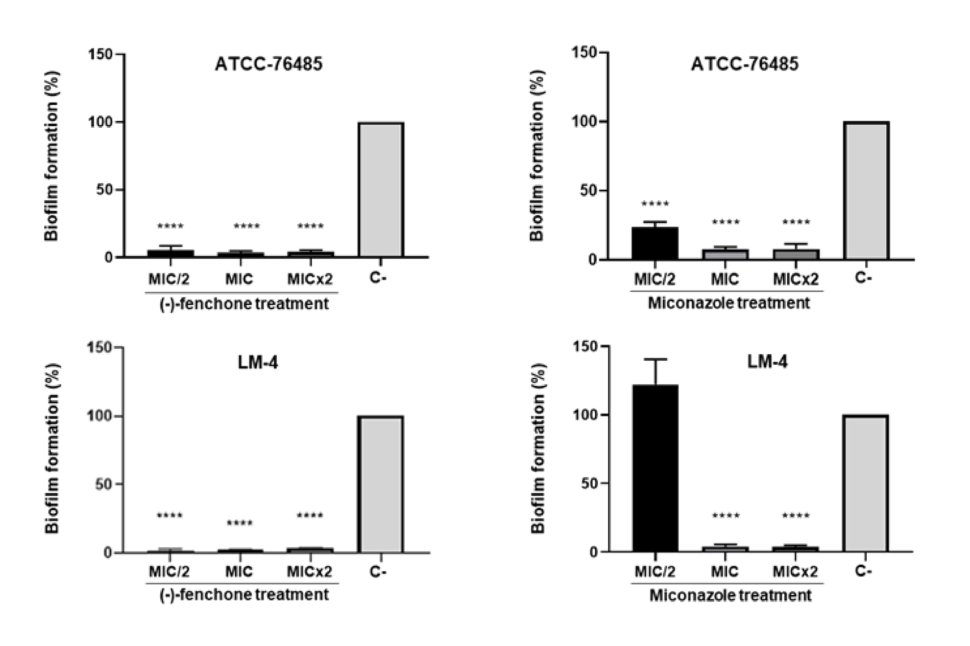
Figure 2 represents the percentages of biofilm formation by strains of C. albicans (ATCC-76485 and LM-4) in the presence and absence of (-)-fenchone and miconazole. A strong inhibitory effect was evidenced for (-)-fenchone, which reduced biofilm growth by over 80% in both strains compared to negative control (p<0.0001). In the presence of miconazole, strains ATCC-76485 and LM-4 exhibited inhibition of biofilm formation at low concentrations (MIC/2 and MIC), respectively. After exposure to miconazole at MIC/2, strain LM-4 showed higher biofilm growth than negative control, but without a statistically significant difference (p>0.05).

Impaired biofilm formation by C. albicans after treatment with (-)-fenchone and miconazole. Bars represent the mean (± standard error) of the percentage of adhered cells relative to the negative control (C-). Fungal cells were exposed to (-)-fenchone or miconazole at different concentrations (MIC/2, MIC, and MIC×2). MIC, Minimum Inhibitory Concentration. ****p<0.0001 compared to the C- (One-way ANOVA, Tukey post hoc test, n=3).
Figure 3 shows the results of the preformed biofilm assay. As evidenced, (-)-fenchone had weak activity in disrupting preformed biofilm at MIC×5. In the groups treated with this substance, the percentage of preformed biofilm remained above 80% of that observed in the negative control. There were no statistically significant differences between the analyzed groups (p>0.05).

Disruption of preformed biofilm by C. albicans after exposure to (-)-fenchone and miconazole (Mic). Bars represent the mean (± standard error) of the percentage of adhered cells relative to the negative control (C-). Fungal cells were exposed to (-)-fenchone or miconazole at MIC×5. MIC, Minimum Inhibitory Concentration. *p<0.05 compared to the C-; **p<0.01 compared to the C- (One-way ANOVA, Tukey post hoc test, n=3).
Hemolytic activity
Cytotoxicity of (-)-fenchone was analyzed through its hemolytic potential for human erythrocytes (blood groups A, B, and O). Results of the percentage of hemolysis caused by this monoterpene, compared to a positive control (1% Triton X-100), are presented in figure 4. The results revealed that (-)-fenchone caused hemolysis in erythrocytes of all three blood types, but significantly less than in the positive control (p<0.05). The hemolytic activity of the product was considered low (<40%) up to the highest concentration evaluated (100 μg/mL) in all blood types.

Hemolytic activity of (-)-fenchone on human erythrocytes (blood types A, B, and O) compared to the positive control (C+, 1% Triton X-100). C-, negative control. Results are expressed as the mean (± standard error). *p<0.05; **p<0.001; ***p<0.0001 (One-way ANOVA, Dunnet post hoc test, n=3).
DISCUSSION
In recent times, the increasing global resistance to antifungals and the growing number of immunocompromised patients or those with bacterial, viral, or opportunistic fungal coinfections (Silva et al. 2021) have significantly driven the search for more effective and less toxic treatment modalities. In this context, natural products derived from plants, such as essential oils and their phytoconstituents, have been extensively investigated, resulting in the identification of many substances with promising antimicrobial activity (Sakkas & Papadopoulou 2017, Silva et al. 2021). One of these highlighted compounds is the monoterpene fenchone, which has demonstrated efficacy even against C. albicans. However, its potential for treating oral candidiasis remains underexplored.
To address this gap, we initially conducted an in silico analysis to evaluate the likelihood of the (-)-fenchone enantiomer exhibiting antifungal activity, as well as its pharmacokinetic and toxicity characteristics, in order to determine the feasibility of investing in this compound. Overall, the results were considered promising, as (-)-fenchone showed a higher probability of antifungal activity compared to inactivity in the online PASS, and an acceptable ADMET profile to be considered a medicament. Furthermore, AmetSAR indicated absence of carcinogenicity and low toxicity for relevant tests, such as HERG and AMES test. Regarding the probable toxicity to zebrafish, it is worth noting that, despite having some genetic similarities with humans, they are different organisms, so the results obtained with this model may not accurately reflect the effects in humans. With regard to possible toxicity to honey bees, crucial for pollination and food production, control measures should be implemented if high toxicity is confirmed, aiming to protect these populations and the environment.
Corroborating our findings, Rehman et al. (2022) reported that fenchone presented an acceptable ADMET profile to be considered a medicine and passed drug-likeness tests according to Pfizer, Veber, and Igan. Additionally, the substance demonstrated 98% intestinal absorption and 63% blood-brain barrier permeability, without evidence of AMES and hepatotoxicity, predicting only skin sensitization.
Since the results of the in silico analysis were promising, we proceeded with biological activity and toxicity assays. Regarding antifungal activity against oral strains of C. albicans, (-)-fenchone demonstrated strong activity. In the literature, several monoterpenes have also demonstrated activity against C. albicans, such as linalool (Dias et al. 2018), camphor (Ivanov et al. 2021), eucalyptol (Gupta et al. 2021, Ivanov et al. 2021), eugenol, and geraniol (Khan et al. 2012).
In a study conducted by Raut et al. (2013), several terpenes presented promising MIC values for the C. albicans ATCC-90028 strain, with thymol, carvacrol, and eugenol standing out, with MICs of 0.25 mg/mL. Da Silva et al. (2012) evaluated the antimicrobial effects of two monoterpenes, α-Pinene and β-Pinene, against C. albicans, and found MICs of 3,125 μg/mL for (+)-α-pinene, and 187 μg/mL for (+)-β-pinene.
Moreover, several essential oils with a high concentration of fenchone have demonstrated good antifungal activity, including against the species C. albicans. Examples include the oils from F. vulgare (Roby et al. 2013, Bassyouni et al. 2019), Plectranthus glandulosus (Ngo-Mback et al. 2019), and Lavandula species (Benali et al. 2023, El Hachlafi et al. 2023, Santos et al. 2024). At this point, it is important to highlight that the antifungal activity of essential oils containing fenchone as a phytoconstituent may be superior to that of the isolated monoterpene. This occurs when other phytoconstituents with similar activity are present, resulting in a synergistic effect and consequently enhancing the antifungal activity of the essential oil (Waller et al. 2017).
For Ngo-Mback et al. (2019), fenchone represents one of the main terpenes responsible for the anticandida activity of essential oils. Zuzarte et al. (2009) highlighted fenchone (MIC = 5 μL/mL) as having superior activity than eucalyptol (MIC = 10 μL/mL) and camphor (MIC> 20μL/mL). In fact, this compound, as well as its isolated enantiomers, have demonstrated good antimicrobial potential (Garzoli et al. 2018). Pessoa et al. (2020), for example, observed a strong inhibitory effect of the (-)-fenchone enantiomer against intestinal of C. albicans strains (MIC = 32-64 μg/mL), as well as C. tropicalis (MIC =32 μg/mL) and C. krusei (MIC = 64 μg/mL). Similarly, Ahmad et al. (2022) evaluated the antifungal activity of the (+)-fenchone enantiomer against C. albicans, and found a significant antimicrobial effect, although at concentrations higher (MIC = 41.6±14.4 mg/mL and MFC = 83.3±28.7 mg /mL) than those found in our study. This difference may indicate that indeed the (-)-fenchone isomer has greater biological activity.
It is important to note that that differences in the exact MIC values between studies can be explained by a series of factors, such as the variable purity of substances, the type of culture medium used, the pH of the medium, the time and temperature of incubation, the use of fungal suspensions of different densities and/or other strains with different sensitivities to chemical substances (Karpiński et al. 2021).
A relevant finding of our study was the sensitivity of miconazole-resistant C. albicans strains (LM-4 and LM-12B) to low concentrations of (-)-fenchone (MIC = 4-8 μg/mL). Therefore, it is plausible that this compound represents an effective therapeutic option for cases of oral candidiasis miconazole-resistant, which justifies the need for future studies to investigate its antifungal potential in vivo and in humans.
Regarding predominantly fungicidal activity, other essential oils with high fenchone concentration have demonstrated such activity against C. albicans at low concentrations, such as F. vulgare with MIC and MFC rates of 170 μg/mL and 203 μg/mL, respectively (Gavanji et al. 2015), and L. stoechas, with MIC and MFC of 0.125% (v/v) (El Hachlafi et al. 2023). In the study by Miranda-Cadena et al. (2021), the monoterpenes carvacrol, cinnamaldehyde, and thymol demonstrated fungicidal activity against oral isolates of Candida spp. Cinnamaldehyde was the most effective agent (MIC = 61.5 mg/L and MFC = 99.0 mg/L, followed by thymol (93.2 mg/L and MFC = 241.2 mg/L) and carvacrol (MIC = 105 mg/L and MFC = 251.0 mg/L). It is important to highlight that predominantly fungicidal activity has the advantage of preventing the perpetuation of mutations and, consequently, the development of resistance phenomena (Kumar et al. 2018, Bhattacharya et al. 2020). On the other hand, it can cause irreversible depletion of specific members of the microbiota. In this context, antimicrobials that inhibit the growth of microorganisms (fungistatics) have the advantage of impacting the microbiota less, as they tend to uniformly inhibit community members, which can regrow once the treatment is discontinued (Maier et al. 2021).
Through analysis with time-kill curves, we observed that exposure to (-)-fenchone resulted in a rapid reduction in the cell population of both strains during the first 60 minutes. This finding corroborates previous discoveries by Da Silva et al. (2012) regarding α-pinene and β-pinene against C. albicans. Furthermore, the fungicidal effect of (-)-fenchone was concentration-dependent and greater in the first 8 hours of the assay. This finding is clinically significant as it may aid in defining dosing intervals in future clinical trials.
Concerning the trend of regrowth at the last time analyzed, it is important to note that this phenomenon is commonly observed in cultures exposed to biocidal substances, as documented in previous studies (López-Rojas et al. 2017, Ranieri et al. 2018), and reported since decades (Hamano et al. 1984). The hypothesis for its occurrence has been the presence of cells that remained viable and, possibly due to drug consumption or degradation in the last time points analyzed, proliferated. This resulted in the formation of a new cell population in quantity close to the negative control. In the context of our study, considering that the tested substance may undergo volatilization, it is plausible that its low concentration in the medium between 8h and 24h contributed to the emergence of the cell population in the last time analyzed.
Regarding the mechanism of action, we observed that (-)-fenchone probably does not act by damaging the cell wall or by directly binding to ergosterol in the membrane. In the literature, several studies propose that monoterpenes act through various mechanisms to induce fungal cell death, one of the main ones being the increase in permeability and rupture of cell membranes, triggered after binding to the phospholipids of the lipid bilayer (Ngo-Mback et al. 2019, Angane et al. 2022, El-Abdali et al. 2022, El Hachlafi et al. 2023). Additionally, monoterpenes may also act through fungistatic mechanisms, such as inhibition of hyphae formation or ergosterol biosynthesis, through, for example, gene inactivation (Ivanov et al. 2021). It is important to emphasize that ergosterol, like the cell wall, represents an ideal target for antifungal agents, as it is not present in human cells, ensuring greater selectivity for the substance (Bhattacharya et al. 2020).
It is necessary to note that miconazole currently represents the topical agent that allows for the treatment of oral candidiasis with greater patient comfort. It generally presents as an adverse effect of local irritation, manifested as itching or burning, in less than 5% of patients (Quindós et al. 2019). Despite being widely used in the treatment of this disease, few studies have sought to improve its efficacy and performance in the face of growing fungal resistance, which is the reason for its choice as a control in this study. Therefore, we performed the checkerboard assay of (-)-fenchone with miconazole and found synergy, including for the LM-4 strain, resistant to miconazole. This is another finding of great relevance from our study, as it suggests that this monoterpene may have overcome the resistance mechanism of the LM-4 strain.
In line with this, it has been proposed in the literature that essential oils and their monoterpenes facilitate the penetration of azole agents into the fungal cell by altering the permeability of their walls and membranes (Bassyouni et al. 2019). This alteration in permeability can result in a reduction in the concentration of drug needed to achieve antifungal activity, as well as convert its fungistatic activity into fungicidal. Moreover, these products can interfere with the functioning of efflux pumps, which are a recognized mechanism of microbial resistance. Therefore, it is possible to suggest that (-)-fenchone inhibited efflux pumps that would be actively transporting miconazole to the external environment (Bhattacharya et al. 2020, Mijatovic et al. 2022).
Reports of synergy between isolated monoterpenes and standard drugs are found in the literature. For example, geraniol and eugenol demonstrated synergism with fluconazole against C. albicans (Khan et al. 2012). However, in the study conducted by Pessoa et al. (2020), the combination of (-)-fenchone with amphotericin B against C. albicans resulted in indifference, in contrast to the synergism observed between (-)-fenchone and miconazole in the present study. Similarly, Da Silva et al. (2012) reported only indifference of (+)-α-pinene and (+)-β-pinene, two monoterpenes, with amphotericin B against C. albicans and other fungal species. According to Mijatovic et al. (2022), synergy can occur when two substances act through different mechanisms of action, which enhances their effects by not competing for the same binding site. Therefore, the synergy observed between (-)-fenchone and miconazole, but not with amphotericin B in literature studies, which acts on the plasma membrane, strengthens the hypothesis that the cell membrane is one of the targets of (-)-fenchone‘s attack on the fungal cell.
Considering that the treatment of oral candidiasis usually involves direct topical application to the lesions and that the effective concentrations obtained in in vitro tests were relatively low, the results of the present study highlight (-)-fenchone as a promising antifungal agent for the therapeutic treatment of this disease, including its forms resistant to miconazole.
The ability to form biofilms has been one of the explanations for the greater involvement of the species C. albicans in the development of oral candidiasis (Miranda-Cadena et al. 2021). Once organized into biofilms, fungal cells become more resistant to environmental adversities and antifungal drugs. Therefore, a product that prevents or reduces the formation of this complex structure may represent an important strategy to prevent infections by this fungus (Ranieri et al. 2018, Lohse et al. 2020, Karpiński et al. 2021). In this context, we observed that (-)-fenchone significantly inhibited biofilm formation from the subinhibitory concentration (MIC/2). This result indicates that (-)-fenchone may represent an effective inhibitor of Candida biofilm formation, reinforcing its potential to prevent and/or treat oral candidiasis.
In the literature, several monoterpenes have been identified as effective inhibitors of biofilm formation by Candida spp. In the review by Karpiński et al. (2021), out of 69 compounds obtained from plants, monoterpenes were considered the main ones with activity against Candida spp. biofilms. Among these, the most active in inhibiting biofilm formation were thymol (3.12 mg/L) and eugenol (12.5 mg/L). Supporting this report, in the study by Raut et al. (2013) several terpenes inhibited the formation of biofilms of the C. albicans ATCC-90028 strain, including at concentrations below the inhibitory level for planktonic cells, such as nerol at MIC/4, farnesol at MIC/2, and carvone/carvol at MIC/16, results that corroborate the subMIC inhibition observed for (-)-fenchone in the present study. Against oral isolates of Candida spp., Miranda-Cadena et al. (2021) reported that carvacrol, cinnamaldehyde, and thymol inhibited the biofilm adhesion phase at 104 mg/L, 97 mg/L and, 157.6 mg/L, respectively.
Additionally, Manoharan et al. (2017) observed that camphor and fenchone at 0.005% significantly inhibited over 80% of biofilm formation by C. albicans. In the study by Da Silva et al. (2012), cited previously, biofilm formation by oral C. albicans was observed at the MIC for (+)-α-pinene (3,125 μg/mL) and at MIC×2 for (+)-β-pinene (374 μg/mL). Finally, in the study by Ahmad et al. (2022), (+)-fenchone inhibited C. albicans biofilm formation from 0.25 mg/mL. As this concentration was higher than that observed for (-)-fenchone in the present study, this finding may indicate that indeed this enantiomer exhibits greater activity than (+)-fenchone.
It is important to note that inhibition of Candida biofilms by terpenes can occur through various mechanisms, such as reducing hyphal formation, which is considered a virulence factor due to its invasive capacity (Manoharan et al. 2017, Ivanov et al. .2021). The conversion of yeast cells into filamentous forms is an important step in the initial phase of biofilm formation, as well as in the pathogenesis of fungal infections and drug resistance, which is where many antibiofilm substances act (Manoharan et al. 2017, Müller-Sepúlveda et al. 2020). In line with this, in the study by Raut et al. (2013), 17 terpenes inhibited hyphal morphogenesis at low concentrations, with emphasis on thymol, carvacrol, and eugenol showing this activity at 31 μg/mL. For these authors, the inhibition of the yeast-to-hyphal transformation in C. albicans represents an important research strategy for the development of biofilm formation inhibitors.
Regarding the disruption of the preformed biofilm, on the other hand, we observed that (-)-fenchone did not demonstrate significant activity at MIC×5. A possible explanation for this result lies in the fact that, as mentioned previously, fungal cells in biofilms develop greater resistance to environmental adversities and antimicrobial substances (Raut et al. 2013, Karpiński et al. 2021). In this aspect, biofilms can both inhibit the activity of antifungal drugs and increase tolerance or resistance to them, resulting in additional clinical challenges in the treatment of fungal infections (Ranieri et al. 2018, Karpiński et al. 2021). Moreover, some authors have used concentrations of 10 to 100 times the MIC to evaluate activity to disrupt preformed biofilms (Sardi et al. 2017). However, in the present study, we chose not to excessively increase the MIC for a safety margin, considering that the concentrations observed in vitro will be further elevated when extrapolated to in vivo and clinical trials, which would increase the risk of toxic effects.
Despite this, studies with other monoterpenes have reported activity against preformed biofilms at concentrations close to inhibitory. In the study by Raut et al. (2013), for example, 10 out of 28 terpenes evaluated demonstrated activity against mature biofilms of C. albicans, including carvacrol, eugenol and thymol, at MIC×4 for most of them, which contrasts with the results observed for (-)-fenchone in our study. Furthermore, eucalyptol, another monoterpene, demonstrated activity against mature and developing biofilms of C. albicans and C. glabrata in the study by Gupta et al. (2021). These authors described different antibiofilm action mechanisms for C. albicans, including biochemical changes and enzymatic activity in the extracellular matrix of the biofilm, generation of reactive oxygen species, disruption of the cell cycle in the G1/S phase, alterations in the gene expression of selected ABC transporters, and hydrolytic and cell wall biosynthesis enzymes.
This divergence of results can be attributed to the use of different substances, as well as different standard and clinical strains in the studies. However, although we did not find a significant effect of (-)-fenchone against preformed biofilms of C. albicans at MIC×5, it is possible that at higher concentrations significant activity could be found. In fact, Miranda-Cadena et al. (2021) observed that carvacrol and thymol eliminated mature biofilms of oral Candida spp. at concentrations of 812.7 mg/L and 1106 mg/L, respectively. These values are considerably higher than the inhibitory concentrations observed in that same study. Therefore, we suggest that further research be conducted in this direction, as, given the promising antifungal and antibiofilm activities, (-)-fenchone has great potential to contribute to the development of new effective and safe therapeutic strategies for miconazole-resistant oral candidiasis.
Regarding the assessment of toxicity through the hemolysis assay, it was found that (-)-fenchone demonstrated low hemolytic activity even at a concentration greater than 10 times the MIC for C. albicans. This result is of great relevance and reinforces the potential of this substance as a future effective and safe drug for the treatment of oral candidiasis. In the literature, there are few studies that have investigated the toxicity of fenchone, with some existing divergences. In the study by Abdelgaleil (2010), (-)-fenchone was considered toxic to adult Theba Pisana and larvae of Spodoptera littoralis. However, it is important to note that, since these are organisms distant from the human species, these results do not necessarily indicate toxicity to humans, with assays using mammalian cells being more appropriate for this purpose. On the other hand, in the study by Bashir et al. (2023), an evaluation of the safety of fenchone was conducted through toxicity assays with rats, and the authors concluded that this compound was safe, thus corroborating the results of our study. Along the same lines, Rolim et al. (2017) observed that a very high concentration of (+)-Fenchone (3,000 μg/mL) was necessary to induce 50% hemolysis, suggesting low toxicity for this enantiomer. From a toxicological standpoint, the same authors found that (+)-Fenchone induced a decrease in AST (aspartate aminotransferase) and ALT (alanine aminotransferase) enzymes, suggesting possible liver injury, but they highlighted that additional studies of liver function and histology would be necessary to elucidate this effect. These discrepancies in the literature may be attributed to methodological variations in the studies, including the toxicity model or the enantiomer used.
In conclusion, this study represents the first report of the relevant antifungal and antibiofilm activities of the monoterpene (-)-fenchone. The results obtained highlight the potential of this natural compound as a promising antifungal agent in the treatment of oral candidiasis, including its miconazole-resistant forms. Therefore, we encourage further investigations aimed at elucidating its mechanisms of action, toxicity, in vivo efficacy, and finally through randomized clinical trials.
ACKNOWLEDGMENTS
This research was supported by the Conselho Nacional de Desenvolvimento Científico e Tecnológico (CNPq), Brazil. The authors thank the operational support provided by the Federal University of Paraíba (UFPB) and the Federal University of Campina Grande (UFCG).
REFERENCES
- ABDELGALEIL SAM. 2010. Molluscicidal and insecticidal potential of monoterpenes on the white garden snail, Theba pisana (Muller) and the cotton leafworm, Spodoptera littoralis (Boisduval). Appl Entomol Zool 45: 425-433.
- AHMAD W ET AL. 2022. Antibacterial, Anticandidal, and Antibiofilm Potential of Fenchone: In Vitro, Molecular Docking and In Silico/ADMET Study. Plants 11: 2395.
- ANGANE M, SWIFT S, HUANG K, BUTTS CA & QUEK SY. 2022. Essential Oils and Their Major Components: An Updated Review on Antimicrobial Activities, Mechanism of Action and Their Potential Application in the Food Industry. Foods 11: 464.
- BALOUIRI M, SADIKI M & IBNSOUDA SK. 2016. Methods for in vitro evaluating antimicrobial activity: A review. J Pharm Anal 6: 71-79.
- BASHIR A, MUSHTAQ MN, YOUNIS W & ANJUM I. 2023. Fenchone, a monoterpene: Toxicity and diuretic profiling in rats. Front Pharmacol 26: 1119360.
- BASSYOUNI RH, WALI IE, KAMEL Z & KASSIM MF. 2019. Fennel oil: A promising antifungal agent against biofilm forming fluconazole-resistant Candida albicans causing vulvovaginal candidiasis. J Herb Med 15: 100227.
- BENALI T ET AL. 2023. Chemical Profiling and Biological Properties of Essential Oils of Lavandula stoechas L. Collected from Three Moroccan Sites: In Vitro and In Silico Investigations. Plants 12: 1413.
- BHATTACHARYA S, SAE-TIA S & FRIES BC. 2020. Candidiasis and Mechanisms of Antifungal Resistance. Antibiotics 9: 312.
- CLEELAND R & SQUIRES E. 1991. Evaluation of new antimicrobials in vitro and in experimental animal infections. Antibiotics in Laboratory Medicine 3: p. 739-787.
- CLSI - CLINICAL AND LABORATORY STANDARDS INSTITUTE. 2008. Reference method for broth dilution antifungal susceptibility testing of yeasts. Approved standard M27-A3, Pennsylvania, United States of America: Clinical and Laboratory Standards Institute.
- DA SILVA AC, LOPES PM, BARROS DE AZEVEDO MM, COSTA DC, ALVIANO CS & ALVIANO DS. 2012. Biological activities of α-pinene and β-pinene enantiomers. Molecules 17: 6305-6316.
- DIAS IJ, TRAJANO ERIS, CASTRO RD, FERREIRA GLS, MEDEIROS HCM & GOMES DQC. 2018. Antifungal activity of linalool in cases of Candida spp. isolated from individuals with oral candidiasis. Braz J Biol 78: 368-374.
- EL-ABDALI Y ET AL. 2022. Lavandula dentata L.: Phytochemical Analysis, Antioxidant, Antifungal and Insecticidal Activities of Its Essential Oil. Plants 11: 311.
- EL HACHLAFI N ET AL. 2023. Phytochemical analysis and evaluation of antimicrobial, antioxidant, and antidiabetic activities of essential oils from Moroccan medicinal plants: Mentha suaveolens, Lavandula stoechas, and Ammi visnaga. Biomed Pharmacother 164: 114937.
- ESCALANTE A, GATTUSO M, PÉREZ P & ZACCHINO S. 2008. Evidence for the mechanism of action of the antifungal phytolaccoside B isolated from Phytolacca tetramera Hauman. J Nat Prod 71:1720-1725.
- FANG J, HUANG B & DING Z. 2021. Efficacy of antifungal drugs in the treatment of oral candidiasis: A Bayesian network meta-analysis. J Prosthet Dent 125: 257-265.
- FIGUEIREDO-JÚNIOR EC, CAVALCANTI YW, LIRA AB, PESSOA HLF, LOPES WS, DA SILVA DR, FREIRES IA, ROSALEN PL, COSTA EMMB & PEREIRA JV. 2021. Phytochemical composition, antifungal activity, in vitro and in vivo toxicity of Syzygium cumini (L.) Skeels leaves extract. B Latinoam Caribe PL 20: 536-555.
- FROST DJ, BRANDT KD, CUGIER D & GOLDMAN R. 1995. A whole-cell Candida albicans assay for the detection of inhibitors towards fungal cell wall synthesis and assembly. J Antibiot 48: 306-310.
- GARZOLI S, BOŽOVIĆ M, BALDISSEROTTO A, SABATINO M, CESA S, PEPI F, VICENTINI CB, MANFREDINI S & RAGNO R. 2018. Essential oil extraction, chemical analysis and anti-Candida activity of Foeniculum vulgare Miller - new approaches. Nat Prod Res 32: 1254-1259.
- GAVANJI S, ZAKER SR, NEJAD ZG, BAKHTARI A, BIDABADI ES & LARKI B. 2015. Comparative efficacy of herbal essences with amphotricin B and ketoconazole on Candida albicans in the in vitro condition. Integr Med Res 4: 112-118.
- GUPTA P, PRUTHI V, KM & POLURI KM. 2021. Mechanistic insights into Candida biofilm eradication potential of eucalyptol. J Appl Microbiol 131: 105-123.
- HADACEK F & GREGER H. 2000. Testing of Antifungal Natural Products: Methodologies, Comparability of Results and Assay Choice. Phytochem Anal 11: 137-147.
- HAFIDH RR, ABDULAMIR AS, VERN LS, ABU BAKAR F, ABAS F, JAHANSHIRI F & SEKAWI Z. 2011. Inhibition of Growth of Highly Resistant Bacterial and Fungal Pathogens by a Natural Product. Open Microbiol J 5: 96-106.
- HAMANO S, TSUJI A, ASANO T, TAMAI I, NAKASHIMA E, YAMANA T & MITSUHASHI S. 1984. Kinetic analysis and characterization of the bacterial regrowth after treatment of Escherichia coli with beta-lactam antibiotics. J Pharm Sci 73: 1422-1427.
- IVANOV M, KANNAN A, STOJKOVIĆ DS, GLAMOČLIJA J, CALHELHA RC, FERREIRA ICFR, SANGLARD D & SOKOVIĆ M. 2021. Camphor and Eucalyptol-Anticandidal Spectrum, Antivirulence Effect, Efflux Pumps Interference and Cytotoxicity. Int J Mol Sci 22: 483.
- IYER KR, ROBBINS N & COWEN LE. 2022. The role of Candida albicans stress response pathways in antifungal tolerance and resistance. iScience 25: 103953.
- KARPIŃSKI TM, OŻAROWSKI M, SEREMAK-MROZIKIEWICZ A, WOLSKI H & ADAMCZAK A. 2021. Plant Preparations and Compounds with Activities against Biofilms Formed by Candida spp. J Fungi 7: 360.
- KHAN MS, MALIK A & AHMAD I. 2012. Anti-candidal activity of essential oils alone and in combination with amphotericin B or fluconazole against multi-drug resistant isolates of Candida albicans. Med Mycol 50: 33-42.
- KUMAR A, ZARYCHANSKI R, PISIPATI A, KUMAR A, KETHIREDDY S & BOW EJ. 2018. Fungicidal versus fungistatic therapy of invasive Candida infection in non-neutropenic adults: a meta-analysis. Mycology 9: 116-128.
- KWASNY SM & OPPERMAN TJ. 2010. Static biofilm cultures of Gram-positive pathogens grown in a microtiter format used for anti-biofilm drug discovery. Curr Protoc Pharmacol 13: 13A.8.
- LEWIS RE, DIEKEMA DJ, MESSER SA, PFALLER MA & KLEPSER ME. 2002. Comparison of Etest, chequerboard dilution and time-kill studies for the detection of synergy or antagonism between antifungal agents tested against Candida species. J Antimicrob Chemother 49: 345-351.
- LOHSE MB, GULATI M, CRAIK CS, JOHNSON AD & NOBILE CJ. 2020. Combination of Antifungal Drugs and Protease Inhibitors Prevent Candida albicans Biofilm Formation and Disrupt Mature Biofilms. Front Microbiol 11: 1027.
- LÓPEZ-ROJAS R, FERNÁNDEZ-CUENCA F, SERRANO-ROCHA L & PASCUAL Á. 2017. In vitro activity of a polyhexanide-betaine solution against high-risk clones of multidrug-resistant nosocomial pathogens. Enferm Infecc Microbiol Clin 35: 12-19.
- MAIER L ET AL. 2021. Unravelling the collateral damage of antibiotics on gut bacteria. Nature 599: 120-124.
- MANOHARAN RK, LEE JH & LEE J. 2017. Antibiofilm and Antihyphal Activities of Cedar Leaf Essential Oil, Camphor, and Fenchone Derivatives against Candida albicans. Front Microbiol 8: 1476.
- MILLSOP JW & FAZEL N. 2016. Oral candidiasis. Clin Dermatol 34: 487-494.
- MIJATOVIC S, STANKOVIC JA, CALOVSKI IC, DUBLJANIN E, PLJEVLJAKUSIC D, BIGOVIC D & DZAMIC A. 2022. Antifungal Activity of Lavandula angustifolia Essential Oil against Candida albicans: Time-Kill Study on Pediatric Sputum Isolates. Molecules 27: 6300.
- MIRANDA-CADENA K, MARCOS-ARIAS C, MATEO E, AGUIRRE-URIZAR JM, QUINDÓS G & ERASO E. 2021. In vitro activities of carvacrol, cinnamaldehyde and thymol against Candida biofilms. Biomed Pharmacother 143: 112218.
- MÜLLER-SEPÚLVEDA A ET AL. 2020. Chemical Characterization of Lavandula dentata Essential Oil Cultivated in Chile and Its Antibiofilm Effect against Candida albicans. Planta Med 86: 1225-1234.
- NAWROT U, NOWICKA J, JUSZCZAK K & GUSIN B. 2005. Susceptibility to antifungal agents of Candida species isolated from paediatric and adult patients with haematological diseases. Mycoses 48: 385-390.
- NCUBE N, AFOLAYAN A & OKOH A. 2008. Assessment techniques of antimicrobial properties of natural compounds of plant origin: current methods and future trends. Afr J Biotechnol 7: 1797-1806.
- NGO-MBACK MNL, FAMEWO EB, MUBARAKALI D, EKE P, THAJUDDIN N, AFOLAYAN AJ, JAZET DONGMO PM & FEKAM BOYOM F. 2019. An investigation of chemical composition and antimicrobial activity of essential oils extracted from Aeollanthus and Plectranthus species. Biocatal Agric Biotechnol 22: 101412.
- ONSARE JG & ARORA DS. 2015. Antibiofilm potential of flavonoids extracted from Moringa oleifera seed coat against Staphylococcus aureus, Pseudomonas aeruginosa and Candida albicans. J Appl Microbiol 118: 313-325.
- PESSOA MLS, SILVA LMO, ARARUNA MEC, SERAFIM CAL, JÚNIOR EBA, SILVA AO, PESSOA MMB, NETO HD, LIMA EO & BATISTA LM. 2020. Antifungal activity and antidiarrheal activity via antimotility mechanisms of (-)-fenchone in experimental models. World J Gastroenterol 26: 6795-6809.
- QUINDÓS G, GIL-ALONSO S, MARCOS-ARIAS C, SEVILLANO E, MATEO E, JAUREGIZAR N & ERASO E. 2019. Therapeutic tools for oral candidiasis: Current and new antifungal drugs. Med Oral Patol Oral Cir Bucal 24: e172-e180.
- RAJASEKHARAN SK, RAMESH S, SATISH AS & LEE J. 2017. Antibiofilm and Anti-β-Lactamase Activities of Burdock Root Extract and Chlorogenic Acid against Klebsiella pneumoniae. J Microbiol Biotechnol 27: 542-551.
- RANGEL M, MALPEZZI EL, SUSINI SM & DE FREITAS JC. 1997. Hemolytic activity in extracts of the diatom Nitzschia. Toxicon 35: 305-309.
- RANIERI MR, WHITCHURCH CB & BURROWS LL. 2018. Mechanisms of biofilm stimulation by subinhibitory concentrations of antimicrobials. Curr Opin Microbiol 45: 164-169.
- RAUT JS, SHINDE RB, CHAUHAN NM & KARUPPAYIL SM. 2013. Terpenoids of plant origin inhibit morphogenesis, adhesion, and biofilm formation by Candida albicans. Biofouling 29: 87-96.
- REHMAN NU, ANSARI MN, SAMAD A & AHMAD W. 2022. In Silico and Ex Vivo Studies on the Spasmolytic Activities of Fenchone Using Isolated Guinea Pig Trachea. Molecules 27: 1360.
- REZA ASMA ET AL. 2021. Antiproliferative and antioxidant potentials of bioactive edible vegetable fraction of Achyranthes ferruginea Roxb. in cancer cell line. Food Sci Nutr 9: 3777-3805.
- ROBY MHH, SARHAN MA, SELIM KAH & KHALEL KI. 2013. Antioxidant and antimicrobial activities of essential oil and extracts of fennel (Foeniculum vulgare L.) and chamomile (Matricaria chamomilla L.). Ind Crops Prod 44: 437-445.
- ROLIM TL ET AL. 2017. Toxicity and antitumor potential of Mesosphaerum sidifolium (Lamiaceae) oil and fenchone, its major component. BMC Complement Altern Med 17: 347.
- ROMAN M, ROMAN DL, OSTAFE V, CIORSAC A & ISVORAN A. 2018. Computational Assessment of Pharmacokinetics and Biological Effects of Some Anabolic and Androgen Steroids. Pharm Res 35: 41.
- SAKKAS H & PAPADOPOULOU C. 2017. Antimicrobial Activity of Basil, Oregano, and Thyme Essential Oils. J Microbiol Biotechnol 27: 429-438.
- SANTOS AA ET AL. 2024. Lavandula dentata L. essential oil: a promising antifungal and antibiofilm agent against oral Candida albicans. Braz J Biol 84: e283646.
- SARDI JCO, POLAQUINI CR, FREIRES IA, GALVÃO LCC, LAZARINI JG, TORREZAN GS, REGASINI LO & ROSALEN PL. 2017. Antibacterial activity of diacetylcurcumin against Staphylococcus aureus results in decreased biofilm and cellular adhesion. J Med Microbiol 66: 816-824.
- SHUI Y, LI J, LYU X & WANG Y. 2021. Phytotherapy in the management of denture stomatitis: A systematic review and meta-analysis of randomized controlled trials. Phytother Res 35: 4111-4126.
- SILVA D ET AL. 2020. (R)-(+)-β-Citronellol and (S)-(-)-β-Citronellol in Combination with Amphotericin B against Candida Spp. Int J Mol Sci 21: 1785.
- SILVA RAD, ANTONIETI FMPM, RÖDER DVDB & PEDROSO RDS. 2021. Essential Oils of Melaleuca, Citrus, Cupressus, and Litsea for the Management of Infections Caused by Candida Species: A Systematic Review. Pharmaceutics 13: 1700.
- SLAVCHEV I, DOBRIKOV GM, VALCHEVA V, UGRINOVA I, PASHEVA E & DIMITROV V. 2014. Antimycobacterial activity generated by the amide coupling of (-)-fenchone derived aminoalcohol with cinnamic acids and analogues. Bioorg Med Chem Lett 24: 5030-5033.
- VALGUS JM. 2003. What’s new in antifungals? Curr Infect Dis Rep 5: 16-21.
- WALLER SB, CLEFF MB, SERRA EF, SILVA AL, GOMES AD, DE MELLO JR, DE FARIA RO & MEIRELES MC. 2017. Plants from Lamiaceae family as source of antifungal molecules in humane and veterinary medicine. Microb Pathog 104: 232-237.
- WHITE RL, BURGESS DS, MANDURU M & BOSSO JA. 1996. Comparison of three different in vitro methods of detecting synergy: time-kill, checkerboard, and E test. Antimicrob Agents Chemother 40: 1914-1918.
- XIAO Y, YUAN P, SUN Y, XU Y, DENG X, WANG X, LIU R, CHEN Q & JIANG L. 2022. Comparison of topical antifungal agents for oral candidiasis treatment: a systematic review and meta-analysis. Oral Surg Oral Med Oral Pathol Oral Radiol 133: 282-291.
- ZUZARTE M, GONÇALVES MJ, CAVALEIRO C, DINIS AM, CANHOTO JM & SALGUEIRO LR. 2009. Chemical composition and antifungal activity of the essential oils of Lavandula pedunculata (Miller) Cav. Chem Biodivers 6: 1283-1292.