Polyphenolic contents, antioxidant, and antimicrobial activities of Saccocalyx satureioides Coss. & Dur. essential oil and methanol extracts
Abstract
The whole plant Saccocalyx satureioides, an endemic medicinal plant in Algeria, was evaluated for its polyphenolic contents, antioxidant and antimicrobial activities. The polyphenolic contents of the plant methanolic extracts ranged from 170.47 to 285.56 (µg GAEs/mg extract) and from 25.39 to 82.68 (µg QEs/mg extract), respectively. High antioxidant activity was registered: DPPH average IC50 = 14.68 µg/mL, and β-carotene average IC50 = 21.08 µg/mL. The essential oil and methanolic extracts from the aerial parts and roots were tested against bacteria and molds. Different methods, including disk diffusion, well diffusion, and microdilution tests for bacteria, were used. The essential oil (February and June harvest periods) value 1.25 µL/mL was the MIC and the MBC for all bacterial strains, except E. coli, K. pneumonia and Ps. aeruginosa. The average fungal inhibition of the methanol extracts ranged from 5.10 to 71.25%, except for F. oxysporum. The essential oil effect was 6.67-83.53% and 17.01-90.57% by the food poisoned and fumigation methods, respectively. The preliminary phytochemical evaluation demonstrated the high polyphenolic contents and effective antioxidant power of the plant extracts. This may have an essential role in the antimicrobial effectiveness and indicate that the plant contains high-quality bioactive molecules in addition to the essential oil.
Key words
Saccocalyx satureioides; essential oil; methanol extract; antioxidant activity; antimicrobial activity
INTRODUCTION
Plants have long been known to treat human ailments. They are still an important source of drugs used in medicine, thanks to their beneficial biological activities (Muntean & Vulpie 2023). Bacteria, fungi, and viruses are known to cause diseases in humans, animals, and plants. Microbial food spoilage causes food waste and loss. Moreover, foodborne disease continuously poses a significant safety concern for public health (Gonelimali et al. 2018).
Various economically important plants are targets of Fusarium species. This pathogenic fungus is well known to generate a variety of mycotoxins (Ajmal et al. 2023). Aspergillus niger and A. ochraceus are more prevalent in stored foods. They were isolated from fruits, oilseeds, and cereals. They have been reported to produce ochratoixin A. Cladosporium cladosporioides is commonly present on plant materials and in the air. It causes decay in various fruits and vegetables. It also causes diseases in humans and animals (Hocking 2006, Ma et al. 2021)
The extensive use of antibiotics to save human health from microbial diseases has resulted in the emergence of drug and multidrug resistance in microbes (Srikacha & Ratananikom 2020). Multidrug resistance and virulence, as well as the potential toxicity of antimicrobial chemicals, enhanced the search for new therapeutic strategies, including the search for bioactive molecules such as polyphenols and essential oils (Veiko et al. 2023, Magri et al. 2023). Phenolic acids and flavonoids have been reported to serve as potent antioxidants, neutralizing free radicals and preventing oxidative stress, while also effectively inhibiting microbial growth (Cosmulescu et al. 2017, Oulahal & Degraeve 2022). Essential oils contain terpenes and terpenoids that are very good at killing microbes by breaking down their cell membranes. These compounds have been particularly effective against a broad range of pathogens, including bacteria and fungi (Masyita et al. 2022, Karnwal & Malik 2024).
The species Saccocalyx satureioides Coss. & Dur. (belonging to the Lamiaceae), known in Algeria as ‘Zaâter’ and ‘Azir-el-Bel’, is an endemic species that grows in the dunes of the Algerian pre-desert area. This plant has upright stems and grows to a height of 20 to 100 cm. Its tiny, ovoid, hispid, lanceolate leaves are ciliate, and it can have red, pink, or white flowers (Quezel & Santa 1963). It is traditionally used to treat hypertension, diabetes, kidney problems, and colon and stomach ailments (Allali et al. 2008, Benaradj & Boucherit 2022); it is also recommended to treat colds (Yabrir et al. 2018) and has Antileishmaia (Mostefa Sari et al. 2020), insecticidal (Sassoui et al. 2020) and corrosion inhibition properties (Benahmed et al. 2016). The leaves, flowers, fruit, and seeds of this plant are the parts usually used. But the use of the plant roots is not reported in traditional medicine.
This work aimed to evaluate the polyphenolic contents, antioxidant activity, and antibacterial and antifungal activities of the essential oil and, for the first time, the methanolic extracts of the aerial parts and roots of Saccocalyx satureioides from the center-east of Algeria.
MATERIALS AND METHODS
Plant material
The plant material of S. satureioides, from the El Houamed region (M’sila), was collected at two periods: the aerial parts (F) and the roots (R) were collected in February 2019, and the aerial parts (J) were collected in June 2020. The plant identification was done by the botanist, Dr. SARRI Djamel, and a voucher specimen (N°: SS2331QS28) was deposited in the herbarium of the Department of Nature and Life Sciences, Faculty of Sciences, M’sila university. After being dried, the leaves and roots were recovered and then stored until use.
Essential oil isolation and obtainment of the methanolic extracts
The plant aerial parts (100 g) were subjected to hydrodistillation for 3 h in a Clevenger-type apparatus. The oil yields, estimated on a dry weight basis (v/w), were 2.8% (EOJ) and 2.55% (EOF). After being dried over anhydrous sodium sulfate, the essential oil (EO) was stored at 4 °C until tested.
The plant methanolic extracts (ME) were obtained by Soxhlet: 50 g of the plant leaves (or roots) were extracted with 500 mL of methanol at 40 °C for 6 hours. Each extract was recovered after concentration by a rotary evaporator, then dried in an oven, weighted, and finally stored at 4° C until its use.
Total phenolic and flavonoid contents
The total phenolic content was determined according to Ruiz-Navajas et al. (2013): 300 µL the methanolic solution of the methanol extract (ME) or the EO was mixed with 1000 µL of the Folin-Cioclateu reagent (10 times diluted in distilled water) and 2000 µL of sodium carbonate (7.5%). After being incubated for 5 min at 50 °C, the absorbance, at 760 nm, was measured against a blank. The total polyphenols were determined as µg gallic acid equivalents (GAE) / mg based on a calibration curve.
The total flavonoid content was evaluated according to Hendel et al. (2016): 1 mL of 2% aluminum chloride (AlCl3) was mixed with 1 mL of the methanolic solution of the extract or the EO. After being incubated for 15 min at ambient temperature, the absorbance, at 415 nm, was measured against a blank. The total flavonoids were determined as µg quercetin equivalents (QE) / mg based on a calibration curve.
Antioxidant Activity Evaluation
DPPH test
Fifty microliters of the extract or EO (at different concentrations in methanol) were added to 2 mL of a 0.004% of DPPH methanolic solution. The mixture was incubated to react in the dark for 30 min. The control does not contain extract or EO. The BHT was used as standard. The absorbance of the samples was read at 517 nm (Tepe 2008). The percentage inhibition was calculated by the following formula:
where Ac is the absorbance of the control; As is the absorbance of the sample. The concentration of the sample providing 50% inhibition (IC50) was calculated from the graph representing the percentage of inhibition against the concentration of the extract or the oil.
β-carotene bleaching test
The ß-carotene/linoleic acid bleaching test was carried out as described by Shukla et al. (2012): an emulsion stock solution (a mixture of 0.5 mg of ß-carotene, 01 mL of chloroform, 25 µL of linoleic acid, 200 mg of Tween 40, and 100 mL of distilled water added after evaporating the chloroform) was prepared. Aliquots (2.5 mL) of this solution were transferred to test tubes each containing 350 µL of different concentrations, extracts or EO, diluted in methanol. The absorbance was measured immediately at 470 nm and the test emulsion was incubated in a water bath at 50 °C for 120 min where the absorbance was measured again. The same procedure was applied to BHT as a positive control, and to methanol instead of the extract as a negative control. The relative antioxidant activity (I%) was evaluated in terms of β-carotene bleaching according to the formula:
Where At and Ct are the absorbance values measured for the test sample and the control respectively, after 120 min of incubation, and C0 is the control absorbance value measured initially. Results were expressed as IC50 values (µg/mL); the concentration required to cause 50% inhibition of β-carotene bleaching.
Antimicrobial activity
Microorganisms
Reference bacterial strains were subjected to the effect of the extracts and the EO: Bacillus subtilis ATCC6633, Escherichia coli ATCC8739, Staphylococcus aureus ATCC6538, Pseudomonas aeruginosa ATCC9027, Proteus mirabilis ATCC 35659, Klebsiella Pneumoniae ATCC 700603.
The fungal isolates to be tested for the action of the extracts and the EO include Aspergillus ochraceus ATCC 3174, Aspergillus niger (isolate AnGr61), Fusarium culmorum (isolate Fc13), Fusarium oxysporum (isolate Fob36), and Cladosporium cladosporioides (isolate PeCc321), available at the microbiology laboratory of the department of Microbiology and Biochemistry, M’sila university.
Preparation of the inocula
-
Bacterial suspension: from 18- to 24-hour cultures, the bacterial suspension (prepared in a sterile saline solution) was well homogenized and adjusted to 0.5 of the McFarland scale opacity.
-
Spore suspension: from 10-day fungal cultures (on potato dextrose agar, PDA, 25 °C), 3 mL of sterile saline (0.05% tween 80 added) were added to each Petri plate. After the culture was scraped, the spores were recovered and adjusted to 104 spores per mL using a hemocytometer.
-
The filamentous inocula were prepared as 6 mm-disks taken from the margins of a 7-day-old culture on PDA medium.
Measurement of the inhibition
The fungal inhibition (I%) was evaluated according to the formula:
Where DC is the diameter of the control colony (mm), and DT is the diameter of the treated colony (mm).
Agar-well diffusion and disk diffusion assays
Agar-well diffusion assay was conducted to test the effect of the MEs on both bacteria and fungi. After the Mueller-Hinton agar medium (MHA) was seeded with bacterial suspension, wells (Ø 6 mm) were done on the medium at 3 points, then each was filled with 20 µL of the ME solution (150, 300, and 450 mg/mL of dimethyl sulfoxide (DMSO)). The DMSO replaced the ME in the controls. After the plates were incubated at 37 °C for 24 hours, the inhibition zone (IZ) diameters were measured. The sensitivity degree was based on the IZ diameter: sensitive (8 mm < IZ ≤ 14 mm), very sensitive (14 < IZ ≤ 20mm) and extremely sensitive (IZ ≥20 mm).
After PDA medium was seeded with 6 mm-fungal-disk, wells (Ø 6 mm) were done on the medium at 3 points, then each was filled with 20 µL of the ME solution (150, 300, and 450 mg/mL of DMSO). The DMSO replaced the ME in the controls. The inoculated plates were incubated at 25 °C. The growth was checked daily by measuring the diameters of the colonies until the 7th day. The percentage inhibition (I%) was calculated by the above formula (Hendel et al. 2021).
Disk diffusion test was conducted to test the effect of the EO on both bacterial and fungal strains. Plates containing MHA and PDA media were seeded with bacterial (0.1 mL of 107 germ/mL) and fungal (0.1 mL of 104 spore/mL) suspensions, respectively. A Whatman-paper disk (Ø 6 mm), placed at the center of the Petri plate was soaked with 10 µL of the EO. The EO was replaced by DMSO in the controls. Ofloxacin (OFX5) was used as positive control. The seeded plates were parafilm sealed and then incubated at 37 °C/ 24 h (bacteria) and 25 °C/24-48 h (fungi), and the IZ diameters were then measured. The sensitivity was described as above.
Determination of the EO bacterial minimal inhibitory concentration (MIC) and minimal bactericidal concentration (MBC)
The EOs were first dissolved in 10% DMSO and serial dilutions in Mueller-Hinton broth medium (MHB) were prepared separately. The MIC values were determined by the micro-dilution method according to Gulluce et al. (2007). Briefly, the 96-well microplates were prepared by adding 5 µL of inoculum (107 germ/mL) and 95 µL of the MHB to each well. The wells were filled with 100 µL of each prepared EO according to the prepared serial dilutions. The final concentrations in the wells were 5 ‒ 0.312 µL/mL. After incubation at 37 °C for 24 h, concentrations in wells with no visible growth were considered MICs. 0.1 mL of each well considered MIC concentration was seeded on MHA and placed for further incubation to determine the MBC values.
Antifungal activity of the essential oil
Vapor contact assay
Following Li et al. (2014), petri plates of PDA medium were inoculated with a 6mm fungal disk at the center, and the petri plate was inverted. A sterile filter paper disk (Ø 9 mm) was deposited at the center of the plate lid and soaked with EO (2.5, 5, 10 μL) and distilled water in controls. The plates were then parafilm-sealed and incubated at 25 °C for 7 days. Fungal colonies were examined daily, and their diameters were measured on the 7th day. The growth inhibition was calculated.
Poisoned food assay
Following Shukla et al. (2012), the ME (in DMSO) or EO (0.05% Tween80) was incorporated into a melted PDA to the final concentrations: 150, 300, and 450 mg/mL for the ME and 50, 10, 300, and 500 ppm for the EO. Controls received DMSO and Tween 80 (0.05%) instead of ME and EO, respectively. Then the plates were parafilm-sealed and incubated at 25 °C for 7 days. Fungal colonies were daily examined, and their diameters were measured on the 7th day. The growth inhibition was calculated.
Statistical analysis
All experiments were conducted in triplicate and data expressed as mean ± SD. The analysis of variance (ANOVA) and Tukey’s multiple comparisons were considered significant at p<0.05. The statistical analysis was carried out with GraphPad Prism 6.05 software.
RESULTS AND DISCUSSION
Extraction yields
The hydrodistillation yielded 2.55 and 3.1% (w/w) of a yellow EO with thyme odor for the February (EOF) and June (EOJ) collections, respectively. The methanol extraction’s yields were 23.07 and 26.1% for February and June aerial parts collections, respectively, and the root extraction yielded 12.3%. According to the studies on this Algerian endemic plant, the EO yield varies from 1.96 to 3.5%, depending on the time and site of collection. This was observed in this study; the time of collection focused on the flowering and non-flowering periods. Others have reported 2.3% (w/w) from the plant collected in June from the north-west (Bendahou et al. 2008) and 2.53% (w/w) from the plant leaves collected in April from the north-center region (Mostefa Sari et al. 2020).
Total phenolic and flavonoid contents
The total polyphenolic and flavonoid contents of the different samples (MER: methanol extract from roots; MEF: methanol extract of February period; MEJ: methanol extract of June period) were determined by the Folin-Ciocalteu and aluminum trichloride methods, respectively. The obtained results (Table I) showed that the phenolic contents ranged from 59.46 to 285.56 µg GAEs/mg extract, and the flavonoid contents were between 31.82 and 82.68 µg QEs/mg extract. The MEJ holds the highest contents of polyphenols and flavonoids. The ascending order of the total phenolic content was: MEF<MER<MEJ and EOJ<EOF for oils; that of the total flavonoid content was: MER<MEF<MEJ and EOJ<EOF. These results indicate a high polyphenolic content of the S. satureioides roots in addition to that of the aerial parts, which is somewhat higher compared to the data presented by others: 45.28 ± 0.7 mg QE/g of dry hydro-ethanolic extract (Mostefa Sari et al. 2020), 171.34 ± 1.43 mg GAE/g, and 18.6 ± 0.46 mg QE/g of dry extract of the hydro-methanolic extract (Mehlous et al. 2020), 244 ± 4 mg GAE/g, and 54 ± 1 mg QE/g from the lyophilized plant infusion (Ziani et al. 2015).
Total phenolic, flavonoid contents, and antioxidant activity (DPPH and β-carotene tests) of the methanolic extracts and the essential oils from S. satureioides.
Antioxidant activity
The stable DPPH free radical is often used to determine the antioxidant power of different plant extracts. Based on this technique, our results (Table I) showed that MEF presented the highest DPPH-free radical scavenging power. The antioxidant capacity of the different methanol extracts, expressed as IC50 values, ranges from 11.26 to 17.49 µg/mL and can be presented in the order MEF > MER> MEJ; the IC50 values of the EOs were 382.26 ± 48.81 and 645.10 ± 15.41 for EOF and EOJ, respectively. It is notable that all the methanolic extracts have better antiradical activity compared to the positive control BHT (IC50 = 59.06 µg/mL). It is obvious that the EOs antioxidant activity values are not statistically different, likewise for the EMJ and EMR extracts.
The β-carotene bleaching under radical effect is another method used to test the antioxidant activity. Based on this technique, our results (Table I) showed that the IC50 values ranged from 13.94 to 29.16 µg/mL for the methanolic extracts, with a significant difference between all values (p<0.05). Similarly, the EOF was more effective (IC50 = 382.26 µg/mL) than the EOJ (IC50 = 645.10 µg/mL).
Our plant, originating from the center-Est region, exhibited higher power in terms of DPPH radical scavenging compared to the same plant from the center and the western regions. From plants of the center region, the methanolic extract showed an IC50 of 30µg/mL (Mehlous et al. 2020), and the EO IC50 values ranged from 1292 to 1880µg/mL depending on the harvesting time (Souadia et al. 2020). However, close values to ours were observed with chloroform and ethyl acetate extracts; those of the DPPH radical scavenging power were 23.93 ± 4.15 and 20.81 ± 3.52 µg/mL, respectively; and those of the β-carotene bleaching test were 40.32 ± 5.32 and 57.12 ± 1.55 µg/mL, respectively (Kherkhache et al. 2020). The aqueous extract of S. satureioides, originating from the south-west region, presented lower antioxidant activity with IC50 values of 236 ± 5 and 256 ± 15 µg/mL for DPPH and β-carotene, respectively (Ziani et al. 2015). Mohamadi et al. (2015) isolated and identified 17 compounds from different S. satureioides extracts. They tested their scavenging DPPH free radical, and these compounds were of higher activity (2.42 ± 0.12 to 66.95 ± 1.01 µg/mL). These isolated phenolic compounds and glycoside derivatives may have an important therapeutic indication.
Antibacterial activity
In this study, the disk diffusion and agar-well diffusion methods were utilized to assess the antibacterial effect of the EO and ME, respectively. The inhibitory effect was measured in terms of the bacterial inhibition zone diameter (IZ).
As shown in Table II, the disk diffusion method allowed us to notice that all the tested bacterial strains were sensitive to S. satureioides EO and the OFX5 antibiotic. On the other hand, the tested bacterial strains were extremely sensitive to the EOJ versus the EOF and OFX5. Besides the high sensitivity to the antibiotic OFX5, it is significant that the bacterial strains presented different degrees of sensitivity to the EOF; Ps. aeruginosa and B. subtilis were sensitive (8 mm < IZ ≤ 14 mm); K. pneumoniae, Pr. Mirabilis, and S. aureus were very sensitive (14 < IZ ≤ 20 mm); and E. coli was extremely sensitive (IZ > 20 mm).
Inhibition zones (mm) showing the antimicrobial activity of S. satureioides essential oils EOJ, EOF, and the antibiotic OFX5.
Table III illustrates the antibacterial activity of S. satureioides methanol extracts in terms of inhibition zone diameter. It is clear to notice that the IZ diameter increases proportionally with the extract concentration. Based on the sensitivity criterion, the overall bacterial strains presented increased sensitivity, and this varies between the tested bacterial strains; Streptococcus aureus seems to be most resistant to all the methanol extracts and at all concentrations tested (statistically, there is no significant difference between the extracts at all concentrations, p<0.05). Bacillus subtilis is more sensitive to MEF and MEJ, then accordingly the bacterial strains Ps. aeruginosa, K. pneumonia, P. mirabilis, and E. coli. The Ps. aeruginosa strain is notably resistant to MER.
Inhibition zones (mm) showing the antibacterial activity of S. satureioides methanol extracts.
According to the efficacy of extract concentrations, the EMJ and the EMR at levels of 300 and 450 mg/mL were the most effective; four of the six tested bacterial strains were very sensitive (14 < IZ ≤ 20 mm). For the other extracts and concentrations, all bacterial strains were weakly to moderately sensitive (9 < IZ ≤ 14 mm), except Ps. aeruginosa. Referring to the statistical analysis, there is no difference between the tested EMF concentrations; the antibacterial effectiveness seems to be similar, except for Ps. aeruginosa.
Table IV summarizes the MIC and MBC values of the S. satureioides EO on the tested bacterial isolates. It can be seen that the MIC values of the two EOs are equal to 1.25 µL/mL for all bacterial strains, except K. pneumoniae, with an EOF MIC of 0.31 µL/mL. Besides K. pneumoniae that has an MBC greater than 5µL/mL, it is notable to qualify the MIC values as MBC values for the EOF. The same applies to the EOJ, with the exception of E. coli (MBC = 2.5 µL/mL), K. pneumonia, and Ps. aeruginosa (MBC > 5 µL/mL each).
Minimal inhibitory concentration (MIC) and minimal bactericidal concentration (MBC) of S. satureioides essential oil.
Because S. satureioides is an endemic plant in Algeria, little research has been done on its antimicrobial properties. Laouer et al. (2006) have reported a significant antibacterial effect of S. satureioides EO (half diluted) on E. coli, B. subtilis, and S. aureus. The antibacterial effect ranged from 32 and 35 mm IZ diameter on B. subtilis and S. aureus to total inhibition of E. coli. Bendahou et al. (2008) mentioned a moderate antibacterial activity of the S. satureioides EO from the west of Algeria. The IZ diameters ranged from 7 to 16.5 mm, with an average diameter of 11 mm on B. subtilis, S. aureus, E. coli, Ps. aeruginosa, Pr. mirabilis, Listeria monocytogenes, Enterococcus faecalis, Klebsiella pneumonia, S. typhimarium, and a particular resistance of Ps. aeruginosa (6 mm). Kherkhache et al. (2018) tested the aerial part extracts (with solvents petroleum ether, chloroform, ethyl-acetate, and n-butanol) on the bacterial strains B. cereus, S. aureus, E. coli, Ps. aeruginosa, Pr. mirabilis, and Enterobacter aerogenes. The ethyl-acetate and n-butanol extracts were the most active against all bacterial strains. A particular resistance was observed in Ps. aeruginosa. Basically, all the above results are in concordance with ours. In our study, we multiplied the test techniques and tested the root extract additionally, with varied concentrations. The S. satureioides EO we tested (from the center-east of Algeria) from the harvested plant at two different stages (non-flowering: February and flowering: June) had a stronger antibacterial effect on all tested bacteria. The EO for the June period was the most effective. Moreover, all the methanolic extracts have antibacterial effects according to the extract type and extract concentration. We note, as in the above studies, the particular resistance of Ps. aeruginosa to EOJ and MER, in addition to K. pneumonia being resistant to both EOs. This bacterium had an EO MIC of 0.31–1.25 µL/mL but a MBC higher than 5 µL/mL. Bacterial sensitivity to plant extracts varies according to bacterial species and the plant extract type. Srikacha & Ratananikom (2020) tested different extract types (hexane, ethanol, and aqueous) from five medicinal plants: Cissampelos pareira, Stemona tuberosa, Barringtonia acutangula, Piper betle, and Artocarpus lacucha, against S. aureus, E. coli, Ps. aeruginosa, and Salmonella typhimurium. The extracts from Cissampelos pareira were totally inactive. The ethanolic extract from Piper betle was effective against all bacterial species; that from the other plants was effective against at least one bacterial species. Hexane and aqueous extracts were inactive. Our previous study on the antimicrobial methanolic extract from Lavandula dentata showed an antibacterial effect against 17 bacterial strains on 18 tested bacterial strains (Hendel et al. 2023). It is noteworthy that the chemical composition of extracts from S. satureioides was conducted in only two studies. Mohamadi et al. (2015) isolated phenolic compounds from chloroform, ethyl acetate, and n-butanol extracts of hydro-ethanol extract from S. satureioides aerial parts. Among these phenolic compounds are vanillin, caffeic acid, and quercetin, which are well known as antimicrobial agents. Likewise, Kherkhache et al. (2018) isolated compounds such as indoles, glycoside alkaloids, flavone aglycones, and one monoterpene glycoside from S. satureioides ethyl acetate extract. This latter has shown strong antibacterial activity. Chen et al. (2023) explored the mechanism of action of vanillin against E. coli O157:H7. After treatment, the results showed damage and depolarization of the membrane, as well as leakage of nucleic acid and protein. Additionally, there was disruption to energy metabolism. In addition to its affection for the microbial membrane and leakage of intracellular components, caffeic acid inhibits fungal mycelial growth and mycotoxin production and has an impact on enzymatic activity related to nucleic acid synthesis, radical generation, and protein synthesis (Khan et al. 2021). Quercetin also inhibits the growth of Gram-positive and Gram-negative bacteria and fungi. Besides its action on the cell membrane and the synthesis of nucleic acids and proteins, it affects mitochondrial function, prevents biofilm formation, and inhibits the growth of various drug-resistant microorganisms (Nguyen & Bhattacharya 2022).
Antifungal activity
The Agar-well diffusion assay was performed to check for the inhibitory effect of the methanol extracts on the fungal mycelial growth. The inhibition values were determined as percentages.
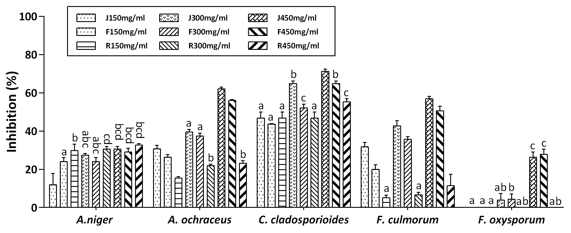
As can be seen from Figure 1, the F. oxysporum mold is the most resistant to all the tested methanol extracts, except for the MEJ and MEF at 450 mg/mL concentrations, with growth inhibition not exceeding 28%. Fusarium culmorum seems to be resistant to all root extract concentrations but sensitive to the other extracts, with an inhibition rate ranging from 20 to 56% (figure 4a). In contrast, C. cladosporioides was the most sensitive to all methanol extracts, with inhibition ranging from 43 to 71%. Aspergillus ochraceus was more resistant to the root methanol extract (MER) compared to the other extracts, and its inhibition was between 15 and 62%. Unlike the other tested strains, A. niger was more sensitive to the MER and was inhibited up to 32%.
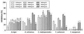
The inhibition of mycelial growth caused by the S. satureioides methanol extracts on the 7th day of incubation. (J:ME June; F: ME February; R: ME Root). No significant difference (p<0.05) between means of the same letter above the histogram bars of each mold.
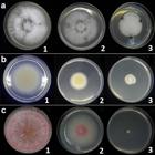
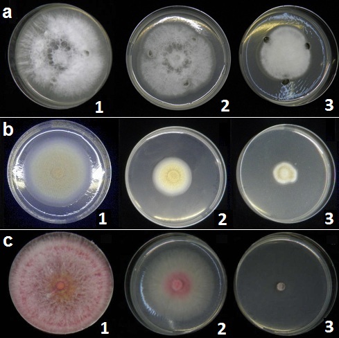
Antifungal effect of S. satureioides on F. culmorum, A. ochraceus, and F. oxysporum. a) Effect of the MEF on F. culmorum (Well-diffusion method): 1) control; 2) 300mg/mL; 3) 450mg/mL. b) Effect of the EOJ on A. ochraceus (Poisoned food method): 1) control; 2) 300ppm; 3) 500ppm. c) Effect of the EOJ on F. oxysporum (Fumigation method): 1) control; 2) 2.5µL; 3) 5 µL.
Regarding the antifungal effect of the extracts, inhibition increases with increasing concentration, depending on the fungal strain. Excluding F. oxysporum, it can be noted that the MEJ presented the highest inhibition degree (from 11 to 71%), followed by the MEF (from 20 to 64%) and MER (from 5 to 55%). Unlike its low inhibitory effect, the MER presented the highest effect on A. niger for all its concentrations (notably with no significant difference, p<0.05).
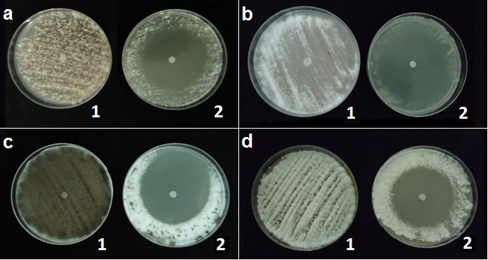
The Disk diffusion assay was used to assess the effect of the S. satureioides EO on the fungal spore germination by means of the measurement of the spore inhibition zone (IZ) diameter. The results presented in Table II indicate the high power of the S. satureioides EO to inhibit spore germination. All the tested fungal spores can be classified as very sensitive to both EOJ and EOF; the IZ diameter varies from 41 to 76mm. It is noteworthy that EOJ is slightly more powerful than EOF. In terms of fungal sensitivity, C. cladosporioides is the most sensitive and F. culmorum is the most resistant. It can be noted that sporulation was strongly affected (Figure 5a-d).

Antifungal effect of S. satureioides essential oil (EOJ) on the fungal spore germination (1: control; 2: test). a) A. ochraceus; b) F. oxysporum; c) A. niger ; d) F. culmorum.
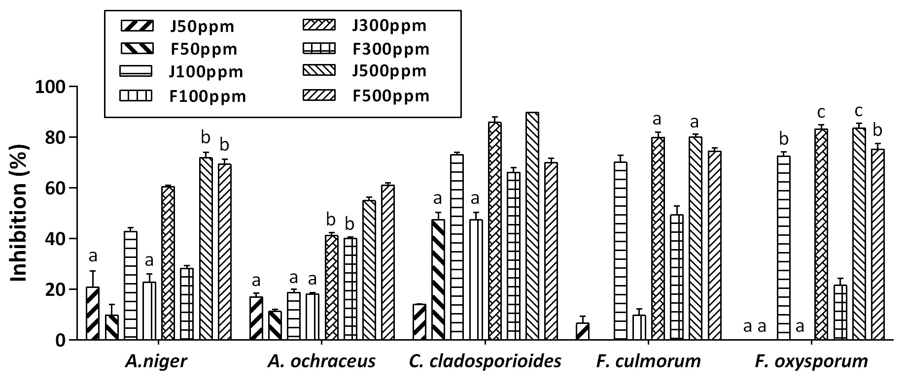
The poisoned food method was used to assess the effect of the incorporated EO on the fungal growth. Four concentrations of each EO were tested (50, 100, 300, and 500 ppm). The results are shown in Figure 2. From this figure, it can be easily seen that the lower EO concentrations (50 ppm) have a weak to no effect on the overall molds. The EOF at 100 ppm has no effect on F. oxysporum. Based on the percentage inhibition values, a progressive increase is proportional to the EO concentration can be seen. The inhibition percentage ranged from 18 to over 80%, and the EOJ was the most effective on all fungal strains compared to the EOF. In a global view, A. ochraceus and A. niger were the least sensitive compared to the other species (Figure 4b). The maximum inhibitory effect of EOJ is clear for the maximal concentration (500 ppm) and varies from 54% on A. ochraceus to 89% on C. cladosporioides. The EOF maximal effect ranged from 61% on A. ochraceus to 75% on F. oxysporum. It is notable that the EOJ concentrations of 300 and 500 ppm are not statistically different in their effect on the Fusarium species (p<0.05). On the other hand, we note a fungal growth delay of 48 h for A. ochraceus and 4 days for the other fungi under the effect of both EOs.

Mycelial growth inhibition registered on the 7th day of incubation. The S. satureioides EOs were applied by the poisoned food method. (J: EO June; F: EO February). No significant difference (p<0.05) between means of the same letter above the histogram bars of each mold.
The EO fumigation test is based on the effect of EO volatilization against the fungal mycelial growth in a close environment. The fungal strains were thus exposed to the progressive volumes of 2.5, 5, and 10 µL. After measurements were done, the results were represented in Figure 3. The inhibition percentage values ranged from nearly 17% to 71% for the two EOs at 2.5 µL. The EOs at 5 and 10 µL presented the same effect on almost all fungal strains, with an inhibition degree ranging from 68 to 90%, except for the EOJ on A. ochraceus. It is clear that Aspergillus species were the least sensitive compared to the other species. It can be noted that the Fusarium species were the most sensitive. The minimal EO volume delayed the growth of all fungi by at least 48 hours. The effect of the EOs was generally manifested by changes in the macroscopic appearance, namely sporulation weakness and pigmentation (Figure 4c).
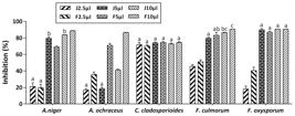
Mycelial growth inhibition registered on the 7th day of incubation. The S. satureioides EOs were applied by the fumigation method. (J: EO June; F: EO February). No significant difference (p<0.05) between means of the same letter above the histogram bars of each mold.
Regarding the antifungal activity, the same studies mentioned above were done with the use of Candida albicans, but there is no clear discussion related to the filamentous fungi A. niger and A. fumigatus in the Laouer et al. (2006) study. In summary, these studies showed inhibitory activity on the C. albicans yeast of about 12 to 23 mm inhibition zones (Bendahou et al. 2008, Kherkhache et al. 2018) and total inhibitory activity on C. albicans, A. niger, and A. fumigatus (Laouer et al. 2006). In a study on the effect of S. satureioides EO on the fungus Ascochyta rabiei, Zerroug et al. (2011) mentioned no growth of the fungus at 3 mg/mL, and reduced growth by 50% at the essential oil concentration of 0.0187 mg/mL. Khaldi et al. (2017) tested the S. satureioides EO from Nâama (west of Algeria) against the mycelial growth of the molds Aspergillus niger, A. flavus, A. ochraceus, Penicillium expansum, Fusarium oxysporum f. sp. albedinis, Alternaria alternata, and Cladosporium sp. using the contact method. They found that all molds were totally inhibited at the concentration of 1/370 (v/v). At 1/1500 (v/v) of the EO (≈ 667 ppm), Aspergillus niger, A. flavus, A. ochraceus, and Penicillium expansum were inhibited to 56-76%, and the other molds were totally inhibited. These results are in concordance with ours at the EO concentration of 500 ppm, so that the mycelial growth inhibition ranged between 61 and 75%. Plant extracts have been shown to have an inhibitory effect on molds. In an in vitro study on the effect of plant extracts on Fusarium species, Kursa et al. (2022) found that ethanolic leaf extracts from Achillea millefolium, Tanacetum vulgare, Artemisia absinthium and Salvia officinalis caused fungal mycelial growth inhibition, and this varied according to extract type and concentration. The highest inhibition coefficient ranged from 63.82% to 83.53% at a plant extract concentration of 20%. Aqueous extracts from medicinal plants Artemisia herba alba, Cotula cinerea, Asphodelus tenuifolius, and Euphorbia guyoniana also showed antifungal action against Fusarium graminearum and Fusarium sporotrichioides at concentrations of 10% and 20% in the culture medium. These plants were found to contain tannins, flavonoids, saponins, steroids, and alkaloids (Salhi et al. 2017). Essential oil (85% eugenol) from clove (Eugenia caryophyllata) showed a total in vitro inhibitory effect on Fusarium oxysporum at concentration of 500 ppm and from 400 to 500 ppm on Aspergillus niger. On tomato fruits, the fungal growth was reduced from 50% to 70% and to 40% for A. niger and F. oxysporum, respectively (Muñoz Castellanos et al. 2020). Some of the most important postharvest fungi, Alternaria alternata, Botrytis cinerea, and Penicillium italicum, were effectively inhibited by aromatic or medicinal plant EOs from Cinnamomum verrum, Syzygium aromaticum, Gautheria fragrantissima, Cupressus sempervirens, Cymbopogon nardus, Pelargonium asperum, Mentha piperita, and Foeniculum vulgare at 0.5 mg/mL. The antifungal effect varied depending on the EO source and fungus resistance (Allagui et al. 2024). Qu et al. (2022) found reduction in the ergosterol and aflatoxin B1 contents, in the thickness of hyphae, and inhibition of the mycelial growth and spore germination, following treatment of Aspergillus flavus with carvacrol concentrations up to 200 µg/mL. Xing-dong & Hua-Li (2014) studied the effect of Zanthoxylum bungeanum EO and its major constituent α-pinene on Fusarium sulphureum. The hyphal normal structure was affected, such as hyphal swelling with more secretions at the surface and cytoplasmic disorder with massive vacuolation and osmiophilic granule formation. The dry rot of potatoes inoculated with F. sulphureum was well controlled, and the EO was more interesting than α-pinene.
We found previously that our S. satureioides EO is composed mainly of thymol (25.6%), α-terpineol (24.6%), borneol (17.4%), and p-cymène (11.4%) (Sassoui et al. 2020). These compounds form 79% of our tested EO and are the basic components of the S. satureioides EO cited in the literature, taking into consideration that this plant is endemic to Algeria (Biondi et al. 2006, Laouer et al. 2006, Bendahou et al. 2008, Aouf et al. 2020, Souadia et al. 2020). Thymol, a phenolic component in EOs, was reported to have an antibacterial effect. Due to its pleasant smell and low toxicity, this compound will be used as a food additive in order to prevent bacterial spoilage (Memar et al. 2017). It is an effective antioxidant and reduces oxidative stress and the risk of associated disorders (Rathod et al. 2021). The α-terpineol is known for its praiseworthy and useful properties, including the antimicrobial activity (Ben Akacha et al. 2024). Natural borneol is well known in traditional Chinese medicine, and it possesses antifungal activity. It has been shown to inhibit the C. albicans germ tube and biofilm formation (Wang et al. 2022). The p-Cymene is a precursor for thymol and carvacrol. It possesses antibacterial and antifungal activities, and it is considered “generally recognized as safe” by the U.S. Food and Drug Administration (Balahbib et al. 2021). These four reputable compounds make S. satureioides EO of high quality and suitable for use in the food and pharmaceutical fields.
CONCLUSIONS
The results of this study showed that S. satureioides is endowed with great antimicrobial activity against bacteria and filamentous fungi, in addition to antioxidant properties. This is the first report on the bioactive effect of the S. satureioides roots. Root extract had an antibacterial effect similar to that of the aerial parts on almost all bacterial strains tested. It also had an antifungal effect, but it was lower than the aerial parts extracts. It presented a particularly high inhibition effect on A. niger at the lowest concentration. We note the resistance of the Fusarium species to the plant extracts, particularly at low concentrations. In contrast, Aspergillus species were less sensitive to the S. satureioides essential oil compared to the other fungal strains. Further advanced analyses and tests are needed to identify and quantify bioactive compounds in these plant extracts, to determine their effect under field conditions, and to verify whether biopreparations may be produced to carry less risk to the environment and to humans.
REFERENCES
- AJMAL M, HUSSAIN A, ALI A, CHEN H & LIN H. 2023. Strategies for controlling the sporulation in Fusarium spp. J Fungi 9: 10.
- ALLAGUI MB, MOUMNI M & ROMANAZZI G. 2024. Antifungal activity of thirty essential oils to control pathogenic fungi of postharvest decay. Antibiotics 13: 28.
- ALLALI H, BENMEHDI H, DIB MA, TABTI B, GHALEM S & BENABADJI N. 2008. Phytotherapy of diabetes in west Algeria. Asian J Chem 20: 2701.
- AOUF A, ALI H, AL-KHALIFA AR, MAHMOUD KF & FAROUK A. 2020. Influence of nanoencapsulation using high-pressure homogenization on the volatile constituents and anticancer and antioxidant activities of Algerian Saccocalyx satureioides Coss. et Durieu. Molecules 25: 4756.
- BALAHBIB A ET AL. 2021. Health beneficial and pharmacological properties of p-cymene. Food Chem Toxicol 153: 112259.
- BEN AKACHA B, MICHALAK M, GENERALIĆ MI, KAČÁNIOVÁ M, CHAARI M, BRINI F, BEN SAAD R, MNIF W, GARZOLI S & BEN HSOUNA A. 2024. Mixture design of α‐pinene, α‐terpineol, and 1, 8‐cineole: A multiobjective response followed by chemometric approaches to optimize the antibacterial effect against various bacteria and antioxidant activity. Food Sci Nutr 12: 574-589.
- BENAHMED M, DJEDDI N, AKKAL S & LAOUAR H. 2016. Saccocalyx satureioides as corrosion inhibitor for carbon steel in acid solution. Int J Ind Chem 7: 109-120.
- BENARADJ A & BOUCHERIT H. 2022. Ethnobotanical study of the plant of medicinal interest Saccocalyx satureioides Coss. & Durieu (Lamiaceae) in the region of Naâma (Algeria). Biodiv Res Conserv 68: 27-34.
- BENDAHOU M, BENYOUCEF M, MUSELLI A, DESJOBERT JM, PAOLINI J, BERNARDINI AF & COSTA J. 2008. Antimicrobial activity and chemical composition of Saccocalyx satureioides Coss. et Dur. essential oil and extract obtained by microwave extraction. Comparison with hydrodistillation. J Essent Oil Res 20: 174-178.
- BIONDI DM, SARI M, ZEDAM A & ROBERTO G. 2006. Essential oil of Algerian Saccocalyx satureioides Coss et Durieu. Flavour Fragr J 21: 546-548.
- CHEN P, LIU Y, LI C, HUA S, SUN C & HUANG L. 2023. Antibacterial mechanism of vanillin against Escherichia coli O157: H7. Heliyon 9: e19280.
- COSMULESCU S, TRANDAFIR I & NOUR V. 2017. Phenolic acids and flavonoids profiles of extracts from edible wild fruits and their antioxidant properties. Int J Food Prop 20: 3124-3134.
- GONELIMALI FD, LIN J, MIAO W, XUAN J, CHARLES F, CHEN M & HATAB SR. 2018. Antimicrobial properties and mechanism of action of some plant extracts against food pathogens and spoilage microorganisms. Front Microbiol 9: 389103.
- GULLUCE M, SAHIN F, SOKMEN M, OZER H, DAFERERA D, SOKMEN A, POLISSIOU M, ADIGUZEL A & OZKAN H. 2007. Antimicrobial and antioxydant properties of essential oils and methanol extract from mentha longifolia L. ssp. Longifolia. Food Chem 103: 1449-1456.
- HENDEL N, DJAMEL S, MADANI S, SELLOUM M, BOUSSAKRA F & DRICHE O. 2021. Screening for in vitro antioxidant activity and antifungal effect of Artemisia campestris L. Int J Agric Environ Food Sci 5: 251-259.
- HENDEL N, LAROUS L & BELBEY L. 2016. Antioxidant activity of rosemary (Rosmarinus officinalis L.) and its in vitro inhibitory effect on Penicillium digitatum. Int Food Res J 23: 1725-1732.
- HENDEL N, SARRI M, SARRI D, SEGHIOUR S, NAPOLI E, SELLOUM M & RUBERTO G. 2023. Phytochemical analysis, antibacterial and antifungal effect of Lavandula dentata L. essential oil and methanol extract. Nat Prod Res 38: 3498-3507. Doi: 10.1080/14786419.2023.2252973.
- HOCKING AD. 2006. Aspergillus and related teleomorphs. In: Blackburn CW (Ed), Food Spoilage Microorganisms. Woodhead Publishing Limited, Cambridge, p. 451-487.
- KARNWAL A & MALIK T. 2024. Exploring the untapped potential of naturally occurring antimicrobial compounds: novel advancements in food preservation for enhanced safety and sustainability. Front Sustain Food Sys 8: 1307210.
- KHALDI A, MEDDAH B, MOUSSAOUI A, SONNET P & BENMEHDI H. 2017. In vitro antifungal activities of essential oil of Saccocalyx satureioides Coss. et Dur. on fungal species development. Appl Biol Sahar Areas 1: 8-15.
- KHAN F, BAMUNUARACHCHI NI, TABASSUM N & KIM YM. 2021. Caffeic acid and its derivatives: antimicrobial drugs toward microbial pathogens. J Agric Food Chem 69: 2979-3004.
- KHERKHACHE H, BENABDELAZIZ I, SILVA AM, LAHRECH MB, BENALIA M & HABA H. 2020. A new indole alkaloid, antioxidant and antibacterial activities of crude extracts from Saccocalyx satureioides. Nat Prod Res 34: 1528-1534.
- KURSA W, JAMIOŁKOWSKA A, WYROSTEK J & KOWALSKI R. 2022. Antifungal effect of plant extracts on the growth of the cereal pathogen Fusarium spp.—An In Vitro Study. Agronomy 12: 3204.
- LAOUER H, AKKAL S, DEBARNOT C, CANARD B, MEIERHENRICH UJ & BALDOVINI N. 2006. Chemical composition and antimicrobial activity of the essential oil of Saccocalyx satureioides Coss. et Dur. Nat Prod Commun 1: 645-650.
- LI Y, FABIANO-TIXIER AS & CHEMAT F. 2014. Essential oils as reagents in green chemistry, Springer, Briefs in Green Chemistry for Sustainability. Doi 10.1007/978-3-319-08449-7_3.
- MA X ET AL. 2021. Assessment of the pulmonary adaptive immune response to Cladosporium cladosporioides infection using an experimental mouse model. Sci Rep 11: 909.
- MAGRI A, CURCI M, BATTAGLIA V, FIORENTINO A & PETRICCIONE M. 2023. Essential oils in postharvest treatment against microbial spoilage of the rosaceae family fruits. Applied Chem 3: 196-216.
- MASYITA A, SARI RM, ASTUTI AD, YASIR B, RUMATA NR, EMRAN TB, NAINU F & SIMAL-GANDARA J. 2022. Terpenes and terpenoids as main bioactive compounds of essential oils, their roles in human health and potential application as natural food preservatives. Food Chem: X 13: 100217.
- MEHLOUS S, BENCHIKH F, BENABDALLAH H, LOUCIF K, KAOUDOUNE C, LAOUER H & AMIRA S. 2020. Evaluation of antioxidant activity and polyphenols content of the hydro-methanolic extract from Saccocalyx satureioides Coss & Dur. J Drug Deliv Ther 10: 188-190.
- MEMAR MY, RAEI P, ALIZADEH N, AGHDAM MA & KAFIL HS. 2017. Carvacrol and thymol: strong antimicrobial agents against resistant isolates. Rev Res Med Microbiol 28: 63-68.
- MOHAMADI S, ZHAO M, AMRANI A, MARCHIONI E, ZAMA D, BENAYACHE F & BENAYACHE S. 2015. On-line screening and identification of antioxidant phenolic compounds of Saccocalyx satureioides Coss. et Dur. Ind Crops Prod 76: 910-919.
- MOSTEFA SARI F, ALLEM R & TOUMI M. 2020. Bioactive compounds of thyme (Thymus fontanesii Boiss. & Reut.) and native savory (Saccocalyx satureioides Coss. & Dur.) Against cutaneous leishmaniasis. Revue Agrobiologia 10: 1739-1748.
- MUÑOZ CASTELLANOS L, OLIVAS NA, AYALA-SOTO J, CONTRERAS CMO, ORTEGA MZ, SALAS FS & HERNÁNDEZ-OCHOA L. 2020. In vitro and in vivo antifungal activity of clove (Eugenia caryophyllata) and pepper (Piper nigrum L.) essential oils and functional extracts against Fusarium oxysporum and Aspergillus niger in tomato (Solanum lycopersicum L.). Int J Microbiol 1 : 1702037.
- MUNTEAN D & VULPIE S. 2023. Antioxidant and antibacterial activity of plant extracts. Antibiotics 12: 1176.
- NGUYEN TLA & BHATTACHARYA D. 2022. Antimicrobial Activity of Quercetin: An Approach to Its Mechanistic Principle. Molecules 27: 2494.
- OULAHAL N & DEGRAEVE P. 2022. Phenolic-rich plant extracts with antimicrobial activity: an alternative to food preservatives and biocides? Front Microbiol 12: 753518.
- QU C, LI Z & WANG X. 2022. UHPLC-HRMS-based untargeted lipidomics reveal mechanism of antifungal activity of carvacrol against Aspergillus flavus. Foods 11: 93.
- QUEZEL P & SANTA S. 1963. Nouvelle flore d’Algérie et des régions désertiques méridionales, tome II. Paris (France): CNRS.
- RATHOD NB, KULAWIK P, OZOGUL F, REGENSTEIN JM & OZOGUL Y. 2021. Biological activity of plant-based carvacrol and thymol and their impact on human health and food quality. Trends Food Sci Technol 116: 733-748.
- RUIZ-NAVAJAS Y, VIUDA-MARTOS M, SENDRA E, PEREZ-ALVAREZ JA & FERNANDEZ-LOPEZ J. 2013. In vitro antibacterial and antioxidant properties of edible films incorporate with thymus moroderi or thymus piperella essential oil. Food Control 30: 386-392.
- SALHI N, MOHAMMED SAGHIR SA, TERZI V, BRAHMI I, GHEDAIRI N & BISSATI S. 2017. Antifungal activity of aqueous extracts of some dominant Algerian medicinal plants. Biomed Res Int 1: 7526291.
- SASSOUI A, HENDEL N, SARRI DJ, SARRI M, FILIPPO M, MAURIZIO B, DONATO R, ANGELO C, ROMAN P & GIOVANNI B. 2020. Essential oils from three Algerian medicinal plants (Artemisia campestris, Pulicaria arabica, and Saccocalyx satureioides) as new botanical insecticides?. Environ Sci Pollut Res Int 27: 26594-26604.
- SHUKLA R, SINGH P, PRAKASH B & DUBEY NK. 2012. Antifungal, aflatoxin inhibition and antioxidant activité of Callistemon lanceolatus (Sm.) Sweet essential oil and its major component 1,8-cineole against fungal isolates from chickpea seeds. Food Control 25: 27-33.
- SOUADIA A, GOURINE N & YOUSFI M. 2020. Variability in phytochemical composition and antioxidant activity of Saccocalyx satureioides essential oils due to harvest period. J Herbs Spices Med Plants 26: 435-446.
- SRIKACHA N & RATANANIKOM K. 2020. Antibacterial activity of plant extracts in different solvents against pathogenic bacteria: An: in vitro: experiment. J Acute Dis 9: 223-226.
- TEPE B. 2008. Antioxidant potentials and rosmarinic acid levels of the methanolic extracts of Salvia virgata (Jacq), Salvia staminea (Montbret & Aucher ex Bentham) and Salvia verbenaca (L.) from Turkey. Bioresour Technol 99: 1584-1588.
- VEIKO AG, OLCHOWIK-GRABAREK E, SEKOWSKI S, ROSZKOWSKA A, LAPSHINA EA, DOBRZYNSKA I, ZAMARAEVA M & ZAVODNIK IB. 2023. Antimicrobial activity of quercetin, naringenin and catechin: flavonoids inhibit Staphylococcus aureus-induced hemolysis and modify membranes of bacteria and erythrocytes. Molecules 28: 1252.
- WANG Y & LIU H & ZHAN F. 2022. Effects of natural borneol on germ tube formation and preformed biofilm activity in Candida albicans. Nat Prod Commun 17(9): 1-5.
- XING-DONG L & HUA-LI X. 2014. Antifungal activity of the essential oil of Zanthoxylum bungeanum and its major constituent on Fusarium sulphureum and dry rot of potato tubers. Phytoparasitica 42: 509-517.
- YABRIR B, TOUATI M, ADLI B, BEZINI E, GHAFOUL M, KHALIFA S & GUIT B. 2018. Therapeutic use of spontaneous medicinal flora from an extreme environment (dune cordon) in Djelfa region, Algeria. J Pharm Pharmacogn Res 6: 358-373.
- ZERROUG MM, LAOUER H, STRANGE RN & NICKLIN J. 2011. The effect of essential oil of Saccocalyx satureioides Coss. Et Dur. on the growth of and the production of solanapyrone A by Ascochyta Rabiei (Pass.) Labr. J Adv Environ Biol 5: 501-506.
- ZIANI BE, CALHELHA RC, BARREIRA JC, BARROS L, HAZZIT M & FERREIRA IC. 2015. Bioactive properties of medicinal plants from the Algerian flora: Selecting the species with the highest potential in view of application purposes. Ind Crops Prod 77: 582-589.