Micro-XRF chemical elementary analysis on the holotype of Sinopterus atavismus Lü, Teng, Sun, Shen, Li, Gao, Liu, 2016 (Pterodactyloidea, Tapejaridae)
Abstract
The Early Cretaceous pterosaur Sinopterus atavismus, from Northeast China, belongs to the Tapejaridae. The furrowed structure of its wing phalanges is relatively primitive, possibly indicative of atavism. New information of Sinopterus atavismus is therefore of great interest in studies of evolution among Tapejaridae, especially for interpreting some anatomical features of the holotype. In this paper, the distribution patterns and characteristics of chemical elements in the holotype specimen were analyzed by non-destructive micro-X-ray fluorescence (micro-XRF). The chemical mapping results show that the element Ca was significantly associated with pterosaur bones, showing residual traces of the left lower part of the humerus; Sr replaced Ca and the distribution was uniform, suggesting that phosphate recrystallization occurred in the specimen; Fe is concentrated in the soft tissue parts of the pterosaur fossil, which may be related to pyritization, and Mn appears as the alteration phase of the original iron-rich phase. The elemental migration and sedimentary environment of the fossil require further study. The XRF elemental imaging study of Sinopterus atavismus is helpful for explorations of the development and evolution of Tapejaridae, and also provides more chemical information on the taphonomy of the Jehol Biota.
Key words
pterosaur; μ-XRF; elementary scan; Jehol Biota; China
INTRODUCTION
There are nine species of Tapejaridae pterosaur have been discovered and reported from the Lower Cretaceous Jiufotang Formation in the western Liaoning of China, which contains the fossils of last stage of Jehol Biota (Wang & Zhou 2003, Li et al. 2003, Lü & Yuan 2005, Lü et al. 2006a, b, 2007, 2016, Liu et al. 2014). The area is currently the only known site of tapejarid fossils in China. All the specimens found belong to Sinopterus of the Sinopterinae subfamily (Lü et al. 2016), and the Jiufotang Formation in this area also includes the suspected Eoazhdarcho liaoxiensis (Lü & Ji 2005). The holotype of Sinopterus atavismus was originally classified as a new species within the genus Huaxiapterus and designated as Huaxiapterus atavismus. This nomenclature is based on the distinct groove-like structure observed on the posterior surface of the second and third wing phalanges (Lü et al. 2016). Later, Zhang et al. (2019) proposed that compared with other pterosaurs, the unique skeletal features of Huaxiapterus atavismus include a sharp turn in the dorsal margin of the premaxillary crest and a downward-sloping straight line formed by the dorsal margin of the premaxilla crest at the rostral end, which was corrected to the genus Sinopterus.
The holotype of Sinopterus atavismus was unearthed from the Jiufotang Formation in Chaoyang City, Liaoning Province. Affected by the destruction of the North China Craton, numerous volcanic basins were formed in the western Liaoning region during the Early Cretaceous (Zhu et al. 2012). The abundant material resources, coupled with frequent tectonic movements, contributed to the preservation of a significant number of fossils within the lacustrine deposits of the Jiufotang Formation (Wang et al. 2013) (Figure 1). Previous studies on the holotype of Sinopterus atavismus were limited by the presence of squashed fossil specimens with messy features and soft tissue residue covering the skeletal structure (Lü et al. 2016, Zhang et al. 2019). Morphological studies are prone to some subjective judgment, leading to disputes and information waste, and lack the relationship between microorganisms and fossil burial preservation. However, the chemical element composition of fossil substances can provide new information for morphological observation.

The distribution of fossils of Sinopterus and a sketched geological map of western Liaoning.
X-ray fluorescence spectroscopy (micro-XRF) can rapidly, yield nondestructive and quantitative studies on the chemical composition of samples on a microscale (Liu et al. 2022). The research of Bergmann et al. (2010) revealed through SRS-XRF that the original chemical composition of the Solnhofen Archaeopteryx in the late Jurassic, and the feathers were not simple impressions but chemical fossils. Since then, spectral analysis techniques, such as XRF, have been employed to conduct extensive research on fossils. In previous studies, Chen et al. obtained the elemental distribution of salamander fossils by using micro-XRF and inferred the elements exchange during the fossil burial (Chen et al. 2023); Wogelius’s work demonstrated that Cu exists in fossils in the form of organometallic compounds, most likely derived from primitive eumelanin (Wogelius et al. 2011); Leal et al. (2018) discovered that the Fe and Zn in the cracks of the fossilized bones of Tapejaridae in northern Brazil were related to fossilized bacterial mats. In this paper, the micro-XRF technique was used to conduct a chemical element distribution scan studies on the holotype specimen XHPM 1009 of Sinopterus atavismus from the Jiufotang Formation. This is the first time that the non-destructive XRF imaging technology has been used to study a fossil specimen of Tapejaridae, and more valuable information on the morphology and taphonomy of fossil bones can be obtained from the distribution patterns of related elements.
MATERIALS AND METHODS
The researched specimen is the holotype of Sinopterus atavismus, specimen number XHPM 1009. The abbreviation XHPM represents the fossil hosting institute Dalian Xinghai Paleontological Museum. The specimen was analyzed using the Bruker M6 Jetstream mobile X-ray fluorescence (XRF) scanner when it was on trail at Dalian Xinghai Paleontological Museum.
The analysis of these specimens by means of X-ray fluorescence was approved by the ethics institutional review board of Dalian Xinghai Paleontological Museum and was conducted under the research permit number. All methods were carried out in accordance with the relevant guidelines and regulations of the Dalian Xinghai Paleontological Museum. Non-destructive XRF analysis posed no risk to the integrity of the specimens.
SYSTEMATIC PALEONTOLOGY
Pterosauria Kaup, 1834
Pterodactyloidea Plieninger, 1901
Tapejaridae Kellner, 1989
Sinopterus Wang and Zhou, 2003
Sinopterus atavismus Lü et al. 2016
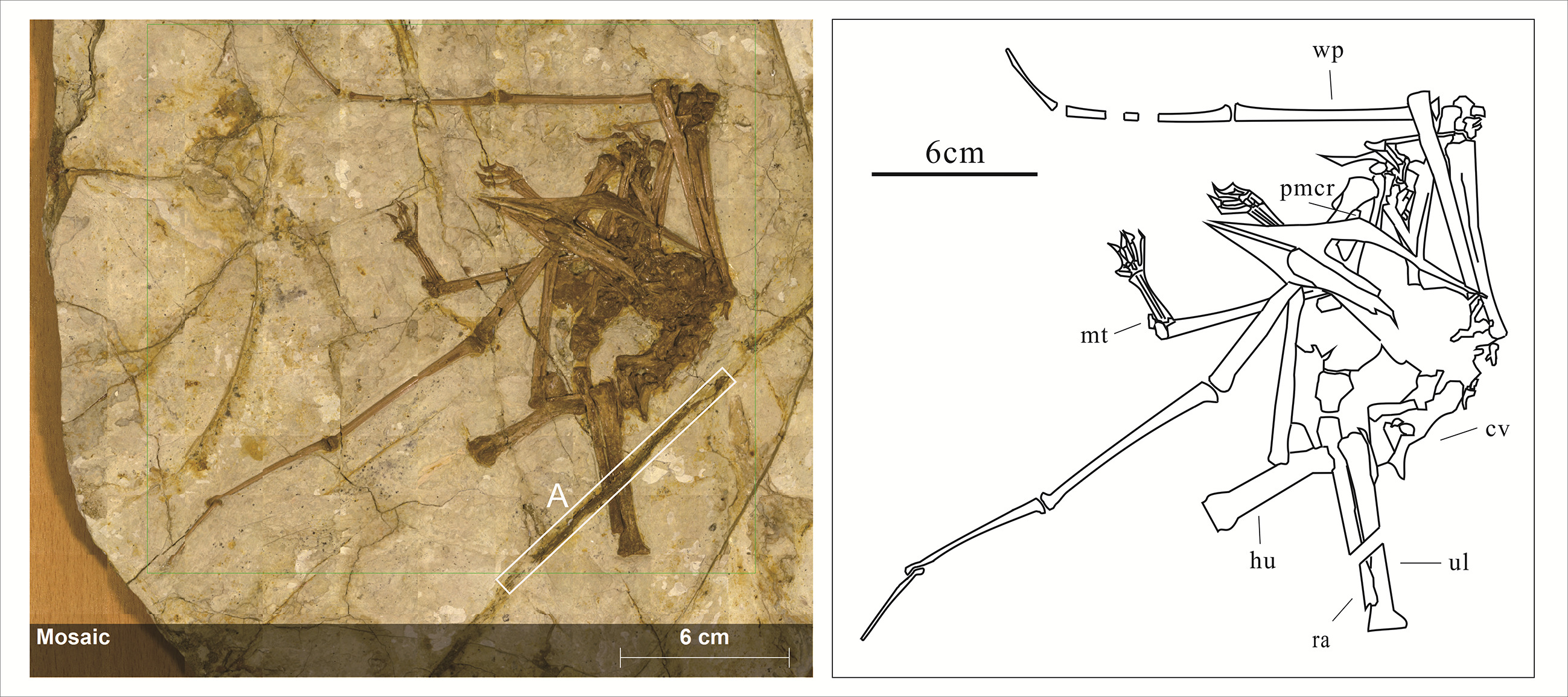
Referred specimen: A skeleton with an intact skull is deposited at the Xinghai Paleontological Museum, Dalian, with the specimen number XHPM 1009 (Figure 2).

Optical image (left) and line drawing (right) of Sinopterus atavismus XHPM 1009. The dark brown part is the fossil skeleton: cv, cervical vertebrae; ul, ulna; ra, radius; wp,wing phalanx; hu, humerus; mt,metatarsal; pmcr, premaxillary crest. The left side of the fossil is marked by cracks and glue residue. The area A marked by the white frame in the optical image is an unknown feature.
Locality and horizon: Chaoyang City, Liaoning Province, China; Jiufotang Formation, Lower Cretaceous.
Comments on preservation: The skeletal position of the specimen is clear, the back of the skull is severely damaged, the nasoantorbital fenestra is healed, and the maxilla and mandibular have no teeth. A sharp turn in the dorsal margin of the premaxillary crest, and the dorsal margin of the snout forms a downward-sloping straight line. The ulna and radius are parallel, and the dorsal vertebrae and sacral vertebrae are covered by other bones. The scapula and coracoid bones were not fused, indicating that the fossil was a young individual.
Micro-x-ray fluorescence spectrometry
This experiment was conducted at the maximum energy setting of 50 kV and 600 μA in Bruker M6. The acquisition conditions were 432 × 390 pixel, and the acquisition time per pixel was 15 ms with a pixel size of 500 µm. The total measurement time was 1 h 60 min.
Following data collection, the element distribution maps and elemental content data (Figures 3,4 and 5) were analyzed and generated using the characterization software provided by Bruker Micro Analytics. In the processing software that comes with μ-XRF, the possible fitting results of specific element combinations can be selected. The fitting results between similar and adjacent peaks of different elements are different, which are significantly different from the peak maps we obtained. The elemental peaks we selected are the combination results obtained through comparison and most in line with the sum spectrum peak map of this sample. The color scheme used in the maps serves solely to illustrate the presence and concentration of elements. In addition, XRF analysis has limitations in detecting light elements, as it is more suitable for characterizing heavy elements and trace elements. Further detection of light elements requires energy dispersive spectroscopy analysis (EDS). For instance, EDS plays a significant role in analyzing the ultrastructure of the fossilized skin of Psittacosaurus by providing distribution information of light elements such as C, O, Na, and Mg (Yang et al. 2024).

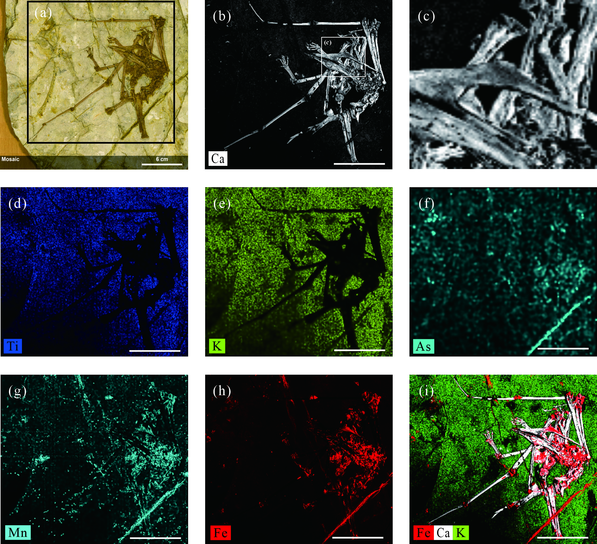
(a) optical image of Sinopterus atavismus XHPM1009, with the black box showing the actual scanning area of the specimen, (b) Micro-XRF false color element distribution map of Ca, (c) The element distribution map of Ca shows the sharp turn in the dorsal margin of the premaxillary crest of the pterosaur, (d)-(h) Micro-XRF false color element distribution maps, and each map corresponds to the following elements: (d) Ti, (e) K, (f) As, (g) Mn, (h) Fe, (i) the combination of the three elements. Red, Fe; White, Ca; Green, K. The scale is 6mm.
(a)-(i) Micro-XRF false color element distribution maps, and each map corresponds to the following elements: (a) Sr, (b) Another form of Sr, (c) Rb, (d) Y, (e) Si, (f) Zn, (g) Zr, (hTi, (i) P.
RESULTS
The scanning sum spectrum (Figure 5) obtained by micro-XRF shows that the scanned area of the specimen was mainly composed of Sr, Zn, Fe, Mn, Ti, Ca, K, P, Si, as well as some other elements. These elements are represented by the corresponding energy peaks of their characteristic X-rays. In the spectrum, In the spectrogram, in order to display the existence of all Kɑ related energy peaks of the elements in the studied specimen, the main scale of the y-axis increases exponentially at a rate of 10.

The element distribution pattern in the element distribution map showed that Ti, Si, Zr and K are uniformly distributed on the fossil matrix except for the Pterosaur and area a. Ca, P, Y and Sr were correlated with the skeleton, among which Ca was the most abundant element, and was the only element that showed residual traces of the left lower part of the humerus, followed by Sr, Y and P.
Fe showed correlation with other biological tissues than skeleton in the pterosaur fossil (Figure 3i), it can be observed that Fe tends to be distributed at the bone junctions and also with the highest concentration in the A region(Figure 1). The distribution areas of Mn and As are similar as Fe, but the distribution range of Mn is slightly larger than that of Fe, and the enrichment degree of As relative to surrounding rock is slightly lower than that of Fe.
In addition, Rb is distributed in the regions except for the pterosaur. The concentration of As was more abundant in the A region. Zn, Ni and other elements were unevenly distributed in the specimen.
DISCUSSION
The enrichment and distribution of elements are of great significance for distinguishing and comparing fossils. The results of this micro-XRF experiment provide more in-depth information on the holotype of Sinopterus atavismus. The composition and exchange of elements can allow us to further speculate on the composition, burial conditions and processes of the specimen, and help to discuss the evolution of Fe and Mn elements during the burial process.
Chemical composition of skeleton
The content of Al, Ti, K and Si elements in the surrounding rock is high, as the host rock of the Sinopterus atavismus holotype is tuffaceous sandstone. The content of As in fosil is slightly higher than surroundig rock, which may be absorbed from the outside by Fe and Mn.
According to the element distribution map, Fe and Mn are enriched in the A area, and the elements are evenly distributed, This is significantly different from the bones of Sinopterus atavismus. The inconsistency with the surrounding rock elements indicates that it is not the bone filler of the pterosaur. Its morphology and distribution patterns of chemical elements are similar to those of podozamites-type plant fossils, and being woody plant also explains the less absorption of Si (Hodson et al. 2005, Cooke & Leishman 2011, Pole et al. 2016). In addition, the enrichment of As indicates that plants absorbed more toxic substances from the soil during its growth and were preserved after burial. Other way to interpret the preferential association of Fe and Mn to the A region is that the minerals that have preserved it are different from those that have preserved the other parts of the fossil. However, based on the shape of the fossils, A region is more in line with the former interpretation.
Burial conditions
Compared with the surrounding rock, Ca and P are obviously enriched in the skeleton of the pterosaur. More fine-grained bone structure has been detected, revealing details of the skeleton fractures at the back of the skull under greater pressure. The anterior part of the skull is well-preserved, it shows a sharp turn at the edge of the premaxillary crest, which more clearly verified that the premaxilla does not have a square crest, representing characteristics of Sinopterus atavismus (Zhang et al. 2019, Lü et al. 2016). Relative to the matrix, Sr and P are slightly less enriched in the skeleton than Ca. And as elements with affinity to apatite, P and Sr almost completely exist in skeleton. The coexistence of Sr and Ca indicates that Ca was replaced by Sr during the lithification process. Therefore, the recrystallization of phosphate in the fossil is very likely to have occurred, which also leads to less organic matter residue in the specimen. In the micro-XRF study of salamander fossils found in the Haifanggou Formation in Daohugou area, Inner Mongolia, Ca exists in the whole fossil including soft tissue (Chen et al. 2023), which is likely related to the local preservational environment.
The S, Cu and Zn shown in the sum spectrum diagram could be explained that originate from the input of hydrothermal fluid. Another explanation is originated from organic materials: the S element may be derived from S-containing substances produced by the degradation of keratin in the skin; Metal elements such as Cu and Zn may come from melanin chelation (Wogelius et al. 2011). Compared with the matrix, the content of Zn in fossils is slightly higher, indicating that at least one melanin chelator exists in the skin of fossils, and Zn is conducive to the preservation of fossils.
Origin of distribution of Fe and Mn elements
The distribution of Fe and Mn in this fossil is in a negative correlation state with the distribution of Ca (Figure 3i), Fe and Mn tend to be distributed at the bone junctions. This might be due to anaerobic microorganisms decomposing the organic matter in soft tissues, causing pyrite mineralization in a reducing environment. Or hemoglobin degrading into ferritin after the organism’s death and transforming into iron-containing minerals, the iron may be directly protecting proteins by blocking active sites recognized by enzymes of degradation, or it may be providing protection indirectly by binding to oxygen (Schweitzer et al. 2014). The Fe content in the bones was not significantly higher than that in the surrounding rock matrix, possibly due to the fact that both the bones and the surrounding rocks were simultaneously modified by hydrothermal fluids and other environmental factors. Or the environmental/diagenetic conditions were limiting for pyritization: such as the bones lacked organic carbon, making it difficult to synthesize sufficient S-containing substances; apatite was difficult to adsorb Fe from the surroundings; and an oxidizing environment existed. Part of the Fe element in the fracture site of the skull may come from Fe-containing minerals that filled the macroscopic pores formed by bone fractures. The concentration of Mn associated with Fe is more widely distributed, which might be because Mn in its oxidized state forms oxides. And these oxides can appear as alteration phases of the original iron-rich mineral phases.
In the micro-XRF study of two near-bird fossils found in the same strata in this region, Ni and Ti elements were associated with the fossilization process of feathers, Ni and Ti are related to the lithification process of feathers (Liu et al. 2024). The distribution of major elements such as Ca, P, Sr, Ti, Si and K is similar to that of Sinopterus atavismus. The information provided by the Sinopterus atavismus holotype fossil needs more in-depth analysis, and the migration of elements and the specific formation of pyrite also require further study.
CONCLUSIONS
The element distribution maps revealed by micro-X-ray fluorescence spectroscopy shows the ontogenetic level and elemental distribution characteristics of the holotype of Sinopterus atavismus, which were replaced by different elements at burial due to the different composition of bones and soft tissues. Most of the major elements of the fossil and its surrounding rocks are originally and uniformly distributed, indicating the stability of the sedimentary environment. The skeletal structure of the fossil could be more clearly observed through the Ca element distribution map, Sr and Ca coexist strongly, Fe and Mn in the fossil are concentrated in the soft tissue, fossils may have undergone recrystallization and pyrite formation during the lithification process..
This study is the first to apply micro-XRF imaging technology to the research of a pterosaur fossil revealing the geological and mineralized characteristics of the pterosaur juveniles of the Jiufotang Formation, and providing useful information for the preservation patterns and overall burial situation of other fossils in the site, future fossil research of the Jehol biota, and future XRF research. In addition, the data disclosed here can also guide the application of this technology to the study of other tapejarid fossil specimens, especially those with soft tissue residues, which has certain significance for exploring the evolutionary relationships within the tapejarids of China.
Acknowledgements
This work was supported financially by the National Natural Science Foundation of China (42288201, 42072017) and National Key Research and Development Program of China (2024YFF0807701); The authors thank Prof. Jinhua Li for supporting the laboratory works in the Key Laboratory of Earth and Planetary Physics, Institute of Geology and Geophysics, Chinese Academy of Sciences (Beijing, China).
References
- BERGMANN U, MORTON RW, MANNING PL, SELLERS WI, FARRAR S, HUNTLEY KG, WOGELIUS RA & LARSON P. 2010. Archaeopteryx feathers and bone chemistry fully revealed via synchrotron imaging. Proc Natl Acad Sci USA 107: 9060-9065.
- CHEN YY, LI XB, TENG FF, CHEN J & REISZ RR. 2023. Micro-XRF mapping study on the taphonomy of a Jurassic larval salamander fossil from Inner Mongolia of China. Atom Spectrosc 44: 24-31.
- COOKE J & LEISHMAN MR. 2011. Is plant ecology more siliceous than we realise? Trends Plant Sci 16: 61-68.
- HODSON MJ, WHITE PJ, MEAD A & BROADLEY MR. 2005. Phylogenetic Variation in the Silicon Composition of Plants. Ann Bot 96: 1027-1046.
- LEAL MEC, PEGAS RV, LIMAVERDE S, BONDE N & KELLNER AWA. 2018. Cervical vertebrae of an enigmatic pterosaur from the Crato formation (lower cretaceous, Araripe Basin, NE Brazil). Geol Soc London Special Publications 455: 195-208.
- LI JJ, LÜ JC & ZHANG BK. 2003. A new sinopterid pterosaur from the Mesozoic of western Liaoning Province, China. Acta Paleontol Sin 42: 442-447.
- LIU DX, ZHOU CF, WANG JQ, LI WG & WEI QW. 2014. New data on the cervical morphology of the Chinese tapejarine. Hist Biol 27: 638-645.
- LIU Y, ZHANG CQ, ZHANG D, LIU T, QIU H, LI QL & LI JH. 2022. Non-destructive micro X-ray fluorescence quantitative analysis of geological materials. Atom Spectrosc 43(5): 378-387.
- LIU YC, JI Q, WANG XR, YANG J, O’CONNOR JK, GUO B, REN TL, YIN TT & WANG YB. 2024. Micro-XRF mapping elucidates the taphonomy of two early cretaceous paravian fossils from western Liaoning, China. Atom Spectrosc 45: 123-131.
- LÜ JC, GAO YB, XING LD, LI ZX & JI Q. 2007. A new species of Huaxiapterus (Pterosauria: Tapejaridae) from the early cretaceous of western Liaoning, China. Acta Geol Sin-Engl Ed 81: 683-687.
- LÜ JC & JI Q. 2005. New azhdarchid pterosaur from the early cretaceous of western Liaoning. Acta Geol Sin-Engl Ed 79: 301-307.
- LÜ JC, JIN XS, UNWIN DM, ZHAO LJ, AZUMA Y & JI Q. 2006a. A new species of Huaxiapterus (Pterosauria: Pterodactyloidea)from the Lower Cretaceous of western Liaoning, China with comments on the systematics of tapejarid pterosaurs. Acta Geol Sin-Engl Ed 80: 315-326.
- LÜ JC, LIU JY, WANG XR, GAO CL, MENG QJ & JI Q. 2006b. New material of pterosaur Sinopterus (Reptilia: Pterosauria) from the early cretaceous Jiufotang formation, western Liaoning, China. Acta Geol Sin-Engl Ed 80: 783-789.
- LÜ JC, TENG FF, SUN DY, SHEN CZ, LI GQ, GAO X & LIU HF. 2016. The toothless pterosaurs from China. Acta Geol Sin 90: 2513-2525.
- LÜ JC & YUAN CX. 2005. New tapejarid pterosaur from western Liaoning, China. Acta Geol Sin-Engl Ed 79: 453-458.
- POLE M, WANG YD, BUGDAEVA EV, DONG C, TIAN M, LI L & ZHOU N. 2016. The rise and demise of Podozamites in east Asia—An extinct conifer life style. Palaeogeogr Palaeoclimatol Palaeoecol 464: 97-109.
- SCHWEITZER MH, ZHENG W, CLELAND TP, GOODWIN MB, BOATMAN E, THEIL E, MARCUS MA & FAKRA SC. 2014. A role for iron and oxygen chemistry in preserving soft tissues, cells and molecules from deep time. Proc R Soc B-Biol Sci 281: 20132741.
- WANG XL & ZHOU ZH. 2003. A new pterosaur (Pterodactyloidea, Tapejaridae) from the Early Cretaceous Jiufotang Formation of western Liaoning, China and its implications for biostratigraphy. Chin Sci Bull 48: 16-23.
- WANG YL, YAO Y, ZHANG CB, ZHAO CY, LU S, ZHAO B & WANG CJ. 2013. Stratigraphic division of the Jiufotang Formation in Kazuo Basin of western Liaoning and related issues. Geol China 40: 1523-1530.
- WOGELIUS RA ET AL. 2011. Trace metals as biomarkers for eumelanin pigment in the fossil record. Science 333: 1622-1626.
- YANG ZX, JIANG BY, XU JX & MCNAMARA ME. 2024. Cellular structure of dinosaur scales reveals retention of reptile-type skin during the evolutionary transition to feathers. Nat Commun 15: 4063.
- ZHANG XJ, JIANG SX, CHENG X & WANG XL. 2019. New material of Sinopterus (Pterosauria, Tapejaridae) from the early cretaceous Jehol Biota of China. An Acad Bras Cienc 91: e20180756. DOI 10.1590/0001-376520192018756.
- ZHU RX, XU YG, ZHU G, ZHANG HF, XIA QK & ZHENG TY. 2012. Destruction of the North China Craton [J]. Sci China Earth Sci 55: 1565-1587.