A simple method for mapping microplastics filter collectors for microscopic analyses
Abstract
Microplastic has stood out as one of the main contaminants derived from plastic wastes due to its slow decomposition rate and dangers to the aquatic environment, the ecosystem, and even human health. However, microplastic fragment detection, identification, removal, and quantification remain a challenge. One of the main methods of removing these fragments is the filtration process using filter membranes such as paper, glass fibers, wool, etc. Among several techniques for localizing and identifying these fragments, optical microscopy is an easily accessible alternative, however, the spatialization to locate these fragments has become a challenge for equipment operators. This work proposes a simple spatialization system using coordinates that reduce operation time to make the localization of microplastic fragments onto collector filters easy. In addition to the use of an optical microscope, a micro-Raman was used to demonstrate the efficiency of this spatialization system even in another type of microscope, reducing the operating time from 2 hours and 45 minutes to approximately 2 minutes and 20 seconds, making the search for the fragment more effective and consequently, a faster analysis process.
Key words
spatialization; microplastic; microscopy; micro-raman
INTRODUCTION
Microplastics (MPs) are plastic fragments, ranging in size from a few micrometers to millimeters (< 5mm). Microparticulated plastics originate from various sources, including the degradation of larger plastics due to exposure to sunlight and weathering, as well as the mechanical fragmentation of plastic products during improper manufacturing, use, and disposal processes, e.g. breakdown of plastic objects, car tires, and clothing (Wang et al. 2021). Additionally, microplastics can be found in personal care products such as facial and body scrubs, which contain plastic microbeads as ingredients (Singh & Mishra 2023). Once introduced into the environment, microplastics can pose serious problems due to their ability to persist for long periods, accumulate in different ecosystems, and be ingested by several organisms (Kasmuri et al. 2022). This contamination by MPs can harm the health of organisms, disrupt marine and terrestrial ecosystems, and potentially impact human health caused by the ingestion of contaminated food (Vethaak & Legler 2021, Gola et al. 2021). Therefore, proper management of plastic waste and reducing the use of single-use plastics are essential measures to mitigate the impacts of MPs on the environment.
The scientific community faces a challenge in detecting, identifying, and removing MPs from the environment, particularly aquatic environments. The literature presents different treatment technologies to remove this contaminant, such as pulser clarifier (Sarkar et al. 2021), electrocoagulation process (Shen et al. 2022), bioremediation by seagrasses and aquatic macrophytes (Masiá et al. 2020), and packed biofilters (Liu et al. 2020). However, due to the ease of the process and experimental handling, the filtration process using conventional filter membranes (paper, glass fibers, wool, etc.) has proven to be one of the main methods of collecting these microplastics from water.
In this context, membrane filtration offers a promising approach for fragment removal as it preserves its original structure, preventing chemical aggregation or polymer degradation. Thus, the precise identification of morphology and chemical composition of the collected microplastics on the filter surface can be a great alternative to studying the origin of this material. Wherein optical microscopes serve as the conventional tool, offering the capability to scan a broad area of filters at magnifications of up to 1000x (Girão 2022, Richter et al. 2023).
Imaging and microscopy analyses have been drawing attention to several areas of research such as biological analysis (Hobson et al. 2021), medical imaging (Ma & Fei 2021), materials science (Venkateshaiah et al. 2020), heritage studies (Cantisani et al. 2022), geologic identification, observation of alloys and metallic materials (Jorge et al. 2021), and environmental analysis (Kankanige & Babel 2020).
The microscope operates on the principle of refraction, wherein the light reflected by the target object passes through a set of lenses, magnifying the object size. To enable that huge range of applications, microscopes have undergone significant advancements over the years since their invention in the 16th century (Herman & Lemasters 1993). However, despite the wide variety of optical and electronic microscopes utilized today, certain samples present a challenge in their spatial localization. This occurs particularly for the analysis of small-sized structures, such as microplastics. Moreover, besides the complexity of locating fragments on a flat surface, the search time for these fragments is considerably high, causing exhausting work for the equipment operator.
Thereby, this work proposes a simple approach aimed at reducing time and facilitating the localization of microstructured samples on a flat surface through microscopic analysis such as micro-Raman. In the present case, microplastic fragments present in filtering membranes were used as target objects.
MATERIALS AND METHODS
The sampling process consisted of filtering a total of 20 liters of water containing microplastics by vaccum filtration, collected in a public supply reservoir located in Presidente Prudente, São Paulo, Brazil. The filtration was carried out in a dual-filtration process, firstly using a cellulosic filter paper acquired from JProlab®, with a diameter of 9.0 cm and a pore size of 14 µm and then, was used a glass fiber filter acquired from Whatman®, with a diameter of 4.7 cm and a pore size of 1.2 µm. This procedure allows the retention of the larger microplastic particles in the first filter with larger pores, as well as removing most of the organic matter present in the solution, allowing only particles smaller than 14 µm to remain in the solution for the second filtration process. In addition to selecting microplastics according to the filter pore size, this procedure makes the filtration process faster, saving time and consequently obtaining better results. Both filters were placed in a Buchner funnel coupled to a 1-liter kitasato flask. For the spatialization platform for microscopic analyses, a glass plate measuring 9.5 x 9.5 cm (for the larger membrane) and 5.5 x 5.5 cm (for the smaller membrane) was used. For the localization of samples, an Optical Microscope (Olympus, CX31) coupled with a digital camera (Olympus, EVOLTE E-330) and a Micro-Raman (Renishaw, InVia) equipped with a Leica optical microscope. The room-temperature Raman spectroscopy measurements were performed using a 50 x objective lens with a numeric aperture of 0.75, under an excitation laser line of 785 nm, with 6 seconds of recording time for 4 accumulations. To evaluate the analysis time, a simple timer was used.
RESULTS AND DISCUSSION
Initially, the filtration process was performed for approximately 30 minutes (1 liter), separating the liquid fraction from the solids suspended in the water containing the microplastics (retained in the filter surface). After the complete filtration process, the membranes were placed in the optical microscope analyses (Figure 1). Since the optical microscope presents a user-friendly interface, it was quickly feasible to find the microplastic fragments using a low amplification. In this first process, for each filter examined, detailed observations were recorded regarding the characteristics of the microplastics found, such as the fragment color, shape, and quantity of fragment.

Optical microscope used for the initial location of microplastic fragments: a) filter positioned in the microscope and, b) image obtained by optical microscope with an increase of 100 x.
As observed, the microscope utilized is equipped with a digital camera (Olympus, EVOLTE E-330) that captures and shows the images on a computer screen. Together with the built-in lighting, a focused lamp was employed to illuminate the upper part of select filters. This additional light source was necessary to enhance the visibility of the samples, as the presence of sediments, organic matter, and pigmentation on the filters rendered the microscope’s light insufficient for observing the retained materials. The aforementioned pigmentation resulted from the presence of ferrous sulfate (Fe2+) in the Fenton reagent (H2O2/Fe2+), utilized in the decomposition process of organic matter (Montagner et al. 2021). Furthermore, among the various magnification available (40x, 100x, 400x, 1000x), the 100x magnification yielded the most optimal results, providing a clear observation of the microplastic fragments present in the filter surface (Figure 2).

Optical microscopy images of a microplastic fiber-like fragment in different magnifications: a) 40x, b) 100x, c) 400x and d) 1000x.
Following the examination of filters under an optical microscope, those exhibiting materials with characteristics resembling plastics were chosen for Micro-Raman analysis. This step is crucial for acquiring precise information regarding the observed materials.
In this study, Raman spectroscopy using micro-Raman equipment was used due to the ability to provide information on the chemical composition and structure of the material analyzed by spectra obtained through the inelastic light scattering from the molecule present in the sample (Le Ru & Etchegoin 2008, Prata et al. 2020). The assistance of the microscope coupled to the spectrometer makes possible to observe the exact point of analysis, making the micro-Raman an important tool for analyzing and identifying microplastic fragments.
When starting the filter scanning operations, some complications arose regarding the spatialization of microplastic fragments. As the microscope of micro-Raman equipment has a high magnification capacity and the process consisted of searching for materials without any spatial reference, it was observed that the time needed to inspect all the filters would be much longer than expected. For instance, when analyzing a 4.7 cm diameter filter, in which it was already known that there were 10 fragments of materials analogous to microplastics, it took about 40 minutes for the first fragment to be found.
In this regard, if proportionality is applied, considering the filter area, the time required to detect the first material in the 9.0 cm diameter filter would be approximately 2 hours and 45 minutes. Another issue that emerged was the inability to reanalyze the filter on another day without having to spend the same amount of scanning time, whether on the same equipment or a different one. Consequently, it was decided to halt the activity and devise a method to overcome this obstacle.
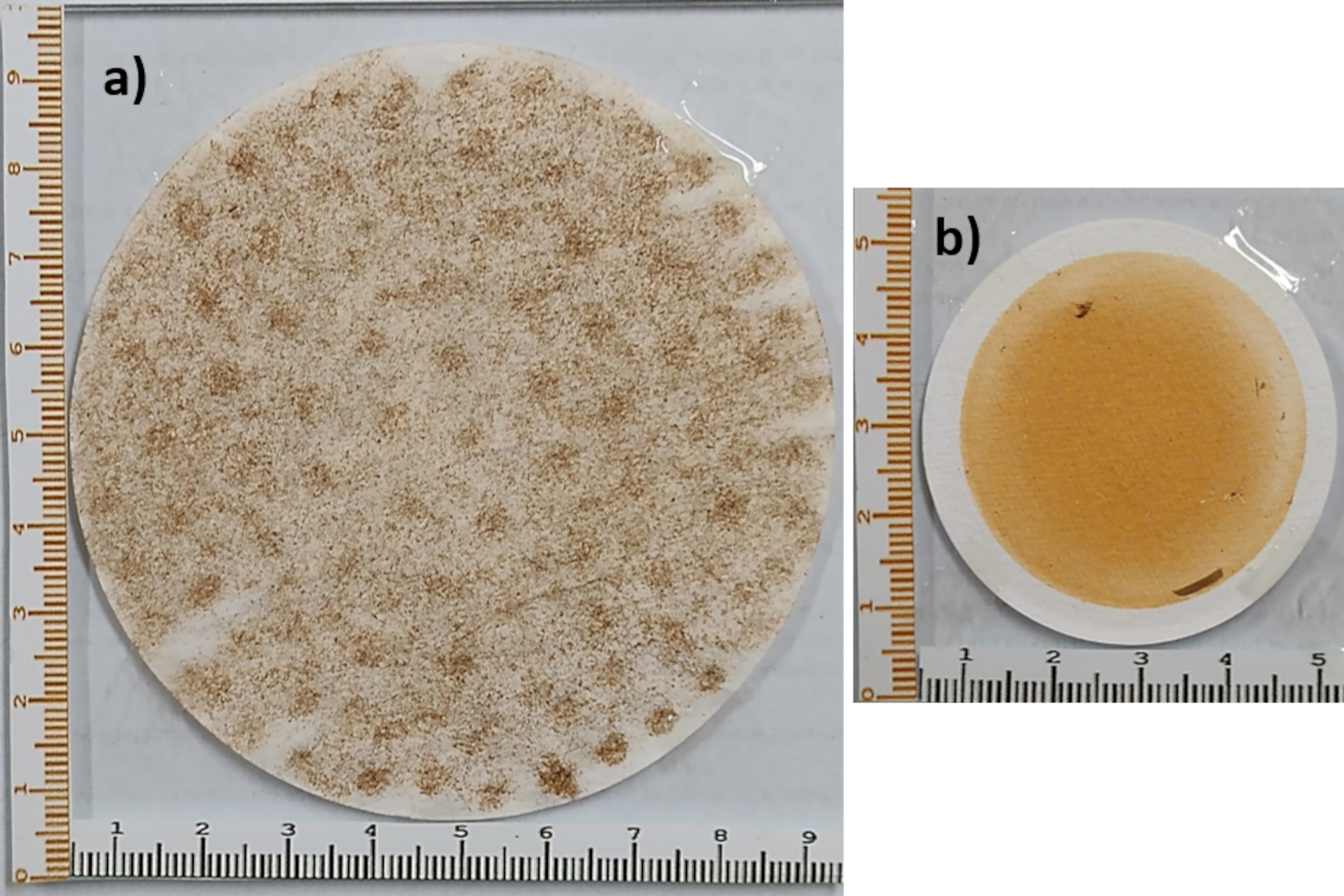
Considering that the main problem was the absence of a system that would make it possible to record the spatial location of the observed microplastics, an attempt was made to develop a system that would make it possible, making the work easier for the operator. Thus, based on the Cartesian principles of the geographic coordinate system, in which the location of any point on the Earth can be obtained by intersecting the meridians with the parallels, the solution found was the use of glass slides with graduations on two axes (Figure 3).

Proposed spatialization system containing a graduated glass plate with coordinates on two axes: a system with a 9.0 cm diameter cellulose filter and a 14 µm pore size (a) and a system with a 4.7 cm diameter glass fiber filter and a 1.2 µm pore size (b).
As can be seen, the glass slides have different sizes (9.5 cm x 9.5 cm and 5.5 cm x 5.5 cm), given that the filters used have different diameters (9.0 cm and 4.7 cm). To facilitate the visualization and minimize the possibility of confusion when noting the localization of the microplastic fragment, it was decided to differentiate the markings with different colors, one axis being red and the other black colors. The graduation of both slides is in millimeter units.
With the filters properly fixed to the glass slides, the scanning was carried out again under the optical microscope so that, for each material found, notes were made regarding the spatializations, e.g., 5.5 cm (red axis) and 4.5 cm (black axis). At the end of the process, the plates were again taken to Micro-Raman to evaluate the effectiveness of the spatialization methodology developed.
The first procedure performed was to establish correlations between the glass slide markings and the Micro-Raman spatialization system. To achieve it, returning to the example mentioned above, the focal light was positioned at the references of the red and black axes to record the values of the X and Y axes of the equipment (Figure 4).

Process to localize the microplastic fragments through micro-Raman using the proposed spatialization system: Initially, the light focus is positioned on one of the axes (a) and then moves until it finds the point of intersection with the second axis (b).
Once the focal light was placed, the process of searching for materials began, which, this time, were in an average time of 2 minutes. This time was maintained regardless of whether the filter had a diameter of 4.7 cm or 9.0 cm. Figure 5 shows the spectrum of a microplastic fiber found on a paper filter using the spatialization procedure developed in the present work. The highlighted Raman peaks are assignments of the vibrational modes of the poly (ethylene terephthalate - PET) chains, as shown by the work of Rizzo et al. (2018).

Raman spectrum of a PET fiber located on the paper filter using the spatialization procedure.
Despite the procedure proved to be advantageous, some observations arose that needed further investigation. A pertinent comment is whether there would be a difference if one technician carried out the spatialization using an optical microscope and another carried out the analysis using micro-Raman. The main justification for the question is because different degrees of training are required to operate these devices since the equipment provides different information. However, when focusing on the skills in localization using an optical microscope or the microscope present in the micro-Raman, the same technician could locate the microplastic fragments, as well as for other types of microscopes. Thus, to evaluate the influence of the operator on the sample localization, the first operator obtained an average time of 2 minutes (± 0.18) to locate each sample in the filter. On the other hand, when analyzing the samples prepared by the second operator, the average time was 2 minutes and 12 seconds (± 0.20), an increase of approximately 17% in localization time. It is important to consider that the times described above refer only to the localization process, not computing the analysis time, that is, how long each sample was under the laser’s incidence. As well known, the time spent analyzing each material depends on different factors, which are not the scope of this work.
Therefore, if strict description criteria are maintained, such as color, shape, and position in relation to the axes, there will be major time reductions regardless of the operator and the laboratory where the analyses will be carried out. This system can contribute to improving the quality of the work, since it reduces the pressure involved, which is generated when searching for materials without any type of reference, including doubts as to whether they have not been lost in some previous process such as filter transportation. In this sense, as already mentioned, if the operator wants to analyze the samples on other equipment, the spatialization will be of great value. Furthermore, the possibility of reanalyzing the sample at different times makes this spatialization system very useful.
An important recommendation in this system refers to the positioning of the glass slides, whether in the spatialization or analysis procedure. In this regard, the operator must ensure that these glass slides are aligned with the base of the equipment, thus avoiding deviations in direction. These deviations can lead to the failure of the operation. Figure 6 shows the correct positioning of the glass slide with the filter and an erroneous positioning of the slide, causing an error of around 4 millimeters on both axes.

Positioning of the glass slides in the analysis equipment: a) correct positioning of the glass slide containing the filter, with the coordinates aligned to the equipment and b) a glass slide with the filter misaligned, causing an error when finding the microplastic fragments.
As can be seen, if the glass slide is not positioned correctly, there will be variations in the spatialization of the materials in the filters. This occurs because the equipment bases move on fixed axes with an angle of 90 degrees. Therefore, when positioning the plate on the devices, it is necessary to make sure that the plates are completely aligned with their bases, as if they are not there will be a change in angle that, consequently, interferes with the location of the materials. To illustrate it, while on the correctly positioned plate (Figure 6a) the material is visibly located at 3.5 cm from the red axis and 3.0 cm from the black axis, on the plate that was placed incorrectly it appears that the material has moved to the left position 3.1 cm from the red axis and 2.6 cm from the black axis (Figure 6b).
According to the previous discussion, the black axis of the glass slide is strictly aligned with the metal base of the micro-Raman. An important detail to be said is that there is no need to align the two axes. This is because given the fact that the base of most devices has a rectangular base with right angles, when aligning one side the other will also be aligned.
CONCLUSIONS
The present work establishes a simple system for spatializing samples on a micrometric scale, in this case, microplastic fragments found in water, microscope techniques, making it easier for the operator to locate the sample and reducing the working time. In this way, the filters were analyzed using an optical microscope and a micro-Raman spectrometer, reducing the time to find the fragments for characterization from approximately 2 hours and 45 minutes to approximately 2 minutes and 20 seconds. Finally, given the results, it is stated that the developed system proved to be completely effective and was therefore incorporated into the analysis routines of the laboratories where the authors are involved, speeding up the localization and consequently the process of analysis and identification of each microplastic fragment found.
Acknowledgements
This study was financed in part by the Coordenação de Aperfeiçoamento de Pessoal de Nível Superior – Brazil (CAPES) – Finance Code 001. This study was financed in part by the Fundação de Amparo à Pesquisa do Estado de São Paulo (FAPESP) (EMU 2014/11410-8).
References
-
CANTISANI E, FRATINI F & PECCHIONI E. 2022. Optical and Electronic Microscope for Minero-Petrographic and Microchemical Studies of Lime Binders of Ancient Mortars. Minerals 12: 41. https://doi.org/10.3390/min12010041.
» https://doi.org/10.3390/min12010041 - GIRÃO AV. 2022. SEM/EDS and Optical Microscopy Analysis of Microplastics. In: Rocha-Santos T, Costa M & Mouneyrac C (Eds), Handbook of Microplastics in the Environment, Springer. https://doi.org/10.1007/978-3-030-39041-9_7.
-
GOLA D, TYAGI PK, ARYA A, CHAUHAN N, AGARWAL M, SINGH SK & GOLA S. 2021. The impact of microplastics on marine environment: A review. Environ Nanotechnol Monit Manage 16: 100552. https://doi.org/10.1016/j.enmm.2021.100552.
» https://doi.org/10.1016/j.enmm.2021.100552 - HERMAN B & LEMASTERS JJ. 1993. Optical Microscopy: Emerging Methods and Applications. Academic Press. ISBN 0123420601.
-
HOBSON CM, AARON JS, HEDDESTON JM & CHEW TL. 2021. Visualizing the Invisible: Advanced Optical Microscopy as a Tool to Measure Biomechanical Forces. Front Cell Dev Biol 9: 706126. https://doi.org/10.3389/fcell.2021.706126.
» https://doi.org/10.3389/fcell.2021.706126 -
JORGE JCF, SOUZA LFG, MENDES MC, BOTT LS, ARAÚJO IS, SANTOS VR, REBELLO JMA & EVANS GM. 2021. Microstructure characterization and its relationship with impact toughness of C–Mn and high strength low alloy steel weld metals – a review. J Mater Res Technol 10: 471-501. https://doi.org/10.1016/j.jmrt.2020.12.006.
» https://doi.org/10.1016/j.jmrt.2020.12.006 -
KANKANIGE D & BABEL S. 2020. Smaller-sized micro-plastics (MPs) contamination in single-use PET-bottled water in Thailand. Sci Total Environ 717: 137232. https://doi.org/10.1016/j.scitotenv.2020.137232.
» https://doi.org/10.1016/j.scitotenv.2020.137232 -
KASMURI N, TARMIZI NAA & MOJIRI A. 2022. Occurrence, impact, toxicity, and degradation methods of microplastics in environment—a review. Environ Sci Pollut Res 29: 30820-30836. https://doi.org/10.1007/s11356-021-18268-7.
» https://doi.org/10.1007/s11356-021-18268-7 - LE RU E & ETCHEGOIN P. 2008. Principles of Surface-enhanced Raman Spectroscopy and Related Plasmonic Effects. Elsevier. ISBN 978-0444527790.
-
LIU F, NORD NB, BESTER K & VOLLERTSEN J. 2020. Microplastics Removal from Treated Wastewater by a Biofilter. Water 12: 1085. https://doi.org/10.3390/w12041085.
» https://doi.org/10.3390/w12041085 -
MA L & FEI B. 2021. Comprehensive review of surgical microscopes: technology development and medical applications. J Biomed Opt 26(1): 010901. https://doi.org/10.1117/1.JBO.26.1.010901.
» https://doi.org/10.1117/1.JBO.26.1.010901 -
MASIÁ P, SOL D, ARDURA A, LACA A, BORRELL YJ, DOPICO E, LACA A, MACHADO-SCHIAFFINO G, DÍAZ M & GARCIA-VAZQUEZ E. 2020. Bioremediation as a promising strategy for microplastics removal in wastewater treatment plants. Mar Pollut Bull 156: 111252. https://doi.org/10.1016/j.marpolbul.2020.111252.
» https://doi.org/10.1016/j.marpolbul.2020.111252 -
MONTAGNER CC, DIAS MA, PAIVA EM & VIDAL C. 2021. Microplásticos: ocorrência ambiental e desafios analíticos. Quím Nova 44: 1328-1352. https://doi.org/10.21577/0100-4042.20170791.
» https://doi.org/10.21577/0100-4042.20170791 -
PRATA JC, PAÇO A, REIS V, COSTA JP, FERNANDES AJS, COSTA FM, DUARTE AC & SANTOS TR. 2020. Identification of microplastics in white wines capped with polyethylene stoppers using micro-Raman spectroscopy. Food Chem 331: 127323. https://doi.org/10.1016/j.foodchem.2020.127323.
» https://doi.org/10.1016/j.foodchem.2020.127323 -
RICHTER S, HORSTMANN J, ALTMANN K, BRAUN U & HAGENDORF C. 2023. A reference methodology for microplastic particle size distribution analysis: Sampling, filtration, and detection by optical microscopy and image processing. Appl Res 2: e202200055. https://doi.org/10.1002/appl.202200055.
» https://doi.org/10.1002/appl.202200055 -
RIZZO P, TROTTA D, MUSTO P & GUERRA G. 2018. Vibrational Spectra of Poly (ethylene terephthalate) Chains in the Mesomorphic Form. Macromol Chem Phys 219(3): 1700362. https://doi.org/10.1002/macp.201700362.
» https://doi.org/10.1002/macp.201700362 -
SARKAR DJ ET AL. 2021. Microplastics removal efficiency of drinking water treatment plant with pulse clarifier. J Hazard Mater 413: 125347. https://doi.org/10.1016/j.jhazmat.2021.125347.
» https://doi.org/10.1016/j.jhazmat.2021.125347 -
SHEN M, ZHANG Y, ALMATRAFI E, HU T, ZHOU C, SONG B, ZENG Z & ZENG G. 2022. Efficient removal of microplastics from wastewater by an electrocoagulation process. Chem Eng J 428: 131161. https://doi.org/10.1016/j.cej.2021.131161.
» https://doi.org/10.1016/j.cej.2021.131161 -
SINGH A & MISHRA BK. 2023. Microbeads in personal care products: An overlooked environmental concern. J Clean Prod 427: 139082. https://doi.org/10.1016/j.jclepro.2023.139082.
» https://doi.org/10.1016/j.jclepro.2023.139082 -
VENKATESHAIAH A, PADIL VVT, NAGALAKSHMAIAH M, WACLAWEK S, ČERNÍK M & VARMA RS. 2020. Microscopic techniques for the analysis of micro and nanostructures of biopolymers and their Derivatives. Polymers 12: 512. https://doi.org/10.3390/polym12030512.
» https://doi.org/10.3390/polym12030512 -
VETHAAK AD & LEGLER J. 2021. Microplastics and human health. Science 371: 672-674. https://doi.org/10.1126/science.abe5041.
» https://doi.org/10.1126/science.abe5041 -
WANG C, ZHAO J & XING B. 2021. Environmental source, fate, and toxicity of microplastics. J Hazard Mater 407: 124357. https://doi.org/10.1016/j.jhazmat.2020.124357.
» https://doi.org/10.1016/j.jhazmat.2020.124357