Analytical challenges and advances in detecting microplastics in human tissue and organ samples
Abstract
Microplastic contamination of biological matrices is a pressing concern in environmental and biomedical research. While their presence has been confirmed in human blood, placentas, lungs, and other tissues, accurate identification remains an analytical challenge. Obstacles include the small size and heterogeneous composition of microplastics, as well as spectral interference from complex organic matter. This review systematically analyzes—following PRISMA 2020 guidelines—the current state of microplastic detection in human tissues, evaluating 218 studies (2000-2024). We focus on the primary techniques: Fourier Transform Infrared (FTIR) and Raman spectroscopy, and Pyrolysis-Gas Chromatography/Mass Spectrometry (Py-GC/MS). Our analysis confirms FTIR as the most widely employed technique, followed by Raman and Py-GC/MS, with each method exhibiting distinct advantages and limitations in sensitivity, specificity, and sample preparation requirements. A critical finding is the widespread lack of robust quality assurance; notably, few studies consistently implemented blank controls and spike-recovery experiments, thereby compromising the reproducibility and reliability of data. In response, we propose a consolidated framework of best practices for sample handling, contamination control, and analytical validation. This review underscores the urgent need for standardized, validated protocols to refine analytical techniques, ensure consistent results, and ultimately support accurate risk assessment of human exposure to microplastics.
Key words
Microplastics; Biological tissues; FTIR; Raman spectroscopy; Py-GC/MS
INTRODUCTION
The issue of microplastic contamination in the environment has attracted interest in academia, resulting in a growing number of published papers in the last few years. Given that this subject relates to various scientific disciplines, the term “microplastics (MPs)” encompasses not just plastics but also any kind of polymer in a size of 1mm to 5 mm. Nanoplastics (NPs) are polymer particles with a size lower than 1 mm. Microplastics are classified as primary and secondary based on their source. Primary microplastics refer to particles of small size that are directly produced by industry, such as those found in cosmetics, toothpaste, detergents, and other products. Secondary microplastics are produced from the degradation and fragmentation of polymer product waste that is disposed of in the environment (Zhang et al. 2025, Kang et al. 2025, Liu & Zheng 2025, Moreira et al. 2025). Kang et al. (2025) reported that soil microplastic contamination is primarily caused by agricultural plastic products and domestic waste, traffic emissions, and plastic materials from civil construction and industry. The authors reported that soils are contaminated with a variety of polymers, including polyethylene (PE), polypropylene (PP), polystyrene (PS), poly(ethylene terephthalate) (PET), poly(vinyl chloride) (PVC), and polyamide (PA), with polyethylene being the most prevalent. Understanding these sources and their movement is crucial for developing effective strategies to mitigate environmental. MPs can enter the human body by ingestion of drinking water and practically all types of beverages and food into the human digestive system. This fact contributes to the importance of quantifying human exposure to MPs in seafood and their implications for human health. MPs can also enter the respiratory tract through polluted air inhalation, which happens in both outdoor and indoor environments. Studies highlight inhalation as a major route of human microplastic exposure, estimating that humans inhale up to 73,000 MPs per year. The explosion by dermal contact is considered a less significant route since the potential of microplastic to penetrate skin is highly influenced by particle size, given that human skin pores typically measure between 10 and 50 μm in diameter. For instance, the use of facial cleansers, toothpaste, and other household products that contain MPs can lead to the penetration of microplastics into skin. Considering their prevalence in daily life, they can pose a potential health risk, which has led to a significant increase in the number of recently published papers that aim to investigate this subject (Zhu et al. 2024, Li et al. 2024a, Zuri et al. 2023).
Özsoy et al. (2024) report the presence of MPs in the human stomach, having extracted 97 particles from 26 cadavers. Among the particles extracted, the majority of them were fibers, fragments, and films. Rotchell et al. (2024) investigate MPs contamination in urine samples from both healthy individuals and those with endometriosis. They found no difference in MPs levels, but the predominant types of polymers did differ. In healthy donor samples, the most prevalent polymers were PE, PS, and PP, whereas in participants with endometriosis, polytetrafluoroethylene (PTFE) and PE were more common. Leslie et al. (2022) identified and quantified PET, PE, PS, and poly(methyl methacrylate) (PMMA) in human blood, revealing that they are bioavailable for uptake into the human bloodstream. Ibrahim et al. (2021) analyzed colectomy samples from eleven adults (mean age 45.7, six males) employing stereo-microscopes and Fourier Transform Infrared (FTIR) techniques. They found MPs in all eleven samples, with the majority of them being filaments or fibers. The polymers identified are polycarbonate, polyamide, and polypropylene. Other studies also detected MPs in human feces through the use of FTIR (Schwabl et al. 2019, Zhang et al. 2021). Li et al. (2024a) stated that current methods for identifying MPs face challenges related to sensitivity and specificity and that organic compounds in biological samples may hinder detection efforts. To enhance the accuracy of research and comprehension of MPs’ behavior within the human body, it is essential to develop innovative analytical methods, increase the sensitivity and specificity of existing techniques, and establish standardized detection protocols.
The concern about the impact of MPs on human health is increasing. Their detection in human tissue and organ samples is needed to provide evidence that they can cross biological barriers and be retained in the human body. This direct evidence of MPs in the human body allows the scientific community to understand how this process happens and provides the essential link between exposure and potential toxicological effects. Therefore, given the multidisciplinary nature and the importance of this subject, this review analyzes the existing literature on microplastic identification in human tissue and organs, as well as invertebrates, seafood, and environmental matrices due to their methodological similarities. The purpose of this review is to explore the existing evidence regarding the detection of MPs, focusing on the methodologies employed, while emphasizing the advantages and drawbacks of these techniques to identify significant gaps in knowledge and suggest opportunities for future research.
Literature review methodology
This review examined papers published in peer-reviewed journals indexed in the Web of Science (WOS), Scopus, and PubMed databases. We utilized papers published in English, omitting book chapters, conference proceedings, and reviews. A comprehensive search for potentially relevant documents was conducted using the following Boolean query: (microplastic*) in Title AND (characterization* OR identification* OR method* OR protocol*) in Topic AND (blood* OR stomach* OR intestine* OR lung* OR umbilic* OR placent* OR tissue* OR heart* OR brain*) in Topic. The timeframe was from 2000 to November 2024.
Paper selection was guided by the PRISMA 2020 framework (Haddaway et al. 2022), as illustrated in Figure 1. The research conducted in Web of Science, Scopus, and PubMed databases produced 543, 550, and 77 documents, respectively, for a combined total of 1,170 documents. Initially, duplicate records (n = 368) were removed using the Bibliometrix package 4.1.3 (Aria & Cuccurullo 2017) within RStudio (version 2024.09.1+394). Following the removal of 368 duplicates, the remaining 802 records underwent a reading screening of titles and abstracts to determine eligibility. Primary inclusion criteria focused on papers detecting microplastics in human health contexts, including human food samples and mammalian organs or tissues. Studies focusing on invertebrates, seafood, and environmental samples were included strictly for the methodological insights they provided relevant to human tissue analysis (e.g., digestion protocols, contamination control, and polymer identification performance); these were not treated as evidence of microplastic occurrence in human organs. Furthermore, papers explicitly naming analytical tests in their titles were also selected to support the methodological focus of this review. As a result, 584 papers were excluded since they did not meet the predefined inclusion criteria. Following the eligibility assessment, data from the 218 included papers were analyzed using the Bibliometrix package in the R programming language.

Data descriptive analysis
Figure 2 illustrates a significant increase in the number of papers during the period from 2014 to 2024, exhibiting an annual growth rate of 60.83%. China is the country with the highest participation in publication. A total of 1483 authors from various nationalities contributed to the 218 eligible papers, with an average of 8.39 authors and 47.86 citations per document. This scenario indicates that this subject has many collaborators and a substantial citation count, demonstrating a high level of interest from the academy. Table I lists the top five most globally cited papers.

(a) Annual production, (b) Main information, and (c) Corresponding Author’s Countries of eligible papers.
Leslie et al. (2022) conduct a study to characterize and semi-quantify five polymers, specifically PMMA, PP, PS, PE, and PET, through the pyrolysis-gas chromatography/mass spectrometry (Py-GC/MS) technique in twenty-two blood samples from anonymized and healthy adults. The microplastic particle size detection was set between 700 and 500,000 nm due to the filtration step and the inner diameter of the blood-drawing needles. The authors stated that PP was not detected in any of the donors, and just one donor was tested for PMMA. A total of 5 donors tested positive for PE, 8 for PS, and 11 for PET. Even though this study carefully planned the sampling and storage of samples, along with rigorous quality control at every procedural step to avoid self-contamination and erroneous identification, the authors observed discrepancies in certain duplicate measurements. Specifically, for the same donor, one duplicate measurement indicated a positive detection of microplastic, whereas the other duplicate did not detect it. The authors attributed this result to the inhomogeneous distribution of MPs in blood, along with the observation that the quantity of particles in the mass is near the detection limit of the technique. This outcome highlights the challenges faced in the identification of MPs in samples.
Ragusa et al. (2021) used Raman microspectroscopy to identify MPs in human placenta samples from six consenting patients with uneventful pregnancies. Although the authors reported a detection of twelve MPs in the four placenta samples, they do not directly identify polymer types but chemical products such as iron hydroxide oxide yellow and copper phthalocyanine. Given the fact additives, such as pigments, are added to polymer products in very low amounts, attributing a polymer detection to the presence of any additive used in formulation is a questionable affirmation that should be avoided considering the importance of this subject.
Jenner et al. (2022) conducted an analysis of thirteen lung tissue samples collected from eleven patients (5 females and 6 males, aged 32–77) using the micro-Fourier transform infrared (μ-FTIR) with a microscope, which has a lower size limit of detection of 3 μm. The study found a total of 39 MPs across 11 of the 13 samples, yielding an average of 3.00 ± 2.55 particles per sample, compared to the 0.53 ± 1.07 particles observed in the blank samples. The study identified twelve types of MPs particles, with PP (23%), PET (18%), and resin (alkyd/epoxy/hydrocarbon, 15%) being the most common. Unfortunately, this study just presented six FTIR spectra in the supplementary material.
Dehaut et al. (2016) made an interesting study that compared six protocols to digest organic matter from mussels, crabs, and fish samples, addressing technological limitations for monitoring microplastic contamination in human-consumption fish and shellfish. Fifteen different plastic polymers were evaluated using microscopic inspection, weighing, pyrolysis, gas chromatography, mass spectrometry, and Raman spectrometry. Five out of six tested protocols resulted in significant degradation of plastic particles or insufficient tissue digestion.
Hwang et al. (2019) assesses the toxicity of PP microplastics in normal cells, immune cells, blood cells, and murine immune cells through a variety of tests. The author milled PP samples and classified the particles into two size groups: approximately 20 μm, comparable to human-derived cells, and 25–200 μm. To enhance dispersion and boost the interaction between the PP and cells, 20 mg of PP was mixed with 200 μL of dimethyl sulfoxide (DMSO) solvent. Additionally, PP in powder form was used directly at concentrations of 0.1, 0.3, 1.5, 3.0, and 4.5 mg/well (milligrams in a multi-well plate). The size of PP particles was characterized using scanning electron microscopy (SEM). The authors state that PP particles exhibited a low cytotoxicity effect in size and concentration. However, at a high concentration, small-sized and dissolved in DMSO, PP particles stimulated the immune system and enhanced potential hypersensitivity by increasing the levels of cytokines and histamines in PBMCs, Raw 264.7, and HMC-1 cells.
Considering the top five cited papers, Py-GC/MS, Raman, FTIR, and microscopy were used to detect microplastic particles in biological samples. In order to identify most techniques used, we carried out an analysis of authors’ keywords because it is useful to identify the frequency of keyword usage and identify trending topics (Köse et al. 2024). The total number of author keywords in the 218 eligible papers is 742; however, some of these are variations of the same word or synonyms, for instance, “mouse and mice”, “polyamide and nylon”, “fibers and fibers”, “microplastic and microplastics” and “polystyrene, polystyrene microplastics, and micro-polystyrene”. Thus, it was necessary to group the synonymous keywords together. Furthermore, keywords associated and correlated with those used for document searches were eliminated due to their high frequencies in order to not input a bias in this analysis. Figure 3 shows the word cloud map and trend topics for the author’s keyword. The word map was generated for the word with a frequency of at least 5, and the trend topics graph was generated for words with a minimum frequency of 5 and a yearly count of 3 words.

Authors’ keywords: (a) Word cloud map (word frequency ≥ 5) and (b) trend topics graph (word frequency ≥ 5 and a minimum of 3 words per year) for the (Source: Biblioshiny).
The most cited polymer is PS, with a frequency of 36 times, while PET follows with 9 mentions and PE with 6. FTIR is the most used technique for characterizing microplastics, with a frequency of 24. Besides, researchers utilize Raman spectroscopy (16), Py-GC/MS (15), and scanning electron microscopy (SEM) (5). Figure 3b illustrates a decline in interest in the Raman technique, alongside an increasing emphasis on Py-GC/MS, while the utilization of FTIR remains solid in research. Additionally, SEM does not appear in the topic trends graphic (Figure 3a), suggesting a potential sporadic use of this technique. The keyword analysis supports the findings from the top five cited papers. To understand the current state of the art regarding the use of Py-GC/MS, Raman, and FTIR in the detection of microplastics in the biological samples, a screening of the 218 papers was conducted to identify the documents that mention those techniques not only in the author’s keywords but also in the title and in the abstract. This screening identified 73 documents, which we then used to generate the data used in this review (Alpaydin et al. 2024, Anderssen et al. 2022, Ao et al. 2023, Besseling et al. 2015, Bobori et al. 2022, Bonifacio et al. 2022, Braun et al. 2021, Campen et al. 2024, Chen et al. 2022, Colombo et al. 2023, Da Costa et al. 2021, 2023, Dantas et al. 2020, Debbarma et al. 2022, Dehaut et al. 2016, Demirelli et al. 2024, Deng et al. 2024, Garcia et al. 2024, Geppner et al. 2023, Giarratano et al. 2022, Guo et al. 2024, Horvatits et al. 2022, Ibrahim et al. 2021, Jenner et al. 2021, 2022, Kaloyianni et al. 2021, Ke et al. 2023, Keys et al. 2022, Krafft et al. 2023, Leonard et al. 2024, Leslie et al. 2022, Li et al. 2023, 2024b, Littman et al. 2020, López-Marcano et al. 2023, Lourenço et al. 2024, Lu et al. 2024, Maganti & Akkina 2023, Martinez-Tavera et al. 2021, Masoero et al. 2024, Michishita et al. 2023, Monteleone et al. 2019, Mu et al. 2023, Naidu 2019, Nigamatzyanova & Fakhrullin 2021, Ogunola et al. 2022, Parobková et al. 2024, Prata et al. 2022, Qiu et al. 2023, Ragusa et al. 2021, Rotchell et al. 2023, 2024, Salvia et al. 2023, Santini et al. 2024, De Simone et al. 2021, Sipps et al. 2023, Sun et al. 2024, Tokunaga et al. 2023, Xu et al. 2024, Wang et al. 2023, 2024a, b, Wu et al. 2023, Zhang et al. 2022a, b, c, d, 2024, Zhong et al. 2024, Zhu et al. 2023, Zitouni et al. 2020).
State of the art, challenges, and opportunities in microplastic detection techniques
MPs are present in the environment and, consequently, can be in the human body. However, identifying these contaminants in biological samples is challenging and requires specified analytical techniques such as FTIR, Raman, and Py-GC/MS. Each technique possesses unique advantages and limitations that significantly impact the accuracy of the results and the interpretation of data. Table II shows a comparison of differences between FTIR, Raman, and Py-GC/MS.
General differences between Fourier Transform Infrared Spectroscopy (FTIR), Raman Spectroscopy, and Pyrolysis-Gas Chromatography-Mass Spectrometry (Py-GC/MS).
For conventional FTIR, the practical lower detection limit is ~15–50 µm, while µ-FTIR enables simultaneous visual imaging, particle sizing, and spectral analysis down to 10–20 µm. Conventional Raman is limited to >20 µm, whereas µ-Raman detects particles as small as 1 µm (Ardini et al. 2025, Thaiba et al. 2023). Both FTIR and Raman face significant limitations with irregular particle shapes and surface irregularities, where reflectance mode introduces refractive errors from roughness that prevent correct signal acquisition, and ATR-FTIR requires intimate crystal contact that irregular/protruding surfaces cannot maintain, compromising spectral quality.
Given these differences, the ideal approach for microplastic identification in biological samples involves combining multiple techniques. The combined use of FTIR and Raman techniques can be useful to increase the accuracy of identification, and Py-GC/MS is especially helpful for analyzing small microplastics and mixtures of them. However, it’s important to be careful when interpreting the data because overlapping signals in FTIR, interference from fluorescence in Raman, and thermal degradation in Py-GC/MS can all affect the results. The development of standardized protocols and improved spectral databases is crucial for ensuring reliable analyses and a deeper understanding of the potential health effects of microplastics in the human body. Furthermore, carefully following specific steps for collecting and preparing samples, along with using quality checks, is important to avoid contamination and unclear results. These topics are presented and discussed in the following subitems.
Sampling, digesting, quality assurance, and control measurement
MPs are frequently found as fragments and fibers in the environment, detected in air, soil, and water, including unexpected places such as the South Pole. The majority of microplastics in the environment are secondary and typically small in size, which challenges their detection, identification, and classification without advanced approaches and instrumentation (Zheng et al. 2024, Zhang et al. 2022, Brander et al. 2020).
To ensure that biological samples stay pure and the results are accurate, strict rules must be followed to prevent the samples from getting contaminated by microplastics during the collection and treatment processes. For example, the sampling methods should incorporate additional information beyond the type of samples that were collected. It is advised that studies include a full description of the sampling location, as well as extra-aleatory samples (blanks) acquired during the sampling for controlling background contamination to avoid false positives. Additionally, a clear explanation of the digestion process should be provided, including how samples are handled, how instruments are cleaned, and what methods are used to check for contamination from the samples themselves. Standardization and thorough documentation of these methods make it easier to compare studies, which helps us learn more about where microplastics are found and how they can affect health. Publishing the protocols used in a clear way is therefore essential for the progress of microplastic detection and the development of effective ways to reduce their impact. Brander et al. (2020) emphasize the importance of standardized sampling and quality control protocols in MPs detection to ensure reliable results. They encourage having accurate documentation and stringent procedures to prevent contamination, facilitate better comparisons of studies, and advance understanding of microplastics’ contamination and impact.
Brander et al. (2020) discuss the principles of quality assurance (QA) and quality control (QC) for MPs sampling, extraction, and identification methods developed by various laboratory groups. The authors propose three strategies to mitigate self-contamination: (a) implementing effective sampling and laboratory protocols; (b) incorporating samples with background checks, such as field and procedural blanks, and applying blank subtraction or adjustments to the sample data; and (c) utilizing sampling blanks to determine the limit of detection (LOD) and quantification methods to evaluate whether the data obtained fall below a threshold established by collecting blank and laboratory blank samples. The United States Environmental Protection Agency (EPA) sets protocols for determining limits of detection (LODs), requiring at least seven blanks for non-plastic, mostly water-soluble pollutants. Further details regarding the use of blank samples in MPs detection protocols are available in the paper by Shruti & Kutralam-Muniasamy (2023).
Furthermore, the use of matrix spikes, which is considered one of the best practices in analytical chemistry, is recommended for inclusion in MPs detection protocols. Spiking is the deliberate addition of a known quantity of a reference compound—often called a spike or spiking solution—to a sample. The purpose is to assess the accuracy, precision, and reliability of an analytical method. This approach is useful for measuring the recovery of the methods used. For that, it is necessary to produce representative standard reference samples with microplastic particles in them that are inserted and analyzed at the same time as the samples for MPs detection. Similar to other methods for other analytes, researchers recommend a recovery rate of 80% or higher (Brander et al. 2020).
To further understand the methodology of microplastic protocols, we posed the following questions to the 73 selected papers: “Does the paper outline how to avoid sample contamination by microplastics during the sampling and digestion steps?” and “Does the paper include blank and spike samples?” Figure 4 shows the answers to the previous questions.

Questions and answers related to sampling and digestion approaches outlined in the 73 selected papers.
When we analyzed the papers that answered “YES” to questions 1 and 2, we observed that many of their strategies to prevent the sample contamination during the collection and digestion steps were remarkably comparable (Alpaydin et al. 2024, Bonifacio et al. 2022, Braun et al. 2021, Chen et al. 2022, Colombo et al. 2023, Da Costa et al. 2023, Dantas et al. 2020, Debbarma et al. 2022, Demirelli et al. 2024, Deng et al. 2024, Giarratano et al. 2022, Horvatits et al. 2022, Ibrahim et al. 2021, Jenner et al. 2021, 2022, Jiménez-Arroyo et al. 2023, Ke et al. 2023, Keys et al. 2022, Leonard et al. 2024, Leslie et al. 2022, Li et al. 2023, 2024b, Littman et al. 2020, López-Marcano et al. 2023, Lourenço et al. 2024, Maganti & Akkina 2023, Martinez-Tavera et al. 2021, Michishita et al. 2023, Monteleone et al. 2019, Naidu 2019, Ogunola et al. 2022, Prata et al. 2022, Qiu et al. 2023, Ragusa et al. 2021, Rotchell et al. 2023, 2024, Salvia et al. 2023, Santini et al. 2024, De Simone et al. 2021, Sun et al. 2024, Wang et al. 2024a, b, Wu et al. 2023, Zhang et al. 2022a, b, c, d, 2024, Zhao et al. 2023, Zhong et al. 2024, Zhu et al. 2023, Zitouni et al. 2020). Therefore, we organized the researchers’ actions into eight sets of protocols that can be used to minimize the sample contamination by microplastics, as illustrated in Figure 5.

Eight strategies for minimizing sample contamination by microplastics during sampling and digestion steps.
Among the 73 papers selected, only 9 indicated that they utilized the spike procedure; two were published in 2016 and 2022 (Dehaut et al. 2016, Leslie et al. 2022), while the remaining seven were published in 2024 (Leonard et al. 2024, Deng et al. 2024, Lu et al. 2024, Parobková et al. 2024, Wang et al. 2024a, b, Zhong et al. 2024). This result suggests that, although the spike approach is not commonly employed in MPs protocol investigations, there is a growing interest in its application within research.
The research conducted by Dehaut et al. (2016), Leslie et al. (2022), Leonard et al. (2024) and Lu et al. (2024) marks important progress in the area of MPs detection and quantification. Dehaut et al. (2016) focused mainly on food samples, applying a digestion method to recover polyamide-6 particles. They introduced ten polyamide-6 particles, approximately 500 mm in size (PA-6, sourced from commercial yellow-fluorescent fine fishing line), into the following samples: cod (Gadus mohua) fillets, saithe (Pollachius virens), and mussel. They carried out this spike procedure in triplicate. Thereafter, all samples were subjected to alkaline digestion (10% w/w KOH solution) heated at 60°C for 24 hours while stirring at 300 rpm. The authors determined the recovery percentage by comparing the count of yellow particles found on filters post-filtration to the number of particles that were initially spiked. Ultimately, the PA-6 particles that were recovered following digestion underwent analysis through Py-GC/MS.
Leslie et al. (2022) conducted an analysis of human blood, adding an extra layer of complexity. Their study aims to find PMMA, PP, PS, PE, and PET in human blood using Py-GC/MS methods, which also involved a careful spike protocol. To do the recovery procedure, eight times the mixture of those five polymer standards is spiked into blood samples at both low and high analyte concentrations. In addition, eight unspiked blood samples were added to the set. The process yielded 24 blood samples for pretreatment and Py-GC/MS analysis. At the same time, eight procedural blank analyses using analytical-grade MilliQ® water were conducted. The authors used these blanks to correct background polymer concentrations that may have been introduced during sample preparation and analysis of the unspiked blood, allowing them to determine which polymers were already present in the blood used in the recovery experiment. After measuring, the authors adjusted the spiked blood samples using data from the unspiked blood samples (without considering the MilliQ® procedural blank), and then they calculated the recoveries for the five different polymers. To determine recovery, the authors divide the measured polymer concentration (adjusted for blank) by nominal spiking concentration multiplied by 100%.
Leonard et al. (2024) also investigated microplastics in blood samples. They used a spike method, as did Leslie et al. (2022); however, they further modified the methodology by using pre-filtered water instead of blood, an alternative approach that allows circumventing the limitation of insufficient samples in spike procedures. The polymers used were polyvinyl chloride (PVC) powder, approximately 200 μm in diameter, and polypropylene (PP), 28 μm in diameter and 200 μm in length. The PVC and PP were added to pre-filtered water at two distinct concentrations (called “low” and “high”). They also introduced in their study ten blank samples collected alongside the blood samples to analyze any background contamination, which consisted of distilled water (pre-filtered) and air from the room. The total number of replications of the spiked sample was three per polymer type and dose. The authors then digested and filtered the spiked samples using the same methodology as the blood samples, except for the type of filter membrane. The number of particles on the whole filter was counted using a stereomicroscope equipped with software.
Lu et al. (2024) proposed a method, which does not require a complex separation process, for detecting and quantifying PET and polybutylene terephthalate (PBT) by analyzing the depolymerization products using gas chromatography-tandem mass spectrometry (GC-MS/MS). The authors collected water samples in glass bottles. The samples (1 L each) were filtered through a 1-μm membrane, lyophilized, and kept at -20°C. PET and PBT particles were spiked into water at three concentrations (10, 100, and 1000 mg L⁻¹) for recovery determination. Six samples per spiking concentration, plus six blanks, were submitted to the method for polymer depolymerization under optimal conditions. The approach of Lu et al. (2024) is efficient, but the specificity for only two types of polymers may limit its applicability in more comprehensive studies. Even though the research by Deng et al. (2024), Wang et al. (2024a, b), Parobková et al. (2024), and Zhong et al. (2024) applied the spike approach in their studies, they did not provide a clear explanation of the methods they followed. The use of the spike approach in protocols for microplastic detection and quantification is important because it provides more accurate and reliable results. By introducing known quantities of microplastics into samples, researchers can assess recovery and correct for potential contamination, increasing the credibility of their findings. This method helps overcome challenges posed by complex biological matrices and provides a standardized approach for comparison across studies, thereby enhancing the area of microplastic research.
Fourier Transform Infrared Spectroscopy (FTIR)
The review of the 73 chosen papers found that about half (49%, or 36 papers) used FTIR, µ-FTIR, or similar methods (like Laser Direct Infrared, LDIR) to study MPs in biological samples. The infrared spectroscopy techniques involve measuring the absorption of infrared radiation by a sample, producing a spectrum that displays the distinctive molecular vibrations of the examined materials (Colthup et al. 2012, Larkin 2017). Most studies used FTIR or µ-FTIR techniques to examine microplastics in various biological samples, especially in mammals or human-related situations (Bobori et al. 2022, Bonifacio et al. 2022, Braun et al. 2021, Campen et al. 2024, Chen et al. 2022, Colombo et al. 2023, Da Costa et al. 2023, Demirelli et al. 2024, Garcia et al. 2024, Ibrahim et al. 2021, Jenner et al. 2021, 2022, Kaloyanni et al. 2021, Keys et al. 2022, Leonard et al. 2024, Li et al. 2023, 2024b, Littman et al. 2020, Lourenço et al. 2024, Maganti & Akkina 2023, Martinez-Tavera et al. 2021, Monteleone et al. 2019, Ogunola et al. 2022, Santini et al. 2024, Sipps et al. 2023, Rotchell et al. 2024, Tokunaga et al. 2023, Wang et al. 2023).
The FTIR technique was mostly employed for the analysis of larger particle samples, for instance, samples with a size higher than 500 µm (Bonifacio et al. 2022, Colombo et al. 2023). However, there are studies that used it for analyzing smaller particles, such as Maganti & Akkina (2023), that use FTIR to analyze particle samples with sizes ranging from 2.02 to 10.7 µm. The μ-FTIR was mainly used for smaller particles, specifically between 5 and 250 µm (Jenner et al. 2021) and 10 to 45 µm (Bobori et al. 2022), but it can also be used for larger particles, including those from 800 to 1,600 µm (Ibrahim et al. 2021) and 12 to 2,475 µm (Jenner et al. 2022). Additional studies employed the LDIR technique (Sun et al. 2024, Deng et al. 2024, Guo et al. 2024, Zhu et al. 2023, Zhao et al. 2023, Qiu et al. 2023). LDIR combines detailed imaging with infrared spectroscopy, allowing for quick and accurate analysis of tiny particles and the mapping of microplastic distribution over large areas. However, the LDIR technique requires performing the particle concentration step after digestion.
Several measurement techniques can be employed during FT-IR analysis: transmission, attenuated total reflection (ATR), reflectance, and absorbance. Among the papers employing FTIR and µ-FTIR techniques, the predominant analysis mode is ATR (Bobori et al. 2022, Bonifacio et al. 2022, Campen et al. 2024, Da Costa et al. 2023, Demirelli et al. 2024, Garcia et al. 2024, Ibrahim et al. 2021, Littman et al. 2020, Ogunola et al. 2022, Sipps et al. 2023, Tokunaga et al. 2023, Wang et al. 2023). This predominance of ATR mode is due to its simplicity relative to conventional transmission and to its effectiveness in polymer identification. Transmission is the second most used mode (Braun et al. 2021, Chen et al. 2022, Keys et al. 2022, Jenner et al. 2021, Leonard et al. 2024, Li et al. 2023, 2024b, Santini et al. 2024, Sipps et al. 2023, Rotchell et al. 2024), followed by reflectance (Campen et al. 2024, Lourenço et al. 2024) and absorbance (Kaloyianni et al. 2021). For the LDIR, papers report trans-reflectance as a mode of analysis (Sun et al. 2024, Zhu et al. 2023, Zhao et al. 2023).
Regarding the set of parameters of FTIR analysis, the total number of scans ranges from 1 to 256, with 64 scans being the most used. The number of scans enhances spectrum quality by reducing random noise and improving the signal-to-noise ratio but also increases spectrum acquisition time. The spectral resolution, or the capacity to discern closely spaced peaks in a spectrum, varied between 1 and 16 cm. The scan range varied from 4000 to 400 cm⁻¹ for FTIR and µ-FTIR techniques and from 1800 to 900 cm⁻¹ for LDIR. Only one study (Masoero et al. 2024) analyzed the range of 13,500-9,300 cm⁻¹, which falls in the near-infrared (NIR) region, using the reflectance mode.
The most common supports for samples for FTIR and µ-FTIR are diamond compression cells, KBr, gold foils, and CaF₂ substrates (Garcia et al. 2024, Jenner et al. 2021, Kaloyianni et al. 2021, Lourenço et al. 2024, Santini et al. 2024, Sipps et al. 2023). For the LDIR technique, the most support used is highly reflective slides, such as high-reflectivity glass or low-emissivity slides (Deng et al. 2024, Guo et al. 2024, Sun et al. 2024, Zhao et al. 2023). Sipps et al. (2023) and Santini et al. (2024) state the use of sample support in FTIR-ATR. However, it is mandatory that it be transparent in the infrared region to avoid any interference in the analysis (Löder & Gerdts 2015, Primpke et al. 2018). Additionally, there are analyses performed on the filtration membrane (Jenner et al. 2022, Keys et al. 2022, Leonard et al. 2024, Monteleone et al. 2019, Rotchell et al. 2024). In this instance, they employed the aluminum oxide membrane (Anodisc), which is transparent in the infrared spectrum (Löder & Gerdts 2015, Primpke et al. 2018).
Most of the reviewed papers used spectrum library software to identify the type of MPs. The most cited library is the Omnic Picta and Omnic Polymer Libraries (Bonifacio et al. 2022, Braun et al. 2021, Campen et al. 2024, Chen et al. 2022, Colombo et al. 2023, Da Costa et al. 2023, Demirelli et al. 2024, Deng et al. 2024, Garcia et al. 2024, Guo et al. 2024, Ibrahim et al. 2021, Jenner et al. 2021, 2022, Keys et al. 2022, Leonard et al. 2024, Littman et al. 2020, Li et al. 2023, 2024b, Lourenço et al. 2024, Martinez-Tavera et al. 2021, Masoero et al. 2024, Ogunola et al. 2022, Qiu et al. 2023, Rotchell et al. 2024, Santini et al. 2024, Sipps et al. 2023, Sun et al. 2024, Tokunaga et al. 2023, Zhao et al. 2023, Zhu et al. 2023). Further, only two papers stated that they manually interpreted the bands in addition to using the library (Li et al. 2023, 2024b). Only three papers used polymer standard samples to compare and identify the type of microplastics (Braun et al. 2021, Kaloyianni et al. 2021, Wang et al. 2023). The match index for comparing the results with the library ranged between 50 and 90%. However, most reported a minimum threshold of 70% to consider an identification reliable. Four studies found that they could identify additives using FTIR, including phthalates, bisphenol A, muscovite, palmitic acid, and others (Leonard et al. 2024, Rotchell et al. 2024, Li et al. 2023, Sipps et al. 2023).
In our opinion, the FTIR can be used to identify microplastics based on the absorption of infrared radiation by specific functional groups in the polymer structure. This technique generates spectra that functions as fingerprints, allowing comparison with reliable spectral libraries for sample identification. Further, it is important to have one’s own standard library of spectra for each specific plastic, changing their color and texture, along with the degree of degradation of the samples, to limit the possibility of receiving false positive and negative results. Despite its widespread use, FTIR has limitations when applied to biological samples. The presence of proteins and lipids can interfere with the spectrum, complicating the distinction between MPs and naturally occurring compounds. Therefore, effective sample pretreatment is required to eliminate these interferences, which can be time-consuming and challenging, especially for smaller particles. Additionally, overlapping spectra of polymers with similar functional groups may reduce the accuracy of identification. MPs smaller than 10 µm are often difficult to detect without using microscopy-coupled FTIR (µ-FTIR), which limits its use in biological samples.
Raman spectroscopy
Researchers use Raman spectroscopy, a nondestructive technique, to examine MPs in environmental and biological samples. The review of the 73 selected papers found that 37% use Raman along with other methods to thoroughly and effectively study MPs. Using Raman with imaging systems allowed researchers to look at the shape of samples and get their spectra at the same time (Dai et al. 2024, Krafft et al. 2023, Parobková et al. 2024).
Regarding the methodological aspects, the selection of the excitation wavelength proved to be a crucial factor in improving the intensity of the Raman signal and reducing fluorescence interference. The wavelengths of 785 nm and 532 nm were the most commonly used (Alpaydin et al. 2024, Chen et al. 2022, Da Costa et al. 2021, Dantas et al. 2020, Dehaut et al. 2016, Geppner et al. 2023, Giarratano et al. 2022, Horvatits et al. 2022, Krafft et al. 2023, Michishita et al. 2023, Mu et al. 2023, Naidu 2019, Parobková et al. 2024, Ragusa et al. 2021, De Simone et al. 2021, Sipps et al. 2023, Wang et al. 2024a, Wu et al. 2023, Zitouni et al. 2020).
The selection of the microscope objective was dependent upon the characteristics of the particles under examination, with the 50× objective being the predominant option for obtaining precise details regarding their morphology and composition. Further, images were captured using magnifications of 10×, 20×, and 100×, depending on the area of interest and particle size (Ao et al. 2023, Dehaut et al. 2016, De Simone et al. 2021, Geppner et al. 2023, Horvatits et al. 2022, Wu et al. 2023).
The spectral analysis range focused mainly on the region of 600–1800 cm-1, known as the “fingerprint” region of the most common synthetic polymers, such as polyethylene (PE), polypropylene (PP), and polystyrene (PS) (Alpaydin et al. 2024, Krafft et al. 2023). Additionally, analysis was also done in the range of 2500–3000 cm-1 to identify other important materials, like pigments associated with microplastics (Ao et al. 2023, Wu et al. 2023). At this point, it is important to pay attention to the fact that it is prudent not to report on the presence of any MPs solely based on the detection of materials that are commonly used as additives, i.e., without identifying the specific type of polymer. Additives are usually added in small amounts to polymer formulations; therefore, it is not expected to detect only the additive.
In most of the studies, the identification of polymer types was done by comparing the experimental Raman spectra with polymer spectral libraries, such as the Open Specy Library, KnowItAll, SLoPP, and SLoPP-E. The adoption of a match quality index above 70% was frequently used as a criterion to validate the identification of polymers (Alpaydin et al. 2024, Wu et al. 2023, Giarratano et al. 2022, Chen et al. 2022).
Raman spectroscopy can analyze microplastics from the environment (like water and sediments) and from living things (such as blood, placenta, lung tissue, and tissues from animals like fish and mussels) right on filter membranes. Even though it is possible to analyze samples with minimal preparation, most of the reviewed studies employed specific preparation protocols for complex biological samples. The procedure involves the alkaline digestion (using KOH or NaOH) of tissues to eliminate organic material. Then, a filtration step is performed to collect and hold onto microplastics on membranes made of materials such as fiberglass, silver, or polycarbonate. This preparation process is recommendable for samples from humans and animals because organic materials could disrupt the Raman signal or create unwanted fluorescence. Another example of the versatility of the Raman technique is highlighted in the analysis of complex matrices, such as synovial fluid. After this sample was treated by enzymatic digestion followed by filtration, it was possible to detect MPs particles smaller than 10 µm (Alpaydin et al. 2024, Chen et al. 2022, Dehaut et al. 2016, Horvatits et al. 2022, Li et al. 2024b, Naidu 2019, Parobková et al. 2024, De Simone et al. 2021).
The most common types of plastics found in studies using Raman spectroscopy were polyethylene (PE), polypropylene (PP), polystyrene (PS), polyester (PET), poly(vinyl chloride) (PVC), and polyamide (PA) (Alpaydin et al. 2024, Da Costa et al. 2021, Li et al. 2024b, Zhang et al. 2024). The fibers, composed mainly of polyester and cellulose (including cotton and rayon), represented a common form of MPs (Chen et al. 2022, Da Costa et al. 2021, Giarratano et al. 2022, Li et al. 2024b). The majority of MPs were in the size range of 1 to 50 μm, with a significant proportion of particles smaller than 10 μm identified in biological tissues (Alpaydin et al. 2024, Ao et al. 2023, Chen et al. 2022, Parobková et al. 2024, Zhang et al. 2024). The predominant forms observed were fibers and fragments (Da Costa et al. 2021, Giarratano et al. 2022, Michishita et al. 2023).
The combination of Raman with other analytical techniques was demonstrated to be an effective strategy for a comprehensive characterization of microplastics. For instance, optical microscopy played an important role in the initial screening and morphological characterization of the particles (Alpaydin et al. 2024, Naidu 2019). The scanning electron microscopy (SEM/EDS and FE-SEM) provided important information about the morphology and elemental composition (EDS), especially for smaller particles and nanoparticles (Ao et al. 2023, Chen et al. 2022, Li et al. 2024b, Wang et al. 2024a).
In our opinion, Raman spectroscopy is a complementary technique to FTIR. It enables reliable detection of microplastics down to approximately 1 µm (µ-Raman), surpassing FTIR in spatial resolution. However, its application in biological samples presents significant challenges due to fluorescence interference and longer measurement times. One of the most common issues is fluorescence interference from biological components, which can obscure the microplastic signal, making detection difficult. Additionally, dark or carbonized polymers strongly absorb laser light, reducing the intensity of their Raman signal. The technique also faces difficulties in distinguishing MPs in complex mixtures, as some spectral profiles may be broad and less defined compared to FTIR.
Py-GC/MS spectrometry
Pyrolysis gas chromatography-mass spectrometry (Py-GC/MS) is a technique that breaks down polymers into volatile fragments for identification (Picó & Barceló 2020). Twelve of the 73 research papers examined in this review used Py-GC/MS to identify MPs, seven of which were published in 2024. This suggests that Py-GC/MS is a relatively newer technique to characterize microplastics than the other ones (Dehaut et al. 2016, Leslie et al. 2022, Ke et al. 2023, Maganti & Akkina 2023, Zhao et al. 2023, Campen et al. 2024, Deng et al. 2024, Garcia et al. 2024, Guo et al. 2024, Wang et al. 2024a, b, Zhong et al. 2024).
In a general view, the target polymers chosen in the 12 selected papers are PE, PP, PS, polyamides (PA6, PA66, and PA12), cellulose acetate (CA), PTFE, polyurethane (PU), PET, PMMA, PC, PVC, SBR, ABS, polycarbonate (PC), poly(lactic acid) (PLA), and PBAT (polybutylene adipate terephthalate). According to Kaal et al. (2023), Py-GC/MS can detect several polymers, but it requires the interpretation of complex pyrolytic degradation paths, catalytic effects, and bias that limit quantitative dependability. Therefore, it is recommended to review the pyrolytic degradation literature in order to identify the most relevant intermediate products for use in the analysis.
The mechanisms of pyrolysis for polymers include chain scission, unzipping, and scission. For example, when PE and PP undergo pyrolysis, the main chain breaks randomly, creating end radicals that form end alkenes and alkanes via hydrogen abstraction. The subsequent chain reactions produce methyl-branched terminal alkenes and dienes for PP and linear alkanes, alkenes, and alkadienes (ranging from C11 to C33) for PE. According to literature (Kaal et al. 2023, Peel et al. 2025), the most abundant products from pyrolysis of PP are 2,4-dimethylhept-1-ene and the three 2,4,6,8-tetramethyl undec-1-enes. For PE, the research recommends using C22, C23, and C24 terminal dienes, alkenes, and alkanes, as the pyrolysis of natural materials also creates these compounds, but with shorter chains (Peel et al. 2025). PMMA, PS, and PA6 largely pyrolyze in a process known as unzipping, which results in their monomer units, that is, methyl methacrylate, styrene, and 3-caprolactam, respectively. However, the pyrolysis of PVC, PET, and natural products also produces styrene, making it unsuitable for identifying PS. In this case, styrene dimer and trimer can be used, for instance, diphenylbutenes, bibenzyl, and α-methylbibenzyl (Kaal et al. 2023, Peel et al. 2025). PET produces benzene, toluene, styrene, naphthalene, and biphenyl. The pyrolysis of PVC produces HCl via side chain elimination, followed by aromatization, with the main product being benzene, followed by naphthalene, toluene, and indene. PVC does not produce any alkanes or alkenes (Kaal et al. 2023, Peel et al. 2025).
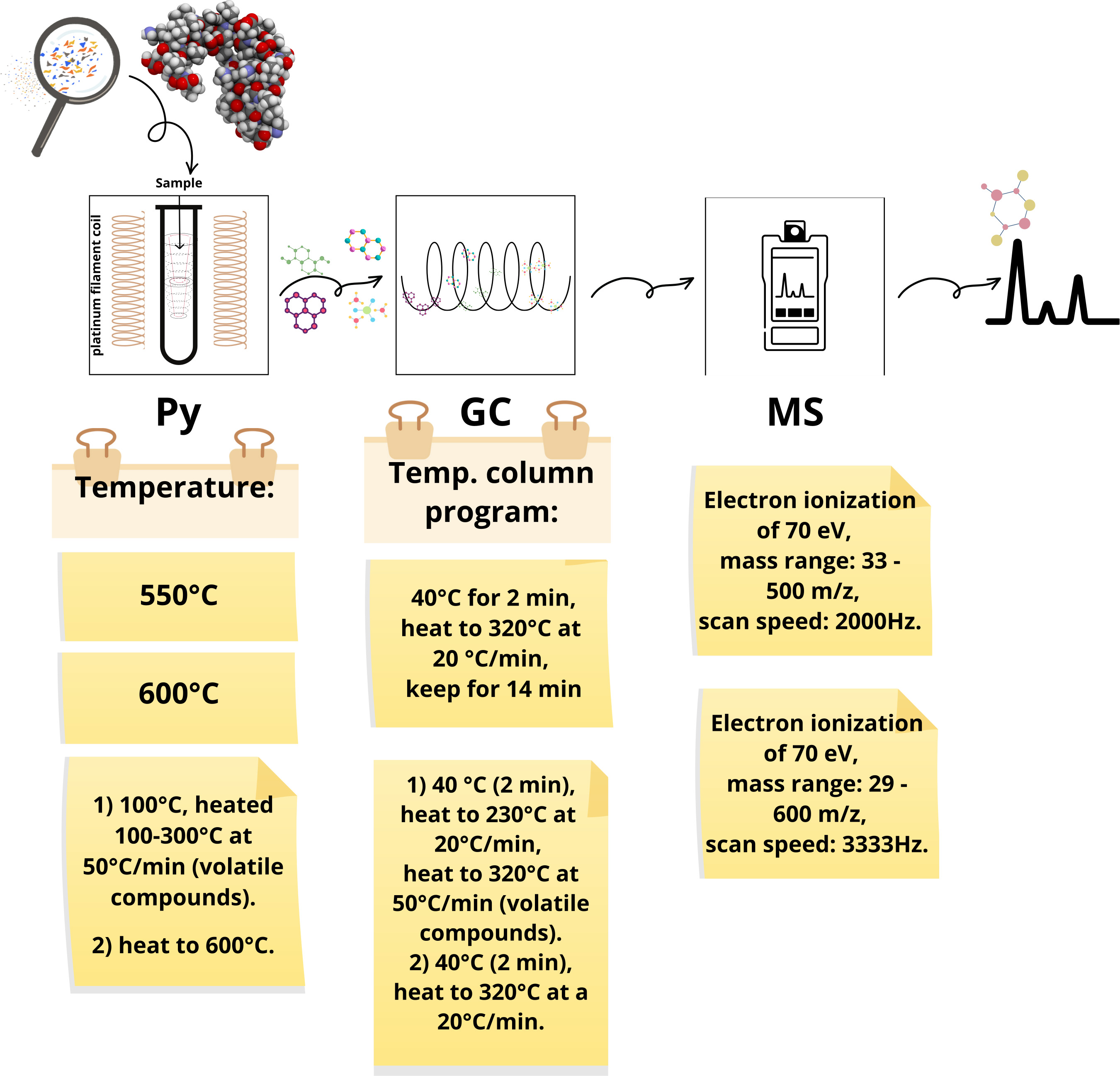
Dehaut et al. (2016) used the temperature of 600°C to pyrolyze the samples and helium as a carrier gas (40 cm s⁻¹). The temperature of the chromatogram column oven started at 40°C for 5 min, increased from 40 to 320°C at a rate of 20°C min⁻¹, and was kept at 320°C for 14 min. In this single-shot analysis, the sample is rapidly fragmented, and the degradation products are separated in the chromatographic column. The mass spectrometer was connected to the GC with the interface temperature at 300°C to avoid any recondensation. The ionization voltage was set to 70 eV, and the mass range was from 33 to 500 m/z with a scanning speed of 2000 Hz. The MPs were identified using F-Search software 4.3 and from the author’s database containing pre-established pyrograms of polymer samples. Further, the similarity of pyrograms of a minimum of 80% was set to certify the identification (Dehaut et al. 2016).
With slight differences in terms of temperatures and heating rates, for example, all the studies show significant similarities in Py-GC/MS protocol analysis to Dehaut et al. (2016). Garcia et al. (2024) stated the importance of carefully defining the pyrolysis temperature, hold time, and carrier gas (helium) flow rate for transporting pyrolysis products to the gas chromatography column. According to Leslie et al. (2022), who did double-shot Py-GC/MS, the monomers produced in the first shot of pyrolysis (100–300°C) cannot be used to identify MPs. The exception is PET, although it is recommended that both the first and second shots be combined. Additionally, Leslie et al. (2022) call attention to the fact that the presence of additive molecules in the first shot does not necessarily mean that MPs are present in a sample. This occurs because, for example, these additives may come from non-plastic sources or may leach from plastic materials to the food chain during their life cycle. Figure 6 shows examples of parameters used in the papers for Py-GC/MS analysis.

In a general view, the identification of polymer types was performed by comparison with databases from software libraries, such as F-Search software (Dehaut et al. 2016, Wang et al. 2024a), Agilent (Zhong et al. 2024, Zhao et al. 2023), LabSolutions software spectral library (Wang et al. 2024b), or through comparison with the author’s database containing standard polymer pyrograms (Maganti & Akkina 2023).
Given the recent application of Py-GC/MS analysis for detecting nano- and microplastics in human tissue, as mentioned in the first paragraph of this section, we extend our search and find papers whose main focus is to critically analyze the use of “Py-GC/MS” for micro- and nanoplastic identification (NPs, particles with size lower than 1 μm), regardless of the origin of the samples (Jing et al. 2025, Rauert et al. 2025).
Rauert et al. (2025) evaluated the efficacy of Py-GC/MS for identifying micro- and nanoplastics in human blood samples. Although they developed a protocol for digesting the samples and mitigating biological interferences, the results show that this technique is not adequate for analyzing PE and PVC in blood samples, since the presence of endogenous components, such as lipids and proteins, produces identical pyrolysis products, leading to false positives. In a pilot study with volunteers, PE interferences remained even after the sample was treated.
Jing et al. (2025) highlight the challenges associated with Py-GC/MS as an emerging technique for analyzing micro- and nanoplastics in various matrices (atmosphere, water, soil, and biological samples). For instance, they pose questions such as the interference of organic matter in the pyrolysis products and quantitative ions. Furthermore, there is a risk of contaminating the pyrolizer and ion source of Py-GC/MS, increasing the uncertainty in measurements. To contribute to the standardization of the protocols of Py-GC/MS, the authors propose four recommendations. The first one is that instead of “disperse,” it is better to “dissolve” the micro- and nanoplastics in an adequate solvent and conduct gradient dilutions to create standard curves. For polymers that present challenging dissolution conditions, it is preferable to use nano-sized plastics suspended in water or ethanol for gradient dilution. The smaller size allows for more precise control over the mass concentration. The second suggestion is to use isothermal thermal desorption to make pyrolysis methods more scalable. This process aims to eliminate residual small molecule interferences and measure volatile organic pollutants in samples. Choosing the appropriate desorption temperature and time is important for ensuring the efficient recovery of target polymers. Additionally, real environmental conditions may lead to biodegradation or combustion of polymers, resulting in stable small molecules that make differentiation via Py-GC/MS difficult. Therefore, integrating thermal desorption with the co-identification of multiple pyrolysis products is essential for effectively characterizing polymers. The third recommendation is to standardize the protocol for quantitative product screening. For that, it is important to optimize the pyrolysis conditions, such as temperature, heating rate, and residence time, to achieve accurate characterization. Since the pyrolysis of micro- and nanoplastics yields multiple products, it is important that the most characteristic pyrolysis products, with nearly 100% recovery as possible, should be used for quantification, while other pyrolysis products serve as qualitative references. One quantitative ion and two/three qualitative ions should correspond to the characteristic ions of the distinct fragments, avoiding common fragments like aromatic hydrocarbons and benzene rings. Further, low molecular weight fragments are often susceptible to matrix interference and should be carefully selected. The authors suggest as a final recommendation the use of multivariate statistics, machine learning, and deep learning for data analysis of Py-GC/MS (Jing et al. 2025).
CONCLUSIONS
The detection of MPs in human tissues and organs represents a critical frontier in environmental health research. Regardless of the technique used, it is imperative that the presence of MPs not be reported solely based on the detection of common additive materials; identification must be substantiated by unequivocal polymer-specific evidence. We highlight the importance of implementing stringent quality assurance and quality control protocols in studies of microplastic identification in tissue and human organ samples to improve the comparability across studies. Strict, well‑documented sampling, digestion, and QA/QC procedures are essential to avoid microplastic contamination and ensure data reliability. We recommend eight strategies for minimizing sample contamination by microplastics during sampling and digestion steps: (a) use cotton lab coats, masks, and gloves; (b) clean all workplaces, materials, and equipment; (c) filter reagents and water before use; (d) use ultrapure water; (e) cover samples, reagents, and devices; (f) adopt plastic-free materials protocols; (g) control the air in the workplace environment; and (h) collect samples to monitor workplace contamination.
FTIR and µ-FTIR techniques are valuable for identifying microplastics. It is essential to clean the equipment prior to analysis. The number of scans significantly affects the quality of the spectrum, with most studies opting for 64. Many studies utilize commercial spectral libraries for microplastic identification. Raman spectroscopy requires careful choice of the excitation wavelength. It is important to highlight that Raman analysis can be affected by the presence of biological sample residue and dark or carbonized polymers. So, in our opinion, Raman should be used with FTIR to determine whether microplastics are present or not.
It is recommended that researchers combine automated libraries with manual interpretation of spectral bands of FTIR and Raman. Additionally, incorporating polymer reference standards and developing an in-house spectral library that addresses variations in color, texture, and degradation states is recommended. During this analysis phase, maintaining a high match index of at least 80% is strongly suggested to minimize the risk of misidentification.
While Py-GC/MS is highly sensitive and can detect MPs, it also has several limitations. Thermal degradation can alter the original polymer structure, potentially leading to misidentifications. Complex biological samples may generate unexpected pyrolysis byproducts, complicating polymer differentiation. Additionally, plastic additives such as plasticizers and flame retardants may be detected, but they cannot always be directly linked to a specific polymer if it is not identified. The accuracy of the results heavily depends on the quality of the spectral database, as improper reference data can lead to erroneous identifications. Therefore, it is essential to develop and implement standardized protocols to ensure consistent, accurate, and reproducible detection of microplastics in biological samples.
Despite the growing number of publications, there are still important gaps to be addressed, especially regarding validation, detection thresholds, and the biological relevance of identified particles. Future efforts must focus on integrating analytical techniques, improving spectral databases, and promoting global standardization of sampling and detection protocols. This can include carrying out studies with a focus on standardization and inter‑laboratory, for instance. Such advancements are essential to ensure robust, reproducible, and reliable results that can support health risk assessments and public policy development related to microplastic exposure in humans.
In our opinion, the detection of plastics in human tissues faces critical technical bottlenecks at the nanoscale. Analytical techniques such as µ-FTIR and Raman spectroscopy allow for reliable particle counting, generally only above ~10 and 1 µm, respectively. Below this threshold, reduced sensitivity, diffraction limits, and biological matrix fluorescence significantly increase the risk of false positives and particle misclassification. Furthermore, analytical results are often reported either as particle counts (particle-based) or total mass (mass-based), which are metrics that are not directly comparable. Thermal methods such as Py-GC/MS can complement spectroscopy by providing qualitative chemical confirmation of the polymers; however, their application in human tissues remains not standardized. For accurate mass quantification, these thermal methods require robust calibration and the formal determination of limits of detection and limits of quantification.
To improve inter-study comparability and reduce false positives, we propose a minimum QA/QC for MPs detection in human tissues that focuses on required controls, documentation, and acceptance criteria rather than prescribing a laboratory protocol (Table III). At minimum, studies should (i) implement and report contamination controls across sampling, processing, and analysis; (ii) include blanks and spikes through the full workflow; (iii) define polymer identification criteria (spectral/library and confirmation rules) and false-positive mitigation steps; and (iv) state method-specific detection limits and size/mass thresholds. Studies not meeting these minimum elements should clearly label results as provisional and interpret polymer assignments cautiously.
Finally, it is important to state that researchers should question their results and consider if findings might be false positives rather than assuming all particles are microplastics. Studies that find no microplastic contamination are also important scientific results and should be reported.
Acknowledgements
The authors would like to thank the Conselho Nacional de Desenvolvimento Científico e Tecnológico (CNPq, 304054/2025-9 – Ana Maria F. de Sousa, and 420661/2023-9 - Mônica Regina C.M. Calderari, Thiago R. Correia, and Debora B. Pereira), Coordenação de Aperfeiçoamento de Pessoal de Nível Superior (CAPES, Financing code 001 - Ananda P.L. Dias and Rafael L. Pinto), and Fundação Carlos Chagas Filho de Amparo à Pesquisa do Estado do Rio de Janeiro (FAPERJ, SEI-260003/006163/2024 - APQ1 - Ana Maria F. de Sousa) for supporting the research.
References
-
ALPAYDIN AÖ ET AL. 2024. Microplastics, as a risk factor in the development of interstitial lung disease-a preliminary study. Environ Pollut 363: 125054. https://doi.org/10.1016/j.envpol.2024.125054.
» https://doi.org/10.1016/j.envpol.2024.125054 -
ANDERSSEN KE, GABRIELSEN GW, KRANZ M & COLLARD F. 2022. Magnetic resonance imaging for non-invasive measurement of plastic ingestion in marine wildlife. Mar Pollut Bull 185: 114334. https://doi.org/10.1016/j.marpolbul.2022.114334.
» https://doi.org/10.1016/j.marpolbul.2022.114334 -
AO J ET AL. 2023. Fast detection and 3D imaging of nanoplastics and microplastics by stimulated Raman scattering microscopy. Cell Rep Phys Sci 4(10): 101623. https://doi.org/10.1016/j.xcrp.2023.101623.
» https://doi.org/10.1016/j.xcrp.2023.101623 -
ARDINI B, PITTURA L, FRONTINI A, BENEDETTI M, GORBI S, REGOLI F, CERULLO G, VALENTINI G & MANZONI C. 2025. Fast detection and classification of microplastics by a wide-field Fourier transform raman microscope. Environ Sci Technol 59(18): 9255-9264. https://doi.org/10.1021/acs.est.5c00165.
» https://doi.org/10.1021/acs.est.5c00165 -
ARIA M & CUCCURULLO C. 2017. Bbibliometrix: An R-tool for comprehensive science mapping analysis. J Informetr 11(4): 959-975. https://doi.org/10.1016/j.joi.2017.08.007.
» https://doi.org/10.1016/j.joi.2017.08.007 -
BESSELING E ET AL. 2015. Microplastic in a macro filter feeder: humpback whale Megaptera novaeangliae Mar Pollut Bull 95(1): 248-252. https://doi.org/10.1016/j.marpolbul.2015.04.007.
» https://doi.org/10.1016/j.marpolbul.2015.04.007 -
BOBORI DC ET AL. 2022. Differentiation in the expression of toxic effects of polyethylene-microplastics on two freshwater fish species: Size matters. Sci Total Environ 830: 154603. https://doi.org/10.1016/j.scitotenv.2022.154603.
» https://doi.org/10.1016/j.scitotenv.2022.154603 -
BONIFACIO PSP, METILLO EB & ROMANO JR EF. 2022. Microplastic in sediments and ingestion rates in three edible bivalve mollusc species in a southern Philippine estuary. Water Air Soil Pollut 23311: 455. https://doi.org/10.1007/s11270-022-05926-w.
» https://doi.org/10.1007/s11270-022-05926-w -
BRANDER SM ET AL. 2020. Sampling and quality assurance and quality control: a guide for scientists investigating the occurrence of microplastics across matrices. Appl Spectrosc 74(9): 1099-1125. https://journals.sagepub.com/doi/10.1177/0003702820945713.
» https://doi.org/10.1177/0003702820945713 -
BRAUN T, EHRLICH L, HENRICH W, KOEPPEL S, LOMAKO I, SCHWABL P & LIEBMANN B. 2021. Detection of microplastic in human placenta and meconium in a clinical setting. Pharmaceutics 13(7): 921. https://doi.org/10.3390/pharmaceutics13070921.
» https://doi.org/10.3390/pharmaceutics13070921 -
CAMPEN M ET AL. 2024. Bioaccumulation of Microplastics in Decedent Human Brains Assessed by Pyrolysis Gas Chromatography-Mass Spectrometry. Research Square. https://doi.org/10.21203/rs.3.rs-4345687/v1
» https://doi.org/10.21203/rs.3.rs-4345687/v1 -
CHEN Q, GAO J, YU H, SU H, YANG Y, CAO Y & LIU H. 2022. An emerging role of microplastics in the etiology of lung ground glass nodules. Environ Sci Eur 34(1): 25. https://doi.org/10.1186/s12302-022-00605-3.
» https://doi.org/10.1186/s12302-022-00605-3 -
COLOMBO CV, FERNÁNDEZ-SEVERINI MD, FORERO-LÓPEZ AD, ARDUSSO MG, RIMONDINO GN, MALANCA FE & BUZZI NS. 2023. Microplastics in commercial seafood: Pleoticus muelleri as a case study in an estuarine environment highly affected by human pressure (Southwestern Atlantic). Environ Res 216: 114738. https://doi.org/10.1016/j.envres.2022.114738.
» https://doi.org/10.1016/j.envres.2022.114738 -
COLTHUP NB, DALY LH & WIBERLEY SE. 2012. Vibrational and rotational spectra. In: Introduction to infrared and Raman spectroscopy. 2nd ed., Elsevier/Academic Press. https://doi.org/10.1016/C2009-0-21628-X
» https://doi.org/10.1016/C2009-0-21628-X -
DA COSTA MB, DOS SANTOS MO, DE FARIAS VIEGAS GM, OCARIS ERY, CANIÇALI FB, DOS REIS COZER C, ZAMPROGNO GC & OTEGUI MBP. 2021. Quantitative evaluation of microplastics in colonies of Phragmatopoma caudata Krøyer in Mörch, 1863 (Polychaeta-Sabellariidae): Analysis in sandcastles and tissues and identification via Raman spectroscopy. Mar Pollut Bull 165: 112127. https://doi.org/10.1016/j.marpolbul.2021.112127.
» https://doi.org/10.1016/j.marpolbul.2021.112127 -
DA COSTA MB, OTEGUI MBP, ZAMPROGNO GC, CANIÇALI FB, DOS REIS COZER C, PELLETIER E & GRACELI JB. 2023. Abundance, composition, and distribution of microplastics in intertidal sediment and soft tissues of four species of Bivalvia from Southeast Brazilian urban beaches. Sci Total Environ 857: 159352. https://doi.org/10.1016/j.scitotenv.2022.159352.
» https://doi.org/10.1016/j.scitotenv.2022.159352 -
DAI H, LI H, QIU W, DENG S, HAN J & AMINABHAVI T. 2024. Nondestructive analysis of plastic debris from micro to nano sizes: A state-of-the-art review on Raman spectroscopy-based techniques. TRAC-Trend Anal Chem 176: 117750. https://doi.org/10.1016/j.trac.2024.117750.
» https://doi.org/10.1016/j.trac.2024.117750 -
DANTAS NC, DUARTE OS, FERREIRA WC, AYALA AP, REZENDE CF & FEITOSA CV. 2020. Plastic intake does not depend on fish eating habits: Identification of microplastics in the stomach contents of fish on an urban beach in Brazil. Mar Pollut Bull 153: 110959. https://doi.org/10.1016/j.marpolbul.2020.110959.
» https://doi.org/10.1016/j.marpolbul.2020.110959 -
DE SIMONE S, PEROŠEVIĆ-BAJČETA A, JOKSIMOVIĆ D, BECCHERELLI R, ZOGRAFOPOULOS DC & MUSSI V. 2021. Study of microplastics and inorganic contaminants in mussels from the Montenegrin Coast, Adriatic Sea. J Mar Sci Eng 9(5): 544. https://doi.org/10.3390/jmse9050544.
» https://doi.org/10.3390/jmse9050544 -
DEBBARMA N, GURJAR UR, RAMTEKE KK, SHENOY L, NAYAK BB, BHUSHAN S, GEETHALAKSHMI V & XAVIER M. 2022. Abundance and characteristics of microplastics in gastrointestinal tracts and gills of croaker fish (Johnius dussumieri) from off Mumbai coastal waters of India. Mar Pollut Bull 176: 113473. https://doi.org/10.1016/j.marpolbul.2022.113473.
» https://doi.org/10.1016/j.marpolbul.2022.113473 -
DEHAUT A ET AL. 2016. Microplastics in seafood: Benchmark protocol for their extraction and characterization. Environ Pollut 215: 223-233. https://doi.org/10.1016/j.envpol.2016.05.018.
» https://doi.org/10.1016/j.envpol.2016.05.018 -
DEMIRELLI E, TEPE Y, OĞUZ U, AYDIN H, KODAT M, TOK DS, SÖNMEZ MG & ÖĞREDEN E. 2024. The first reported values of microplastics in prostate. BMC Urology 24(1): 106. https://doi.org/10.1186/s12894-024-01495-8.
» https://doi.org/10.1186/s12894-024-01495-8 -
DENG C, ZHU J, FANG Z, YANG Y, ZHAO Q, ZHANG Z, JIN Z & JIANG H. 2024. Identification and analysis of microplastics in para-tumor and tumor of human prostate. EBioMedicine 108: 105360. https://doi.org/10.1016/j.ebiom.2024.105360.
» https://doi.org/10.1016/j.ebiom.2024.105360 -
GARCIA MA ET AL. 2024. Quantitation and identification of microplastics accumulation in human placental specimens using pyrolysis gas chromatography mass spectrometry. Toxicol Sci 199(1): 81-88. https://doi.org/10.1093/toxsci/kfae021.
» https://doi.org/10.1093/toxsci/kfae021 -
GEPPNER L, RAMER G, TOMASETIG D, GRUNDHÖFER L, KÜSS J, KAUP M & HENJAKOVIC M. 2023. A novel enzymatic method for isolation of plastic particles from human blood. Environ Toxicol Pharmacol 104: 104318. https://doi.org/10.1016/j.etap.2023.104318.
» https://doi.org/10.1016/j.etap.2023.104318 -
GIARRATANO E, DI MAURO R, SILVA LI, TOMBA JP & HERNÁNDEZ-MORESINO RD. 2022. The Chubut River estuary as a source of microplastics and other anthropogenic particles into the Southwestern Atlantic Ocean. Mar Pollut Bull 185: 114267. https://doi.org/10.1016/j.marpolbul.2022.114267.
» https://doi.org/10.1016/j.marpolbul.2022.114267 -
GUO X ET AL. 2024. Discovery and analysis of microplastics in human bone marrow. J Hazard Mater 477: 135266. https://doi.org/10.1016/j.jhazmat.2024.135266.
» https://doi.org/10.1016/j.jhazmat.2024.135266 -
HADDAWAY NR, PAGE MJ, PRITCHARD CC & MCGUINNESS LA. 2022. PRISMA2020: An R package and Shiny app for producing PRISMA 2020-compliant flow diagrams, with interactivity for optimised digital transparency and Open Synthesis. Campbell Syst Rev 18: e1230. https://doi.org/10.1002/cl2.1230.
» https://doi.org/10.1002/cl2.1230 -
HORVATITS T, TAMMINGA M, LIU B, SEBODE M, CARAMBIA A, FISCHER L, PÜSCHEL K, HUBER S & FISCHER EK. 2022. Microplastics detected in cirrhotic liver tissue. EBioMedicine 82: 104147. https://doi.org/10.1016/j.ebiom.2022.104147.
» https://doi.org/10.1016/j.ebiom.2022.104147 -
HWANG J, CHOI D, HAN S, CHOI J & HONG J. 2019. An assessment of the toxicity of polypropylene microplastics in human derived cells. Sci Total Environ 684: 657-669. https://doi.org/10.1016/j.scitotenv.2019.05.071.
» https://doi.org/10.1016/j.scitotenv.2019.05.071 -
IBRAHIM YS ET AL. 2021. Detection of microplastics in human colectomy specimens. JGH Open 5(1): 116-121. https://doi.org/10.1002/jgh3.12457.
» https://doi.org/10.1002/jgh3.12457 -
JENNER LC, ROTCHELL JM, BENNETT RT, COWEN M, TENTZERIS V & SADOFSKY LR. 2022. Detection of microplastics in human lung tissue using μFTIR spectroscopy. Sci Total Environ 831: 154907. https://doi.org/10.1016/j.scitotenv.2022.154907.
» https://doi.org/10.1016/j.scitotenv.2022.154907 -
JENNER LC, SADOFSKY LR, DANOPOULOS E & ROTCHELL JM. 2021. Household indoor microplastics within the Humber region (United Kingdom): Quantification and chemical characterisation of particles present. Atmos Environ 259: 118512. https://doi.org/10.1016/j.atmosenv.2021.118512.
» https://doi.org/10.1016/j.atmosenv.2021.118512 -
JIMÉNEZ-ARROYO C, TAMARGO A, MOLINERO N, REINOSA JJ, ALCOLEA-RODRÍGUEZ V, PORTELA R, BAÑARES MA, FERNÁNDEZ JF & MORENO-ARRIBAS MV. 2023. Simulated gastrointestinal digestion of polylactic acid (PLA) biodegradable microplastics and their interaction with the gut microbiota. Sci Total Environ 902: 166003. https://doi.org/10.1016/j.scitotenv.2023.166003.
» https://doi.org/10.1016/j.scitotenv.2023.166003 -
JING S, WANG Y, CHEN Y, CHEN Y, GONG X & WANGER TC. 2025. Standardizing pyrolysis gas chromatography mass spectrometry for nanoplastics and microplastics detection to advance environmental research. Npj Emerg Contam 1(1): 2. https://doi.org/10.1038/s44454-025-00001-5.
» https://doi.org/10.1038/s44454-025-00001-5 -
KAAL J, GOÑI-URTIAGA A, WENIG P, VELIU M, MORENO-JIMÉNEZ E, PLAZA C & PANETTIERI M. 2023. Simultaneous molecular fingerprinting of natural organic matter and synthetic polymers (PE, PET, PP, PS, and PVC) using analytical pyrolysis. Anal Appl Pyrolysis 175: 106159. https://doi.org/10.1016/j.jaap.2023.106159.
» https://doi.org/10.1016/j.jaap.2023.106159 -
KALOYIANNI M ET AL. 2021. Toxicity and functional tissue responses of two freshwater fish after exposure to polystyrene microplastics. Toxics 9(11): 289. https://doi.org/10.3390/toxics9110289.
» https://doi.org/10.3390/toxics9110289 -
KANG Q, ZHANG K, DEKKER SC & MAO J. 2025. Microplastics in soils: A comprehensive review. Sci Total Environ 960: 178298. https://doi.org/10.1016/j.scitotenv.2024.178298.
» https://doi.org/10.1016/j.scitotenv.2024.178298 -
KE D ET AL. 2023. Occurrence of microplastics and disturbance of gut microbiota: a pilot study of preschool children in Xiamen, China. EBioMedicine 97: 104828. https://doi.org/10.1016/j.ebiom.2023.104828.
» https://doi.org/10.1016/j.ebiom.2023.104828 -
KEYS BC, GRANT ML, RODEMANN T, MYLIUS KA, PINFOLD TL, RIVERS-AUTY J & LAVERS JL. 2022. New methods for the quantification of ingested nano-and ultrafine plastics in seabirds. Environ Sci Technol 57(1): 310-320. https://doi.org/10.1021/acs.est.2c06973.
» https://doi.org/10.1021/acs.est.2c06973 -
KÖSE SK, AYDOĞDU G, DEMIR E & KIRAZ M. 2024. Looking backward toward the future: A bibliometric analysis of the last 40 years of meningioma global outcomes. Medicine 103(32): e39241. https://doi.org/10.1097/md.0000000000039241.
» https://doi.org/10.1097/md.0000000000039241 -
KRAFFT C, POPP J, BRONSERT P & MIERNIK A. 2023. Raman Spectroscopic Imaging of Human Bladder Resectates towards Intraoperative Cancer Assessment. Cancers (Basel) 15(7): 2162. https://doi.org/10.3390/cancers15072162.
» https://doi.org/10.3390/cancers15072162 - LARKIN P. 2017. Basic principles. In: Infrared and Raman spectroscopy: principles and spectral interpretation. 2nd ed., Elsevier, 268 p.
-
LEONARD SV, LIDDLE CR, ATHERALL CA, CHAPMAN E, WATKINS M, CALAMINUS SD & ROTCHELL JM. 2024. Microplastics in human blood: Polymer types, concentrations and characterisation using μFTIR. Environ Int 188: 108751. https://doi.org/10.1016/j.envint.2024.108751.
» https://doi.org/10.1016/j.envint.2024.108751 -
LESLIE HA, VAN VELZEN MJ, BRANDSMA SH, VETHAAK AD, GARCIA-VALLEJO JJ & LAMOREE MH. 2022. Discovery and quantification of plastic particle pollution in human blood. Environ Int 163: 107199. https://doi.org/10.1016/j.envint.2022.107199.
» https://doi.org/10.1016/j.envint.2022.107199 -
LI Y, CHEN L, ZHOU N, CHEN Y, LING Z & XIANG P. 2024a. Microplastics in the human body: A comprehensive review of exposure, distribution, migration mechanisms, and toxicity. Sci Total Environ 946(10): 174215. https://doi.org/10.1016/j.scitotenv.2024.174215.
» https://doi.org/10.1016/j.scitotenv.2024.174215 -
LI Z ET AL. 2023. Investigation of Microplastics (≥ 10 μm) in Meconium by Fourier Transform Infrared Microspectroscopy. Toxics 11(4): 310. https://doi.org/10.3390/toxics11040310.
» https://doi.org/10.3390/toxics11040310 -
LI Z, ZHENG Y, MAIMAITI Z, FU J, YANG F, LI ZY, SHI Y, HAO LB, CHEN JY & XU C. 2024b. Identification and analysis of microplastics in human lower limb joints. J Hazard Mater 461: 132640. https://doi.org/10.1016/j.jhazmat.2023.132640.
» https://doi.org/10.1016/j.jhazmat.2023.132640 -
LITTMAN RA ET AL. 2020. Coastal urbanization influences human pathogens and microdebris contamination in seafood. Sci Total Environ 736: 139081. https://doi.org/10.1016/j.scitotenv.2020.139081.
» https://doi.org/10.1016/j.scitotenv.2020.139081 -
LIU J & ZHENG L. 2025. Microplastic Migration and Transformation Pathways and Exposure Health Risks. Environ Pollut 368: 125700. https://doi.org/10.1016/j.envpol.2025.125700.
» https://doi.org/10.1016/j.envpol.2025.125700 - LÖDER MG & GERDTS G. 2015. Methodology used for the detection and identification of microplastics—a critical appraisal. In: Marine Anthropogenic Litter. In: Bergmann M, Gutow L & Klages M (Eds), Springer, Cham, p. 201-227. https://doi.org/10.1007/978-3-319-16510-3_8.
-
LÓPEZ-MARCANO JJ, GARCÍA-MARCANO EA & FERMÍN IM. 2023. Microplastics in the sardine Sardinella aurita (Teleostei: Clupeidae) in Sucre, Venezuela. Rev Biol Trop 71: e55101. http://dx.doi.org/10.15517/rev.biol.trop..v71i1.55101.
» https://doi.org/10.15517/rev.biol.trop..v71i1.55101 - LOURENÇO LFA ET AL. 2024. Microplastics in the olfactory bulb of the human brain. JAMA Netw Open 7(9): 1-11. Available at: https://observatorio.fm.usp.br/handle/OPI/79744.
-
LU L, TONG J, WANG H, CHE H, LI Y & TIAN X. 2024. Discrimination and simultaneous quantification of poly (ethylene terephthalate) and poly (butylene terephthalate) microplastics in environmental samples via gas chromatography-tandem mass spectrometry. Microchim Acta 191(7): 388. https://doi.org/10.1007/s00604-024-06464-4.
» https://doi.org/10.1007/s00604-024-06464-4 -
MAGANTI SS & AKKINA RC. 2023. Detection and characterisation of microplastics in animal feed. Online J Anim Feed Res 13(5): 348-356. https://dx.doi.org/10.51227/ojafr.2023.50.
» https://doi.org/10.51227/ojafr.2023.50 -
MARTINEZ-TAVERA E, DUARTE-MORO AM, SUJITHA SB, RODRIGUEZ-ESPINOSA PF, ROSANO-ORTEGA G & EXPÓSITO N. 2021. Microplastics and metal burdens in freshwater Tilapia (Oreochromis niloticus) of a metropolitan reservoir in Central Mexico: Potential threats for human health. Chemosphere 266: 128968. https://doi.org/10.1016/j.chemosphere.2020.128968.
» https://doi.org/10.1016/j.chemosphere.2020.128968 -
MASOERO G, BARBERA S, KAIHARA H, MABROUKI S, PATRUCCO SG, ABID K & TASSONE S. 2024. Rapid detection of microplastics in feed using near-infrared spectroscopy. Acta IMEKO 13(2): 1-6. https://doi.org/10.1016/j.saa.2024.125617.
» https://doi.org/10.1016/j.saa.2024.125617 -
MICHISHITA S, GIBBLE C, TUBBS C, FELTON R, GJELTEMA J, LANG J & FINKELSTEIN M. 2023. Microplastic in northern anchovies (Engraulis mordax) and common murres (Uria aalge) from the Monterey Bay, California USA-Insights into prevalence, composition, and estrogenic activity. Environ Pollut 316: 120548. https://doi.org/10.1016/j.envpol.2022.120548.
» https://doi.org/10.1016/j.envpol.2022.120548 -
MONTELEONE A, SCHARY W, FATH A & WENZEL F. 2019. Validation of an extraction method for microplastics from human materials. Clin Hemorheol Microcirc 73(1): 203-217. https://doi.org/10.3233/ch-199209.
» https://doi.org/10.3233/ch-199209 -
MOREIRA MF, LEAL CG & POMPEU PS. 2025. Trends and gaps in microplastics research in Tropical freshwater ecosystems. An Acad Bras Cienc 97: e20241229. https://doi.org/10.1590/0001-3765202520241229.
» https://doi.org/10.1590/0001-3765202520241229 -
MU J, WANG Y, WANG M, ZHANG D & LIU M. 2023. Identification of reliable reference genes for gene expression studies in mouse models under microplastics stress. Ecotoxicol Environ Saf 252: 114569. https://doi.org/10.1016/j.ecoenv.2023.114569.
» https://doi.org/10.1016/j.ecoenv.2023.114569 -
NAIDU SA. 2019. Preliminary study and first evidence of presence of microplastics and colorants in green mussel, Perna viridis (Linnaeus, 1758), from southeast coast of India. Mar Pollut Bull 140: 416-422. https://doi.org/10.1016/j.marpolbul.2019.01.024.
» https://doi.org/10.1016/j.marpolbul.2019.01.024 -
NIGAMATZYANOVA L & FAKHRULLIN R. 2021. Dark-field hyperspectral microscopy for label-free microplastics and nanoplastics detection and identification in vivo: A Caenorhabditis elegans study. Environ Pollut 271: 116337. https://doi.org/10.1016/j.envpol.2020.116337.
» https://doi.org/10.1016/j.envpol.2020.116337 -
OGUNOLA SO, REIS-SANTOS P, WOOTTON N & GILLANDERS BM. 2022. Microplastics in decapod crustaceans sourced from Australian seafood markets. Mar Pollut Bull 179: 113706. https://doi.org/10.1016/j.marpolbul.2022.113706.
» https://doi.org/10.1016/j.marpolbul.2022.113706 -
ÖZSOY S, GÜNDOGDU S, SEZIGEN S, TASALP E, IKIZ DA & KIDEYS AE. 2024. Presence of microplastics in human stomachs. Forensic Sci Int 364: 112246. https://doi.org/10.1016/j.forsciint.2024.112246.
» https://doi.org/10.1016/j.forsciint.2024.112246 -
PAROBKOVÁ V, HOLUB D, KIZOVSKÝ M, KALČÍKOVÁ G, ROZMAN U, URÍK M & KAISER J. 2024. Raman microspectroscopy and laser-induced breakdown spectroscopy for the analysis of polyethylene microplastics in human soft tissues. Heliyon 10(18): e37844. https://doi.org/10.1016/j.heliyon.2024.e37844.
» https://doi.org/10.1016/j.heliyon.2024.e37844 -
PEEL RH, LLOYD CE, ROBERTS SJ, NAAFS BDA & BULL ID. 2025. Quantification of microplastic targets in environmental matrices using pyrolysis-gas chromatography-mass spectrometry. Environ Sci: Adv 4(1): 159-171. https://doi.org/10.1039/d4va00269e.
» https://doi.org/10.1039/d4va00269e -
PICÓ Y & BARCELÓ D. 2020. Pyrolysis gas chromatography-mass spectrometry in environmental analysis: Focus on organic matter and microplastics. TRAC-Trend Anal Chem 130: 115964. https://doi.org/10.1016/j.trac.2020.115964.
» https://doi.org/10.1016/j.trac.2020.115964 -
PRATA JC, SILVA ALP, DA COSTA JP, DIAS-PEREIRA P, CARVALHO A, FERNANDES AJS & ROCHA-SANTOS T. 2022. Microplastics in internal tissues of companion animals from urban environments. Animals 12(15): 1979. https://doi.org/10.3390/ani12151979.
» https://doi.org/10.3390/ani12151979 -
PRIMPKE S, WIRTH M, LORENZ C & GERDTS G. 2018. Reference database design for the automated analysis of microplastic samples based on Fourier transform infrared (FTIR) spectroscopy. Anal Bioanal Chem 410: 5131-5141. https://doi.org/10.1007/s00216-018-1156-x.
» https://doi.org/10.1007/s00216-018-1156-x -
QIU L, LU W, TU C, LI X, ZHANG H, WANG S & ZHOU Y. 2023. Evidence of microplastics in bronchoalveolar lavage fluid among never-smokers: a prospective case series. Environ Sci Technol 57(6): 2435-2444. https://doi.org/10.1021/acs.est.2c06880.
» https://doi.org/10.1021/acs.est.2c06880 -
RAGUSA A, SVELATO A, SANTACROCE C, CATALANO P, NOTARSTEFANO V, CARNEVALI O & GIORGINI E. 2021. Plasticenta: First evidence of microplastics in human placenta. Environ Int 146: 106274. https://doi.org/10.1016/j.envint.2020.106274.
» https://doi.org/10.1016/j.envint.2020.106274 -
RAUERT C, CHARLTON N, BAGLEY A, DUNLOP SA, SYMEONIDES C & THOMAS KV. 2025. Assessing the efficacy of pyrolysis–gas chromatography–mass spectrometry for nanoplastic and microplastic analysis in human blood. Environ Sci Technol 59(4): 1984-1994. https://doi.org/10.1021/acs.est.4c12599.
» https://doi.org/10.1021/acs.est.4c12599 -
ROTCHELL JM, AUSTIN C, CHAPMAN E, ATHERALL CA, LIDDLE CR, DUNSTAN TS & GUINN BA. 2024. Microplastics in human urine: Characterisation using μFTIR and sampling challenges using healthy donors and endometriosis participants. Ecotoxicol Environ Saf 274: 116208. https://doi.org/10.1016/j.ecoenv.2024.116208.
» https://doi.org/10.1016/j.ecoenv.2024.116208 -
ROTCHELL JM, JENNER LC, CHAPMAN E, BENNETT RT, BOLANLE IO, LOUBANI M & PALMER TM. 2023. Detection of microplastics in human saphenous vein tissue using μFTIR: A pilot study. PLoS One 18(2): e0280594. https://doi.org/10.1371/journal.pone.0280594.
» https://doi.org/10.1371/journal.pone.0280594 -
SALVIA R, RICO LG, BRADFORD JA, WARD MD, OLSZOWY MW, MARTÍNEZ C & PETRIZ J. 2023. Fast-screening flow cytometry method for detecting nanoplastics in human peripheral blood. MethodsX 10: 102057. https://doi.org/10.1016/j.mex.2023.102057.
» https://doi.org/10.1016/j.mex.2023.102057 -
SANTINI S, EXPOSITO N, SIERRA J, CINCINELLI A & ROVIRA J. 2024. Set up and validation of a method to analyse microplastics in stool and small intestine samples. MethodsX 12: 102777. https://doi.org/10.1016/j.mex.2024.102777.
» https://doi.org/10.1016/j.mex.2024.102777 -
SCHWABL P, KÖPPEL S, KÖNIGSHOFER P, BUCSICS T, TRAUNER M, REIBERGER T & LIEBMANN B. 2019. Detection of various microplastics in human stool: a prospective case series. Ann Intern Med 171(7): 453-457. https://doi.org/10.7326/m19-0618.
» https://doi.org/10.7326/m19-0618 -
SHRUTI VC & KUTRALAM-MUNIASAMY G. 2023. Blanks and bias in microplastic research: Implications for future quality assurance. Trends Environ Anal Chem 38: e00203. https://doi.org/10.1016/j.teac.2023.e00203.
» https://doi.org/10.1016/j.teac.2023.e00203 -
SIPPS K, PATI S, OCHOA L, CHAN J, AUGUSTE C, ARBUCKLE-KEIL G & FAHRENFELD NL. 2023. Quantitative and qualitative impacts of nitric acid digestion on microplastic identification via FTIR and Raman spectroscopy, implications for environmental samples. Anal Bioanal Chem 415(27): 6809-6823. https://doi.org/10.1007/s00216-023-04960-9.
» https://doi.org/10.1007/s00216-023-04960-9 -
SUN J, SUI M, WANG T, TENG X, SUN J & CHEN M. 2024. Detection and quantification of various microplastics in human endometrium based on laser direct infrared spectroscopy. Sci Total Environ 906: 167760. https://doi.org/10.1016/j.scitotenv.2023.167760.
» https://doi.org/10.1016/j.scitotenv.2023.167760 -
THAIBA BM, SEDAI T, BASTAKOTI S, KARKI A, ANURADHA KC, ACHARYA S, KANDEL B, GIRI B & NEUPANE BB. 2023. A review on analytical performance of micro-and nanoplastics analysis methods. Arab J Chem 16(5): 104686. https://doi.org/10.1016/j.arabjc.2023.104686.
» https://doi.org/10.1016/j.arabjc.2023.104686 -
TOKUNAGA Y, OKOCHI H, TANI Y, NIIDA Y, TACHIBANA T, SAIGAWA K & HAYAMA SI. 2023. Airborne microplastics detected in the lungs of wild birds in Japan. Chemosphere 321: 138032. https://doi.org/10.1016/j.chemosphere.2023.138032.
» https://doi.org/10.1016/j.chemosphere.2023.138032 -
XU H, DONG C, YU Z, HU Z, YU J, MA D & XIE Y. 2024. First identification of microplastics in human uterine fibroids and myometrium. Environ Pollut 360: 124632. https://doi.org/10.1016/j.envpol.2024.124632.
» https://doi.org/10.1016/j.envpol.2024.124632 -
WANG J, TIAN H, SHI Y, YANG Y, YU F, CAO H & LIU M. 2023. The enhancement in toxic potency of oxidized functionalized polyethylene-microplastics in mice gut and Caco-2 cells. Sci Total Environ 903: 166057. https://doi.org/10.1016/j.scitotenv.2023.166057.
» https://doi.org/10.1016/j.scitotenv.2023.166057 -
WANG J, XIE LG, WU XF, ZHAO ZG, YANG HY & SUN HM. 2024a. Identification and quantification of micro–nano-plastics in polypropylene-bottled injections. Heliyon 10(15): e35101. https://doi.org/10.1016/j.heliyon.2024.e35101.
» https://doi.org/10.1016/j.heliyon.2024.e35101 -
WANG T, YI Z, LIU X, CAI Y, HUANG X, FANG J & GUO S. 2024b. Multimodal detection and analysis of microplastics in human thrombi from multiple anatomically distinct sites. Ebiomedicine 103: 105118. https://doi.org/10.1016/j.ebiom.2024.105118.
» https://doi.org/10.1016/j.ebiom.2024.105118 -
WU D, FENG Y, WANG R, JIANG J, GUAN Q, YANG X & LUO Y. 2023. Pigment microparticles and microplastics found in human thrombi based on Raman spectral evidence. J Adv Res 49: 141-150. https://doi.org/10.1016/j.jare.2022.09.004.
» https://doi.org/10.1016/j.jare.2022.09.004 -
ZHANG C, ZHANG G, SUN K, REN J, ZHOU J, LIU X & HUANG H. 2024. Association of mixed exposure to microplastics with sperm dysfunction: a multi-site study in China. EBioMedicine 108: 105369. https://doi.org/10.1016/j.ebiom.2024.105369.
» https://doi.org/10.1016/j.ebiom.2024.105369 -
ZHANG H, ZHANG S, DUAN Z & WANG L. 2022a. Pulmonary toxicology assessment of polyethylene terephthalate nanoplastic particles in vitro. Environ Int 162: 107177. https://doi.org/10.1016/j.envint.2022.107177.
» https://doi.org/10.1016/j.envint.2022.107177 -
ZHANG N, LI YB, HE HR, ZHANG JF & MA GS. 2021. You are what you eat: Microplastics in the feces of young men living in Beijing. Sci Total Environ 767: 144345. https://doi.org/10.1016/j.scitotenv.2020.144345.
» https://doi.org/10.1016/j.scitotenv.2020.144345 -
ZHANG Q, WANG X, CHEN Y, SONG G, ZHANG H, HUANG K, LUO Y & CHENG N. 2025. Discovery and solution for microplastics: New risk carriers in food. Food Chem 471: 142784. https://doi.org/10.1016/j.foodchem.2025.142784.
» https://doi.org/10.1016/j.foodchem.2025.142784 -
ZHANG T, YANG S, GE Y, WAN X, ZHU Y, LI J & LIANG G. 2022b. Polystyrene nanoplastics induce lung injury via activating oxidative stress: Molecular insights from bioinformatics analysis. Nanomater 12(19): 3507. https://doi.org/10.3390/nano12193507.
» https://doi.org/10.3390/nano12193507 -
ZHANG X, HE Y, XIE Z, PENG S, XIE C, WANG H & LIU Y. 2022c. Effect of microplastics on nasal and gut microbiota of high-exposure population: Protocol for an observational cross-sectional study. Medicine 101(34): e30215. https://doi.org/10.1097/MD.0000000000030215.
» https://doi.org/10.1097/MD.0000000000030215 -
ZHANG X, WANG H, PENG S, KANG J, XIE Z, TANG R & LIU Y. 2022d. Effect of microplastics on nasal and intestinal microbiota of the high-exposure population. Front Public Health 10: 1005535. https://doi.org/10.3389/fpubh.2022.1005535.
» https://doi.org/10.3389/fpubh.2022.1005535 -
ZHAO Q, ZHU L, WENG J, JIN Z, CAO Y, JIANG H & ZHANG Z. 2023. Detection and characterization of microplastics in the human testis and semen. Sci Total Environ 877: 162713. https://doi.org/10.1016/j.scitotenv.2023.162713.
» https://doi.org/10.1016/j.scitotenv.2023.162713 -
ZHENG X, FENG Q & GUO L. 2024. Quantitative analysis of microplastics and nanoplastics released from disposable PVC infusion tubes. J Hazard Mater 465: 133246. https://doi.org/10.1016/j.jhazmat.2023.133246.
» https://doi.org/10.1016/j.jhazmat.2023.133246 -
ZHONG Y, YANG Y, ZHANG L, MA D, WEN K, CAI J & HUANG Z. 2024. Revealing new insights: Two-center evidence of microplastics in human vitreous humor and their implications for ocular health. Sci Total Environ 921: 171109. https://doi.org/10.1016/j.scitotenv.2024.171109.
» https://doi.org/10.1016/j.scitotenv.2024.171109 -
ZHU L, ZHU J, ZUO R, XU Q, QIAN Y & LIHUI AN. 2023. Identification of microplastics in human placenta using laser direct infrared spectroscopy. Sci Total Environ 856: 159060. https://doi.org/10.1016/j.scitotenv.2022.159060.
» https://doi.org/10.1016/j.scitotenv.2022.159060 -
ZHU Y, CHE R, ZONG X, WANG J, LI J, ZHANG C & WANG F. 2024. A comprehensive review on the source, ingestion route, attachment and toxicity of microplastics/nanoplastics in human systems. J Environ Manag 352: 120039. https://doi.org/10.1016/j.jenvman.2024.120039.
» https://doi.org/10.1016/j.jenvman.2024.120039 -
ZITOUNI N, BOUSSERRHINE N, BELBEKHOUCHE S, MISSAWI O, ALPHONSE V, BOUGHATASS I & BANNI M. 2020. First report on the presence of small microplastics (≤ 3 μm) in tissue of the commercial fish Serranus scriba (Linnaeus 1758) from Tunisian coasts and associated cellular alterations. Environ Pollut 263: 114576. https://doi.org/10.1016/j.envpol.2020.114576.
» https://doi.org/10.1016/j.envpol.2020.114576 -
ZURI G, KARANASIOU A & LACORTE S. 2023. Microplastics: Human exposure assessment through air, water, and food. Environ Int 179: 108150. https://doi.org/10.1016/j.envint.2023.108150.
» https://doi.org/10.1016/j.envint.2023.108150
Edited by
-
Handling editor
Maria Inês Tavares
The metadata generated during the current study is available from the corresponding author upon request.